PATHOPHSY IMM
 Call Kai
 Learn
Learn Practice Test
 Spaced Repetition
 Match
Match Flashcards
 Knowt Play
1/87

There's no tags or description
Looks like no tags are added yet.
Name | Mastery | Learn | Test | Matching | Spaced | Call with Kai | Chat |
|---|
No analytics yet
Send a link to your students to track their progress
88 Terms
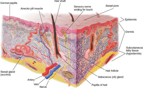
In the epidermis what kind of tissue is there?
stratified squamous epithelial cells
What level of the skin houses the glands.
dermis
What are the 3 levels of the skin? Superficial to deep
epidermis, dermis and subcutaneous
what are the nails composed of?
keratin
Common signs and symptoms of integumentary diseases include
Skin lesion (Lee-zhun). A lesion is a very broad term meaning any discontinuity or abnormality of tissue. Lesions can be hard, soft, flat, raised, large, small, reddened, crusted, fluid-filled, or pus-filled, to name only a few characteristics (Figure 18–2).
Pain.
Pruritus (proo-RYE-tus) or itching.
Edema (swelling).
Erythema (ER-ih-THEE-mah) or skin redness.

A-papule
B-plaque
C-macule
D-patch
E-scale
F-crust
G-wheal
H-cyst
I-pustule
J-vesicle
K-bulla
L-ulcer
M-fissure
Biopsy might be used in diagnosing
nodules and chronic lesions.
Dermascopy is used to
better visualize lesions.
Culture and sensitivity are effective in
determining the presence of bacterial infections
Blood tests are helpful, especially if there is concern about a
systemic infection or metabolic disorder or metabolic disorder.
Diagnosis and identification of fungal and parasitic infections can be determined by
using cultures and microscopic smear examinations.
Patch tests are often done to diagnose allergies by
applying allergens to the skin and observing for reactions.
Common diseases include
infections; metabolic, hypersensitivity, and idiopathic disorders; and tumors and can be categorized according to cause.
What are characteristics of Viral skin diseases
Viral skin diseases can be acute or chronic. Acute viral diseases commonly affect children and usually resolve spontaneously, but many viral infections become lifelong with periods of remission and exacerbation (flaring up).
What are the 4 types of herepes?
Herpes simplex type 1
Herpes genitalis (herpes simplex type 2)
Herpes varicella
Herpes zoster
What is Herpes simplex type 1? Signs, causes?
Commonly called fever blisters and cold sores because febrile conditions and the common cold often bring about an exacerbation. The vesicles commonly appear around the lips and nose (Figure 18–3). Lesions appearing around the lips can be further identified as herpes labialis (labia = lip), and those occurring in conjunction with a fever can be further identified as herpes febrilis.

Herpes genitalis (herpes simplex type 2)
Causes?
Commonly called genital herpes. This is a highly contagious disease and is spread by direct contact. Genital herpes can be a sexually transmitted disease, but transmission is not limited to sexual contact. Autoinoculation with the hands is also possible by touching the lips and then the genitals and vice versa.

Herpes varicella? Common name?
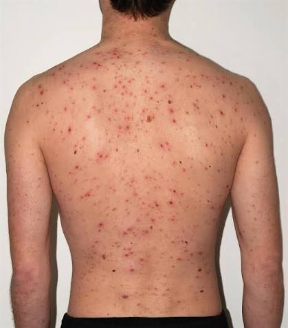
Commonly called chicken pox. This is an acute, highly contagious childhood disease
Herpes zoster? Common name? Symptoms and signs? Vaccine?
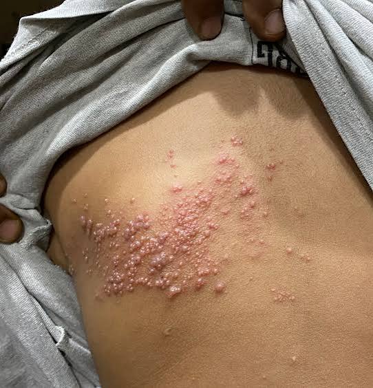
Commonly called shingles. The virus that causes chicken pox in children causes zoster in adults. It is characterized by painful lesions that follow the course of a spinal nerve. Zostavax® is the vaccine for shingles, but it is recommended only for adults over age 60
What is the symptom of herpes?
Herpes is characterized by inflammation of the skin and clusters of fluid-filled vesicles. The infection is painful, embarrassing, and often recurrent.
What is the diagnosis process for herpes?
Diagnosis is made by observation of vesicles, positive viral culture, and blood testing for herpes antibodies.
What is the treatment process for Herpes?
The virus is treatable but remains in the affected individual’s body for life. Some type of balance between the host and the virus exists, with periods of viral remission and exacerbation. The virus exacerbates, or flares up, often during times of decreased immunity as occurs with stress. Valacyclovir, acyclovir, and famciclovir are approved to treat herpes genitalis but are also used for oral herpes. Penciclovir cream can also be prescribed for oral herpes.
Common Drugs for Integumentary Disorders
Antibiotics-Drugs used to treat skin infections
Antifungals-Drugs used to treat fungus infections
Antihistamines-Drugs used to reduce the symptoms from allergies and contact dermatitis
Anti-inflammatories-Drugs used to reduce inflammation
Antivirals-Drugs used for herpes and shingles
Enzyme inhibitors-Drugs used for atopic dermatitis
Immunosuppressants-Drugs used to treat severe cases of psoriasis and eczema
Retinoids-Drugs used to treat skin disorders like acne or psoriasis
Biologies-Drugs used to treat psoriasis
Anesthetics-Drugs used to decrease pain in/on the skin
Parasiticides-Drugs used to treat mites or lice
Common warts, who gets them? Spread? Symptoms?
Predominantly appear on the hands and fingers of children (Figure 18–4). These lesions are contagious and are spread by scratching and direct contact. Although unsightly, they are usually painless and harmless and often disappear spontaneously. Common warts occurring in adults should be called to the attention of a physician to ensure that they are not skin cancers.

Plantar warts? Where? Contain what?
Appear on the sole of the foot. This wart usually grows inward, is smooth on the sole of the foot, and feels like a hard lump. Plantar warts contain small, clotted blood vessels that appear like dark splinters inside the wart and give it a cauliflower-like appearance (Figure 18–5). This wart commonly causes pain with walking; thus, surgical removal is often the treatment of choice.

Genital warts?caused?
A sexually transmitted disease. They are highly
contagious and often need to be removed surgically.

What is the etiology of verruca?
. A verruca is caused by the papillomavirus affecting the keratin cells of the skin, causing cellular hypertrophy.

Verruca symptoms?
The only symptom is a painless, often rough-surfaced skin lesion appearing on any surface of the body, but primarily on the fingers and hands.
What is the treatment for verruca?
reatment depends on the location of the verruca. Warts on the fingers and hands are commonly treated topically with over-the-counter medications. The best topical medications are those containing salicylic acid such as Trans-Ver-Sal, Sal-Acid Plaster, or Sal-Plant Gel. Genital and plantar warts are commonly removed surgically. Verrucae are often resistant to treatment, and recurrence is frequent.
Prevention for verruca?
The best prevention is to avoid skin-to-skin contact with those infected with verruca.
Impetigo description and etiology?
Description. Impetigo is a highly contagious skin disease. It is one of the most common skin infections of children and usually affects the face and hands.
Etiology. Impetigo is caused by Streptococcus and Staphylococcus bacteria.

Impetigo symptoms and diagnosis?
Symptoms. Impetigo is characterized by the appearance of vesicles and pustules (PUS-tyouls; small pus-filled lesions) that rupture, producing a yellow crust over the lesions (Figure 18–6). Impetigo occurs more readily in those with poor hygiene, anemia, and malnutrition.
Diagnosis. Diagnosis is confirmed by symptoms and a positive bacterial culture of the infected lesion.

Impetigo treatment and prevention?
Treatment. Treatment includes washing and drying the affected area several times a day and applying antibiotic ointment. More serious conditions might also require oral antibiotics.
Prevention. Prevention is aimed at good personal hygiene, including frequent handwashing. Those with anemia and malnutrition need treatment to cure those conditions also.

Folliculitis description and etiology?
Description. Folliculitis is inflammation and infection of the hair follicle and can occur anywhere on the skin.
Etiology. Folliculitis usually starts when hair follicles are damaged by shaving or friction from clothing. The damaged hair follicle then becomes infected with Staphylococcus bacteria.
Folliculitis symptoms and diagnosis?
Symptoms. Common symptoms include rash, itching, and the formation of pimples or small pustules surrounding the hair (Figure 18–7) that can also open, drain, and crust over. This condition commonly occurs in young men and affects the neck, groin, thighs, buttocks, beard, and scalp.
Diagnosis. Diagnosis is based on physical examination of pustules and condition of the skin. Cultures can reveal bacterial or fungal infection.

Folliculitis treatment and prevention?
Treatment. Warm, moist compresses help ease the pain and promote drainage of the pustules. Daily cleansing of the area with an antiseptic cleanser and application of antibiotic or antifungal creams for several weeks usually cure the condition. Severe or chronic cases might need additional treatment with oral antibiotics.
Prevention. Preventive measures include reducing friction from clothing, keeping skin clean and dry, avoiding bathing with dirty or contaminated washcloths, and avoiding shaving the area until the infection is healed.
Abscess, Furuncle, Carbuncle description and etiology?
Description. There are some differences in these lesions. An abscess is a localized collection of pus occurring in any tissue of the body, including the skin. Abscesses commonly occur around sites of trauma, embedded foreign material such as splinters, and hair follicles (Figure 18–8).
A small abscess occurring in the tissues of the skin is a furuncle, commonly called a boil. Furuncles generally occur around a hair follicle and can develop during an acute case of folliculitis. Boils can develop in any hairy area of the body, with common sites including the skin of the neck, back, and buttocks.
Carbuncles are larger abscesses and involve several interconnected furuncles. These lesions arise in a cluster of hair follicles and have multiple drainage sites. Needless to say, carbuncles are much larger than furuncles and are less common.
Etiology. These lesions are commonly caused by the pyogenic, normal flora bacteria, Staphylococcus. Predisposing factors for these lesions include a lowered immunity due to the presence of other diseases and poor personal hygiene.

Abscess, Furuncle, Carbuncle symptoms and diagnosis?
Symptoms. Abscess, furuncle, and carbuncle are all characterized by inflammation, infection, and the formation of a capsule to wall off and prevent the spread of infection. All of these encapsulated lesions are extremely painful, usually develop a soft spot or come to a head, and need to be opened or surgically drained.
Diagnosis. Diagnosis is based on history and physical examination of the lesion.

Abscess, Furuncle, Carbuncle treatment and prevention?
Treatment. Warm, moist compresses usually relieve pain and promote spontaneous drainage. If spontaneous opening and drainage do not occur, surgical opening and drainage might be necessary. Antibacterial or antifungal medications are usually prescribed to treat infection.
Prevention. Nothing can prevent these lesions, although using antibacterial soaps can help reduce bacterial count on the skin and thus aid in prevention.

Cellulitis description and etiology?
Description. Cellulitis is a diffuse, or spreading, inflammation of the skin and subcutaneous tissue (Figure 18–9). It commonly appears on the lower legs but can affect any part of the body.
Etiology. Cellulitis is a bacterial infection commonly caused by Streptococcus and Staphylococcus. These bacteria are common bacteria of the skin (normal flora). Cellulitis often appears in open areas of the skin and can be the extension of a wound, ulcer , insect bite, blister, burn, or other skin infection.
Cellulitis symptoms and diagnosis?
Symptoms. Cellulitis is characterized by pain, redness, swelling, warmth, and tenderness of the involved skin. Other symptoms might include headaches, fever, or chills. In advancing cases, red streaks can develop and travel from the affected area.
Diagnosis. Medical history and physical examination of the involved area are helpful in diagnosis. If the leg is involved, an ultrasound can be performed to rule out deep vein thrombosis.

Cellulitis treatment and prevention?
Treatment. Cellulitis is generally treated successfully with oral antibiotics. Analgesics for pain and resting the affected limb or affected area also can be part of the treatment plan. In extreme cases, intravenous antibiotics might be needed. Any cellulitis involving the face can be dangerous because this has the potential of spreading into the sinuses of the skull. If pain becomes severe, necrotizing fasciitis might have developed, which will require emergency surgical treatment.
Prevention. Good handwashing, proper cleansing, and care of open areas of the skin lower the risk of cellulitis. Deep, dirty, and open wounds need prompt medical treatment to prevent cellulitis.

Erysipelas description and etiology?
Description. Erysipelas is an acute infection of the dermis that extends into underlying fat tissue. It can affect the face, especially in children and older adults, but also affects the arms and legs (Figure 18–10).
Etiology. Most cases are due to Streptococcus, specifically group A Streptococcus. These bacteria can come from the skin or from the affected individual’s throat or nasal passages and can enter the skin through any open area such as surgical incisions, ulcers, and minor trauma.

Erysipelas symptoms and diagnosis?
Symptoms. Symptoms include fatigue, chills, fever, headaches, and vomiting. The infected skin develops a red, warm, hard, and painful rash showing a consistency similar to an orange peel. Swelling develops rapidly and exhibits sharply demarcated, raised edges.
Diagnosis. A physical examination of the classic orange-peel rash and affected skin assists in diagnosis. Tests can determine that this skin condition is not herpes zoster or contact dermatitis.

Erysipelas treatment and prevention?
Treatment. Antibiotics given orally or intravenously usually resolve the condition, but it often takes weeks for the skin to return to normal. In some cases, bacteria can infect the blood, leading to endocarditis and osteomyelitis.
Prevention. Prevention includes maintaining healthy skin, avoiding injuries to the skin, and promptly and completely treating streptococcal infections, including strep throat.

Lyme disease description and etiology?
Description. the most common tick-borne disease in the United States.
Etiology. Lyme disease is caused by the Borrelia burgdorferi bacterium and is transmitted to humans by the bite of an infected deer or blacklegged tick.
Lyme Disease symptoms and diagnosis?
Symptoms. The bacteria can affect any organ, causing a variety of symptoms and possibly delaying diagnosis. Symptoms can include flu-like symptoms, arthritis, malaise, chills, and fever. A characteristic bull’s-eye skin rash is a common sign (Figure 18–11). The bull’s eye is a reddened circle with a lighter center and can appear days to weeks after the infected bite.
Diagnosis. Lyme disease is diagnosed by a history confirming possible exposure to infected ticks and a physical examination revealing positive symptoms. Positive blood testing for antibodies confirms the diagnosis.

Lyme Disease treatment and prevention?
Treatment. Most cases can be treated successfully with a few weeks of antibiotics. If left untreated, the disease can cause arthritis and various neurologic and cardiovascular complications.
Prevention. Prevention of Lyme disease is aimed at preventing tick bites by using insect repellent; wearing long-sleeved shirts, long pants, and socks; and tucking the pants into the socks and boots when hiking or camping in grassy or wooded areas. Showering and inspecting the skin immediately after outside activities can also help prevent bites.
Methicillin-Resistant Staphylococcus Aureus (MRSA)
description
Description. Methicillin-resistant Staphylococcus aureus (MRSA) is a strain of bacteria that is resistant to the antibiotics commonly used to treat staphylococcal infections (Figure 18–12). This infection usually affects the elderly and those with other disease conditions and occurs in hospitals, nursing homes, and other health care settings and is known as health care–associated MRSA. More recently, MRSA is appearing in healthy people who might share personal items, such as athletes and students. These individuals are often in a community of people. In this case, MRSA is responsible for skin, tissue, and lung infections and is called community-associated MRSA.
Methicillin-Resistant Staphylococcus Aureus (MRSA) etiology?
. S. aureus is commonly found on the skin of individuals, is usually harmless, and does not cause illness. This presence of bacteria without illness is called being colonized. Individuals who are colonized with MRSA can easily pass these bacteria to others. MRSA represents a group of bacteria that have developed a resistance to antibiotics, which is a natural survival method of bacteria. However, humans have helped build this resistance by excessive and unnecessary use of antibiotics.Staphylococcaceae is one of the families of bacteria that have built such a resistance that currently only a few drugs are effective to kill them.
Methicillin-Resistant Staphylococcus Aureus (MRSA) symptoms?
Symptoms. Symptoms of MRSA, like other staph bacteria, often start with small red bumps that resemble pimples or boils. These can quickly cause deep abscesses or become blood-borne. As a blood-borne infection, staph bacteria can invade all organs of the body, including bones, heart, and lungs. Symptoms of sepsis include a rash over most of the body along with fever, chills, headaches, joint pain, and shortness of breath. Sepsis with MRSA can be life-threatening, and infection requires immediate medical attention.

Methicillin-Resistant Staphylococcus Aureus (MRSA) diagnosis and treament?
Diagnosis. Diagnosis is determined by culture and drug sensitivity testing of wound and nasal secretions for MRSA.
Treatment. Some cases of MRSA do not need treatment with antibiotics. Cleaning the area and washing with antibacterial soap may be effective. Oral antibiotics may be effective for mild to moderate cases of MRSA. Severe cases often need treatment with an expensive medication, vancomycin, or a combination of medications which must be given intravenously.
Even though these medications are currently effective, there are signs that some MRSA bacteria are building resistance to several medications, including clindamycin and vancomycin.
Methicillin-Resistant Staphylococcus Aureus (MRSA) prevention?
Avoiding those with active infection is helpful along with maintaining a healthy lifestyle to keep natural immunity levels high. Other activities include frequent handwashing and carrying hand sanitizer for use when handwashing is not possible. Do not share personal items such as towels, clothing, combs, and eating utensils. Keep any open wounds covered and protected. Do not share or overuse antibiotics.
Prevention in health care facilities requires complete sanitation of all surface areas, fabrics, linens, and equipment in patient areas.
What are the different types of Tinea……(6)
Tinea corporis — Ringworm on smooth skin (arms, legs, torso). Causes red, ring‑shaped patches. Often spread from pets, especially cats. Common in children.
Tinea pedis — Athlete’s foot. The most common type. Spreads through contaminated floors, showers, towels. Causes itching, burning, peeling, and painful cracks between toes.
Tinea cruris — Jock itch. Affects the groin in adult men. Worsens with heat, sweat, tight clothing, and physical activity.
Tinea unguium — Nail fungus. Creates white patches, thickening, brittleness. Hard to treat because the fungus hides under the nail.
Tinea capitis — Scalp infection causing hair loss. Mostly in children.
Tinea barbae — Beard-area infection (“barber’s itch”). Shaving helps control it.
Tinea (Ringworm)
etiology
Symptoms
diagnosis
treatment
Prevention
Etiology
Caused by various fungi that infect the outer layers of skin, hair, or nails.
Symptoms

Itching
Cracking
Peeling or weeping skin
Ring-shaped rashes (depending on type)
Diagnosis
Based on appearance
Confirmed by microscopic exam of skin scrapings showing fungal elements
Treatment
Keep the area clean and dry
Use antifungal creams, liquids, or powders consistently over time
Severe or persistent cases may require oral antifungals such as ketoconazole or fluconazole
Recurrence is common, making long-term care important
Prevention
Keep skin healthy, clean, and dry
Avoid tight clothing
Avoid high-risk areas like community showers, locker rooms, and hot tubs
Wear sandals, cotton socks, and allow shoes to fully dry between uses
Tinea corporis?
Ringworm on smooth skin (arms, legs, torso). Causes red, ring‑shaped patches. Often spread from pets, especially cats. Common in children.

Tinea pedis?
Athlete’s foot. The most common type. Spreads through contaminated floors, showers, towels. Causes itching, burning, peeling, and painful cracks between toes.

Tinea cruris?
Jock itch. Affects the groin in adult men. Worsens with heat, sweat, tight clothing, and physical activity.
Tinea unguium?
Nail fungus. Creates white patches, thickening, brittleness. Hard to treat because the fungus hides under the nail.

Tinea capitis? Tinea barbae?
Tinea capitis — Scalp infection causing hair loss. Mostly in children.
Tinea barbae — Beard-area infection (“barber’s itch”). Shaving helps control it.

Candidiasis
etiology
symptoms
mouth
diaper
nails
finger/toes
vagina
diagnosis
treatment
prevention
The core idea: Candidiasis is an overgrowth of Candida yeast, normally harmless, that becomes infectious when body conditions change. It can be mild and superficial or severe and systemic.
What candidiasis is
A fungal (yeast) infection also called thrush.
Can affect skin, mouth, nails, vagina, or become systemic and dangerous.
Caused by Candida, a fungus that normally lives on the body without causing harm.
Etiology (Cause)
Candida becomes infectious when it overgrows.
Antibiotics are a major trigger because they kill helpful bacteria that keep yeast in balance.
More common in:
People with diabetes mellitus
Those on immunosuppressive medications
Individuals with long-term water exposure (dishwashers, bartenders, waitstaff)
Symptoms (depend on location)
Mouth (Thrush) — white patches on cheeks and tongue, common in infants.
Diaper area — red, inflamed, sometimes scaly rash.
Nails (Candidal onychomycosis) — nail infection; around the nail is paronychia.
Between fingers/toes — itchy skin, blisters, pustules.
Vagina — causes vaginitis (covered in another chapter).
Diagnosis
Microscopic exam of yeast
Positive culture
Blood tests if infection becomes systemic
Treatment
(General information only — for symptoms, a healthcare professional should evaluate.)
Most cases respond to topical antifungal creams, vaginal antifungals, or oral medications.
Severe or systemic infections may require long-term IV antifungal therapy.
Yeast infections can be persistent and sometimes become chronic.
Prevention
Keep skin clean, dry, and intact.
Avoid unnecessary antibiotics.
Reduce prolonged moisture exposure


Pediculosis
Description
Head
Body
Pubic
etitlogy
symptoms
diagonoes
treatment
prevention
The core idea: Pediculosis is an infestation of lice—tiny insects that live on the human body and feed on blood. Different types affect different areas and spread easily through close contact or shared items.
Types of Pediculosis
Head lice — Common in school‑aged children and families. Spread through close contact or shared hair items.
Body lice — Seen in individuals with poor hygiene or limited access to clean clothing. Can spread disease (historically typhus).
Pubic lice — Also called crabs. Spread through sexual contact. Cause intense genital itching and can spread to eyelashes or eyebrows.
Etiology (Cause)
Lice spread through direct contact with an infested person.
Also spread by sharing combs, brushes, towels, clothing, bedding, or furniture.
They affect all socioeconomic groups—anyone can get them.
Symptoms
Visible lice on the scalp, body folds, or pubic area depending on type.
Nits (eggs) attached to hair shafts in head lice.
Severe itching from lice feeding on blood.
Crawling insects may be seen on skin or clothing.
Diagnosis
Made by direct observation of lice or nits on the body, hair, or clothing.
Treatment
(General information only — for symptoms, a healthcare professional should evaluate.)
Medicated shampoos or treatments for hair and body.
Petroleum jelly may be used on eyelashes for pubic lice.
Wash or dry‑clean clothing and bedding in hot water for at least 20 minutes.
Clean and treat furniture to remove any remaining lice.
All lice and eggs on body, clothing, bedding, and furniture must be eliminated to fully eradicate infestation.
Prevention
Avoid contact with infested individuals.
Do not share hair tools, clothing, bedding, or towels.
Maintain good hygiene and regular cleaning of personal items.

Scabies?
Description
Cause
symptoms
diagnosis
treatment
prevention
What scabies is
A mite infestation, not an insect.
The female mite burrows into the skin, lays eggs, and creates tunnels.
Eggs hatch in 3–5 days, and the life cycle continues on the skin surface.
Etiology (Cause)
Caused by the eight‑legged mite Sarcoptes scabiei.
Mites survive 48–72 hours off the human body.
Spread through prolonged skin‑to‑skin contact, especially:
Hugging
Sharing beds
Parent‑child contact
Sexual contact (most common in sexually active young people)
Not spread by:
Brief touch or handshakes
School settings, sports, or community showers
Animals (pet mites cause temporary itching but do not infect humans)
Symptoms
Intense itching, especially at night
Burrows: thin, slightly raised, grayish‑white lines
Vesicles and pustules from hypersensitivity to the mite, its feces, and eggs
Common sites:
Webs between fingers and toes
Wrists
Armpits
Under breasts
Belt line
Groin and genital areas
Scabies mites are not visible to the naked eye, but burrows and reactions are.
Diagnosis
Made by microscopic examination of skin scrapings.
Female mites appear as a tiny black dot at the end of a burrow.
Treatment
(General information only — for symptoms, a healthcare professional should evaluate.)
Apply lindane cream (Kwell®) or similar scabicidal medication to the entire body, left on for 8–14 hours.
All infected individuals in the household must be treated to prevent reinfestation.
Itching may continue for 3–4 weeks even after successful treatment.
Environmental cleaning:
Wash clothing, bedding, towels in hot water or dry clean.
Treat furniture, carpets, rugs.
Items that can’t be washed (stuffed animals, hats, shoes, pillows) should be:
Frozen overnight, or
Sealed in bags for 1–2 weeks (mites die after a week without food)

Acne Vulgaris?
Despictipons
Cause
symptoms
diagnosis
treatment
prevention
The core idea: Acne vulgaris is inflammation of the sebaceous (oil) glands and hair follicles, leading to clogged pores, blackheads, whiteheads, and pimples. It’s the most common form of acne, especially during puberty.
What acne vulgaris is
An inflammatory condition of sebaceous glands and hair follicles.
Characterized by comedones:
Blackheads — open clogged pores oxidized at the surface
Whiteheads — closed clogged pores
Most common in teens due to hormonal changes.
Etiology (Cause)
Exact cause unknown, but strongly linked to puberty and increased androgen hormones, which enlarge and activate oil glands.
Contributing factors:
Heredity
Food allergies
Endocrine disorders
Not caused by: poor hygiene, lack of sleep, sexual activity, chocolate, soda, or fried foods.
Not contagious.
Symptoms
Excess sebum clogs pores → comedones.
Blackheads form when sebum oxidizes.
Whiteheads/pimples form when bacteria infect trapped sebum.
Can range from mild to severe.
Helpful management habits:
Frequent cleansing with antibacterial soap
Avoiding heavy makeup
Using over‑the‑counter acne creams/gels
Avoiding tight clothing that traps heat
Showering after exercise
Avoiding squeezing comedones (prevents deeper infection)
Gentle extraction of comedones; draining of cysts/pustules when needed
Diagnosis
Based on history and physical exam of sebaceous lesions.
Treatment
(General information only — for symptoms, a healthcare professional should evaluate.)
Mild acne: cleansing + OTC treatments.
Severe acne: dermatologist‑guided regimen including:
Prescription cleansers
Oral antibiotics (e.g., tetracycline)
Steroids
Retinoic acid/Retin‑A®
Severe cases may leave permanent scarring.
Symptoms usually improve after puberty, with or without treatment.
Prevention
Consistent skin‑care habits used for treatment also help prevent acne.

Seborrheic Dermatitis?
description
cause
symptoms
diagonois
treatment
prevention
What seborrheic dermatitis is
A common dermatitis involving sebaceous glands.
Not harmful, not contagious.
In infants → cradle cap (usually clears by 12 months).
In adults → dandruff.
Etiology (Cause)
Exact cause unknown.
Likely influenced by heredity and stress.
More common in individuals who:
Are obese
Live in weather extremes
Have acne or other skin disorders
Have Parkinson’s disease, stroke, head injury
Have impaired immunity (e.g., HIV)
Are recovering from major medical stress (e.g., myocardial infarction)
Are confined long‑term (e.g., nursing homes)
Characterized by increased sebum production, causing inflammation in areas with many oil glands.
There is no cure, only management.
Symptoms
Affects:
Scalp
Eyebrows
Eyelashes
Behind the ears
Sides of the nose
Mid‑chest
Skin appears:
Reddened
Covered with greasy, yellowish scales
Eyebrows/eyelashes → dry, dirty‑white scales
Nose area → red and itchy
Chest → red, greasy lesions
Itching may or may not be present.
Diagnosis
Based on physical exam and the location + appearance of lesions.
Treatment
(General information only — for symptoms, a healthcare professional should evaluate.)
Scalp:
Start with over‑the‑counter medicated shampoos.
If ineffective → prescription-strength shampoos.
Severe cases → steroid lotions or creams.
Non‑scalp areas:
Keep skin clean and dry.
Use antifungal or anti‑itch medications.
Prescription therapy if widespread or unmanageable.
Prevention
Reduce severity by controlling risk factors (stress, other skin conditions, environmental triggers).
Treat flare‑ups promptly to prevent worsening.

Sebaceous Cyst?
description
cause
symptoms
diagnosis
treatment
prevention
What a sebaceous cyst is
A closed sac of trapped sebum (oil).
Common locations: scalp, neck, groin.
A special type, pilonidal cyst, forms around a hair near the tailbone (sacrococcygeal area).
Etiology (Cause)
Occurs when a sebaceous gland becomes blocked, trapping sebum under the skin.
Often associated with acne or swollen hair follicles.
Appears as a slow‑growing, painless lump.
Symptoms
Main symptom: visible lump under the skin.
If infected:
Skin becomes red, warm, tender
Pain may increase
Possible drainage
Diagnosis
Made by physical examination of the cyst.
Treatment
(General information only — for symptoms, a healthcare professional should evaluate.)
No treatment needed unless infected.
Warm, moist compresses help relieve pain and promote drainage.
Incision and drainage may be done, but cysts often recur.
Surgical removal is the only permanent solution.
Prevention
No guaranteed prevention.
Keeping skin clean and healthy may reduce risk.

What are hypersensitivity disease?
Hypersensitivity diseases are those caused by an immune reaction within the body. Frequently, the cause is unknown, and treatment is symptomatic.
Eczema?
description
cause
symptpons
diagnosis
treatment
prevention
What eczema is
A type of dermatitis (skin inflammation).
Not dangerous, not contagious, and often not curable, but manageable.
Called atopic dermatitis because it occurs in people with atopic tendencies—a genetic predisposition to allergies. Etiology (Cause)
Strongly linked to genetics and allergies.
Common in infants, often triggered by foods like milk or orange juice.
Can be caused or worsened by:
Heredity
Other diseases
Environmental irritants
Allergies
Removing the offending food often improves infant eczema.
Symptoms
In adults, eczema causes:
Dry, leathery skin
Itching
Redness
Vesicles (small blisters)
Pustules
Scales
Crusting
Symptoms may appear alone or in combination.
Flares triggered by stress, humidity, and temperature changes.
Diagnosis
Based on clinical exam and patient history.
Treatment
(General information only — for symptoms, a healthcare professional should evaluate.)
Goal: reduce frequency and severity of flare‑ups.
Common treatments:
Topical cortisone creams
Antihistamines
Sedatives for severe itching
Avoid sunlight if eczema is light‑sensitive.
Prevention
Eczema itself cannot be prevented.
Symptoms can be reduced by avoiding irritants and managing triggers.

Urticaria?
description
cuase
symptoms
diagnosis
treatment
prevention
Urticaria
Description. Commonly called hives or nettle rash, this is a vascular reaction of the skin.
Etiology. Urticaria is caused by contact with an external irritant such as insect bites, pollen, or plants. Urticaria also can be caused by internal irritants such as food, drugs, and contrast dye.
Symptoms. Urticaria is characterized by slightly elevated lesions that are redder or paler than the surrounding skin and are associated with severe itching. The elevated areas are called wheals (WEELs) or hives (Figure 18–22). Scratching or rubbing the hypersensitive area can lead to formation of larger or additional wheals.
Diagnosis. Diagnosis is made on the basis of physical examination of the characteristic wheal.
Treatment. Treatment includes antihistamines and avoidance of the allergen. (See Chapter 5, “Immune System Diseases and Disorders,” for more information.)
Prevention. Avoiding exposure to the allergen and avoiding hot baths, showers, or exposure to the sun after a recent episode are preventive measures. Exposure to heat can cause the hives to

Contact Dermatitis
description
cause
symptoms
diagnosis
treatment
prevention
What contact dermatitis is
A skin inflammation caused by touching an allergen or irritant.
Can be acute (short-term) or chronic (long-term).
Not contagious, but often uncomfortable.
Etiology (Cause)
Triggered by contact with substances such as:
Cosmetics
Laundry products
Plants (e.g., poison ivy)
Jewelry
Paints
Drugs
Plastics
Sometimes the exact allergen is hard to identify or impossible to avoid completely.
Symptoms
Can range from:
Small, red, localized rashes
To large vesicular (blistering) lesions covering the body
A classic example is poison ivy.
Rash location often hints at the cause (e.g., under jewelry, under clothing, or on sun‑exposed skin).
Diagnosis
Can be challenging.
Helpful methods:
Location clues (rash under a watch, necklace, etc.)
Use test — applying a suspected substance to a different skin area to check for reaction
Patch test — placing patches with common allergens on the skin to observe reactions
Treatment
(General information only — for symptoms, a healthcare professional should evaluate.)
Topical anti‑itch medications (camphor, menthol)
Antihistamines like diphenhydramine (Benadryl®) — may cause drowsiness
Cool baths for comfort
Most important: stop contact with the allergen — treatment won’t work if exposure continues
Prevention
Avoid the allergen whenever possible
If exposed, wash immediately with soap and water
Use barrier creams
Wear protective clothing when dealing with known irritants

What is scleroderma? Cause? Signs and symptoms?
Scleroderma
Scleroderma (SKLEHR-oh-DER-mah; sclero = hardening, derma = skin) is a chronic autoimmune disorder characterized by hardening, thickening, and shrinking of the connective tissues of the body, including the skin (Figure 18–24). It is thought that this autoimmune reaction begins with the skin and connective tissues, attracting lymph cells that stimulate the production of collagen, leading to the disorder. More information can be found in Chapter 5, “Immune System Diseases and Disorders.”

What are Idiopathic diseases?
Idiopathic diseases of the skin have no known cause, but often tend to be familial.
Psoriasis
Cause
symptoms
diagnosis
treatment
prevention
Cause: The cause is unknown, but some hereditary basis does exist. Stress, infection, skin trauma, and sunlight tend to cause an exacerbation of the condition.
Symptoms. Psoriasis is characterized by red, raised lesions with distinct borders and silvery scales (Figure 18–25). These lesions generally occur on the elbows, knees, and scalp.
Diagnosis. Diagnosis is usually made by physical examination of the skin condition. A skin biopsy might be performed to determine the exact type of psoriasis.
Treatment. Treatment includes medications to control itching, creams containing coal tar, creams to remove the scaling (salicylic acid), ultraviolet (UV) light treatments, steroids, and prescription medications for vitamin D or vitamin A. Several prescription medications specifically for psoriasis are also available. Oatmeal baths can also be helpful to loosen the scales.
Prevention. There is no way to prevent psoriasis, but certain activities can reduce flare-up of symptoms. These activities include keeping the skin moist and avoiding cold climates, skin scratches, stress, infection, and smoking.

Rosacea
description
cause
symptoms
diagnosis
treatment
prevention
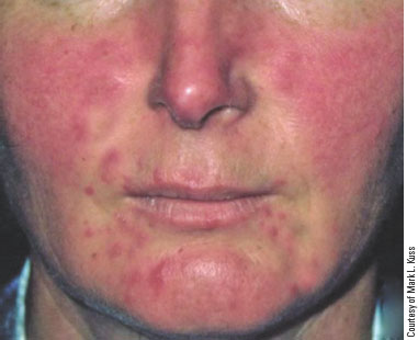
Description. Rosacea is a chronic skin condition characterized by inflammation and redness of the forehead, nose, cheeks, and chin but is not dangerous or life-threatening (Figure 18–26). The individual’s facial skin appearance can lead to psychological damage related to loss of self-esteem.
Etiology. The cause of rosacea is unknown, but individuals affected blush easily and tend to be fair-skinned, female, and between the ages of 30 and 50. Rosacea involves enlargement of the blood vessels just under the skin and might be associated with other skin conditions such as seborrhea and acne vulgaris.
Symptoms. The facial skin appears red with swelling or skin eruptions similar to acne. Other symptoms include:
A red, bulbous nose.
Spider-like blood vessels called telangiectasia of the face.
A burning or stinging sensation of the face.
Bloodshot, irritated, watery eyes.
Diagnosis. Diagnosis is commonly made by physical examination of the skin condition.
Treatment. There is no known cure. Symptoms might be controlled by identifying triggers that cause the condition to become worse. Avoiding sun exposure, prolonged exertion in hot weather, stress, spicy foods, alcohol, and hot beverages might reduce symptoms.
Antibiotic ointments applied to the face might control skin eruptions, and laser surgery might reduce the redness. Surgical reduction of the enlargement of the nose might be preferred to improve appearance.
Prevention. There is no known prevention.
Keloid
description
cause
symptoms
diagnosis
treatment
prevention
Description. A keloid (KEE-loid) is a raised, firm, irregularly shaped mass of scar tissue that develops following trauma or surgical incision. (See Chapter 4, “Inflammation and Infection,” for more information.)
Etiology. Keloids are an overgrowth of collagen during connective tissue repair; they are more common in Blacks (Figure 18–28).
Symptoms. Keloids can be unsightly but are generally considered harmless.
Diagnosis. Diagnosis is easy and consists of a physical examination of the keloid.
Treatment. Surgical removal of keloids is usually not effective because it often results in the formation of another keloid. Radiation, injecting the lesion with steroids, and cryotherapy might be helpful in reducing the size of a keloid.
Prevention. There are no known preventive measures.

ACTINIC KERATOSIS
description
cause
symptoms
diagnosis
treatment
prevention
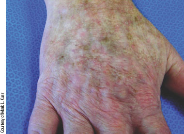
Actinic keratosis is a rough, scaly skin patch caused by long-term sun (UV) exposure, appearing on areas like the face, ears, scalp, and hands; it feels like sandpaper, may be pink or red, and can itch or be tender. It is diagnosed by a doctor examining the spot and sometimes doing a biopsy. Treatment includes freezing, medicated creams, photodynamic therapy, or minor removal procedures. Prevention focuses on daily sunscreen, protective clothing, avoiding tanning beds, and limiting sun exposure.
Basal Cell Carcinoma
description
cause
symptoms
diagnosis
treatment
prevention
Description. Basal cell carcinoma is the most common type of skin cancer. It is most common in fair-skinned, blonde, and blue- or gray-eyed individuals. Basal cell carcinoma is a slow-growing, locally invading tumor that does not metastasize. This is not to say that if left untreated it is not dangerous. Tumors near the eyes and mouth can invade these spaces and cause much concern. Tumors on the nose, lip, and ear can lead to the loss of these tissues.
Etiology. As with most cancers, the cause of basal cell carcinoma is unclear but does seem to be the result of a mixture of genetic and environmental factors. The most common identified environmental factor is excessive exposure to the sun.
Symptoms. The appearance of this tumor varies, appearing as a raised nodule with a depressed or dented center; a smooth, shiny bump that is pink to pearly white in color; or a nonhealing lesion that bleeds easily (Figure 18–31).
Diagnosis. Diagnosis is confirmed by biopsy.
Treatment. Treatment of basal cell carcinoma is surgical removal.
Prevention. Basal cell carcinomas that are related to sun exposure can be prevented by avoiding the strong midday sun, using sunscreen year-round, and covering up with protective clothing if exposure is necessary.


Squamous Cell Carcinoma
description
cause
symptoms
diagnosis
treatment
prevention
Description. Squamous cell carcinoma is less common than basal cell carcinoma, but it tends to grow more rapidly and become metastatic.
Etiology. This tumor, like basal cell carcinoma, tends to occur on the sun-exposed skin of those with fair complexion. It most commonly appears on people over age 50. As a general rule, basal cell carcinoma occurs on the face above the lip line, and squamous cell carcinoma occurs below the lip line. This tumor is often preceded by another skin lesion such as actinic keratosis, chronic ulcers, sinus tracts, or scars.
Symptoms. Squamous cell carcinoma can appear as a firm, red nodule with crusts or a slightly elevated plaque (Figure 18–32). The nodule is usually located on the face, arm, neck, or hands but can appear in other areas. A sore that does not heal or bleeds easily might be a symptom of this cancer.
Diagnosis. Diagnosis is confirmed by skin biopsy.
Treatment. Squamous cell skin cancer has a high rate of cure if caught early. Treatment depends on location and size of the tumor and if there is metastasis. Wide surgical excision with radiation treatments and follow-up for at least five years for signs of recurrence is often the recommended treatment.
Prevention. Reducing sun exposure, examining the skin frequently for suspicious growths or changes in existing skin lesions, and seeking immediate treatment for these are preventive measures.

Malignant Melanoma
description
cause
symptoms
diagnosis
treatment
prevention
Description. Malignant melanoma (melan = black, oma = tumor) is the most serious type of skin cancer. It occurs more commonly in men and is responsible for the majority of skin cancer deaths.
Etiology. Malignant melanoma is due to an uncontrolled growth of pigment, or skin-coloring cells, called melanocytes. Growth of this tumor is caused by genetic and environmental factors, primarily sun exposure. Malignant melanoma rarely occurs before the age of 20 and can be related to a severe childhood sunburn.
Symptoms. This tumor is usually tan, brown, or dark brown in color (Figure 18–33). Often, it arises in a mole and causes a change in size and color of the mole. Malignant melanoma metastasizes quickly and is highly malignant. It spreads into the lymph nodes and can metastasize to all organs of the body.
Treatment. Treatment depends on the degree of spread and might include wide surgical excision, radiation, and chemotherapy. Prognosis depends on the degree of spread when discovered, but approximately 20% of those diagnosed with this tumor die from effects of metastasis.
Prevention. Preventive measures include reducing sun exposure, examining the skin frequently for suspicious growths or changes in existing skin lesions, and seeking immediate treatment.

Kaposi’s Sarcoma
description
cause
symptoms
diagnosis
treatment
prevention
Description. Prior to the discovery of AIDS, Kaposi’s sarcoma (KAP-oh-seez sar-KOH-mah) was relatively rare. The development of this neoplasm has increased in conjunction with the increase in the AIDS-affected population.
Etiology. The relationship between Kaposi’s sarcoma and AIDS is not fully understood. Usually, this tumor is not highly malignant except in the case of AIDS, in which it tends to be widespread and is often the cause of death in affected individuals.
Symptoms. This sarcoma is a malignant vascular skin tumor characterized by bluish-red cutaneous patches that grow under the skin, most often on the face and legs. About one-third of the time, they show up in the lining of the nose, mouth, and throat and can lead to pain and difficulty with eating and swallowing. The patches are usually composed of blood and cancer cells and often cause no symptoms. Tumors developing on the toes, feet, and legs often increase in number and size and spread upward. If the cancer spreads to the digestive tract or lungs, bleeding can result (Figure 18–34).
Diagnosis. Diagnosis is made on the basis of a physical examination of the skin revealing painless, flat, bluish-red lesions that do not itch or drain. A skin biopsy is definitive.
Treatment. The most important advancement in treating Kaposi’s sarcoma has been the development of drugs to control HIV infection and AIDS. Specific Kaposi’s treatments include liquid nitrogen, chemotherapy, and radiation. As with AIDS, there is no cure for Kaposi’s.
Prevention. Because most cases of Kaposi’s are related to HIV infection, taking preventive measures to avoid HIV infection will usually prevent Kaposi’s.

What is Albinism?
A hereditary disorder characterized by a decrease or total absence of pigment in the skin, hair, and eyes. Individuals affected with albinism have pale skin, white hair, and pale blue or pink eyes. These individuals suffer from extreme sunburn if adequate protection is not provided.

What is Vitiligo?
Characterized by destruction of melanocytes in small or large patches of skin (Figure 18–35). This condition can be due to an immune disorder.

What is melasma?
Characterized by dark patches of skin on the face, especially the cheeks (Figure 18–36) and common in pregnant females and those taking birth control pills. It is commonly called the mask of pregnancy. Melasma usually disappears after delivery or discontinuation of birth control pills.

First-Degree Burns
Description
First-degree burns are fairly common and are characterized by pain, skin redness, and swelling. First-degree burns involve only the epidermis and are often the result of sunburn (Figure 18–40). Healing generally occurs within a week, followed by peeling of the damaged epidermis.

Second-Degree Burns
Description
econd-degree burns are also called partial-thickness burns; they involve the epidermis and dermis and are characterized by extreme pain, redness, blisters, and open wounds (Figure 18–41). Second-degree burns usually heal in 2 to 3 weeks. If the burned area becomes infected, a second-degree burn can progress into a third-degree wound.

Third-Degree Burns
description
symptoms
treatment
Description. Third-degree burns are also called full-thickness burns; they involve the epidermis and entire dermis, exposing layers of fat, muscle, and bone. Tissue burned to the third degree is painless because the nerves in the dermis have been destroyed (Figure 18–42). This is not to say that individuals with third-degree burns do not have pain; there is extreme pain, but the pain is due to a layering of degrees of burn, with first- and second-degree areas surrounding the third-degree areas.
Symptoms. This burn is characterized by charred and broken tissue layers. The affected individual can exhibit signs and symptoms of shock.
Treatment. Third-degree burns often need tissue grafting to heal. Scarring and deformity are common with third-degree tissue damage.

Decubitus (Pressure) Ulcer
description
cause
prevention
Description. Decubitus (dee-KYOU-bih-tus) ulcer is a pressure injury commonly called a bedsore or pressure sore (Figure 18–44). The term decubitus actually means the act of lying down or the position of lying down.
Etiology. Decubitus ulcers commonly affect the bony areas of the body such as the heels, sacrum, elbows, and head of individuals who spend prolonged amounts of time in bed. Increased pressure in these areas slows blood flow, thus leading to tissue ischemia and necrosis.
Prevention. Pressure sores can be avoided by frequent turning and repositioning to decrease tissue pressure and allow blood flow to the tissues. Massaging the affected area also can improve circulation.

What is Elephantiasis?
Elephantiasis is characterized by hypertrophy of the skin and subcutaneous tissue, giving it an elephant-like appearance. Inflammation of the lymphatic system also leads to fluid accumulation in the legs, causing them to become enlarged. Elephantiasis is caused by a parasitic worm that enters the lymphatic system and causes obstruction of drainage and accumulation of fluids. This disease is most commonly seen in tropical areas, such as central Africa, and is spread by mosquitoes and bloodsucking flies.
