BILD 2
 Call Kai
 Learn
Learn Practice Test
 Spaced Repetition
 Match
Match Flashcards
 Knowt Play
1/179

There's no tags or description
Looks like no tags are added yet.
Name | Mastery | Learn | Test | Matching | Spaced | Call with Kai | Chat |
|---|
No analytics yet
Send a link to your students to track their progress
180 Terms
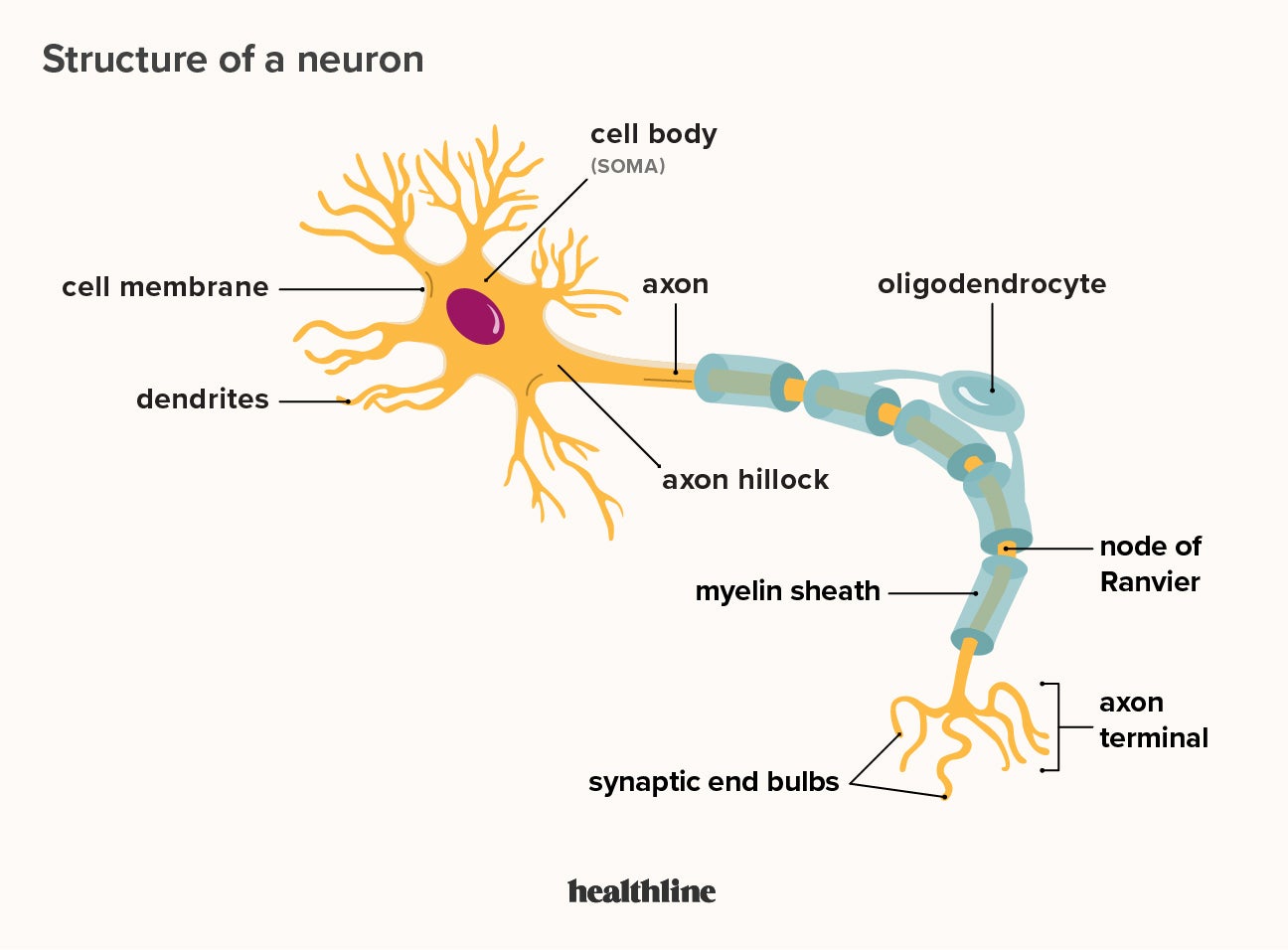
What is a neuron’s basic structure and function?
Dendrite: receive signal from other neurons
Soma: Cell body
Axon Hillock: signal generates/action potential starts
Axon: signal travels across
Myelin Sheath: Insulates axon, sends signal faster, oligodendrocytes (CNS) or Schwann cells
Node of ranvier: gaps between the myelin sheath, where the action potential will jump to
axon terminal: the signal exits, neurotransmitter are
What are other types of cells in the nervous system besides the neuron?
Glial cells: support neurons, induce formation of blood brain barrier, repair brain injuires, can specialize into oligodendrocytes
Ganglia: simple cluster of neurons wehre information integration takes place in the PNS
Where are differences btwn the CNS and PNS
CNS
brain and spinal cord
oligodendrocytes
integration of information
PNS
Everything else, sensory nerves
carries information to and from CNS (Afferent Division and Efferent Division)
What is the basic pathway of neurons processing information
Input —> Integration → output
Internal and external stimuli —> sensory receptors —> afferent division —> CNS —> Efferent Division —> Autonomic Nervous system or Motor system—> output
How is information transmitted/process?
through electrical signals
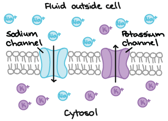
neurons get excited —> alter membrane permeability through channels —> change in membrane potential —> signal
What is membrane potential
voltage difference btwn the inside and outside of the cell
the separation of opp charges across the plasma membrane
measured in mV
uses leak channels and voltated gated

What is resting membrane potential
voltage diff when the cell is at rest
influenced by a permability of Na+ and K+
More K+ channels than Na+
Na+ is outside the cell naturally
K+ is inside the cell naturally
regulated by the sodium potassium ATPase pump
uses leak ion channels and ion pumps
What are the two types of membrane potential changes
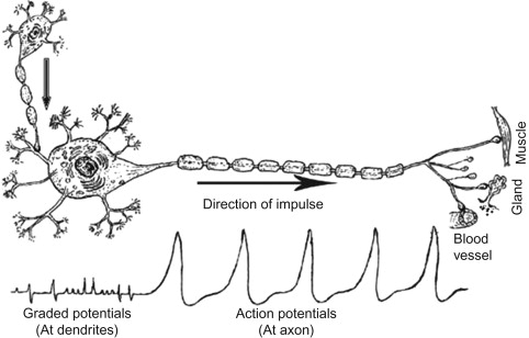
Graded potential
short distance signals
depolarization and hyperpolarization
decays with distance from source
magnitude depends on strength of stimulus
no refactory period
a stimulus acts on a neuron, causing graded potential where ligand gated channels open and ions enter, this ion movement (if strong enought) reaches a threshold and action potential starts
ligand gated
Action Potential
long distance
all or nothing; constant magnitude w/o losing strength
does not decay
absolute refactory and relative refactory period
voltage gated
What is depolarization, repolarization, and hyperpolarization
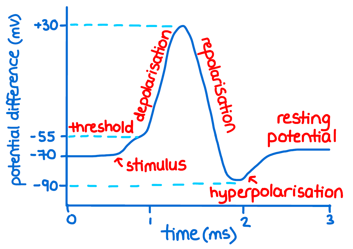
Depolarization: membrane potential becomes less negative (more +)
Repolarization: returns to resting potential
Hyperpolarization: membrane potential becomes more negative
What is the basic outline of an action potential
Resting potential —> stimulus —> threshold potential —> Na+ voltage ion gates open, Na+ gets into the cell —> K+ voltage gates open —> Na+ gates inactive —> hyperpolarization —> resting potential, K+ closes
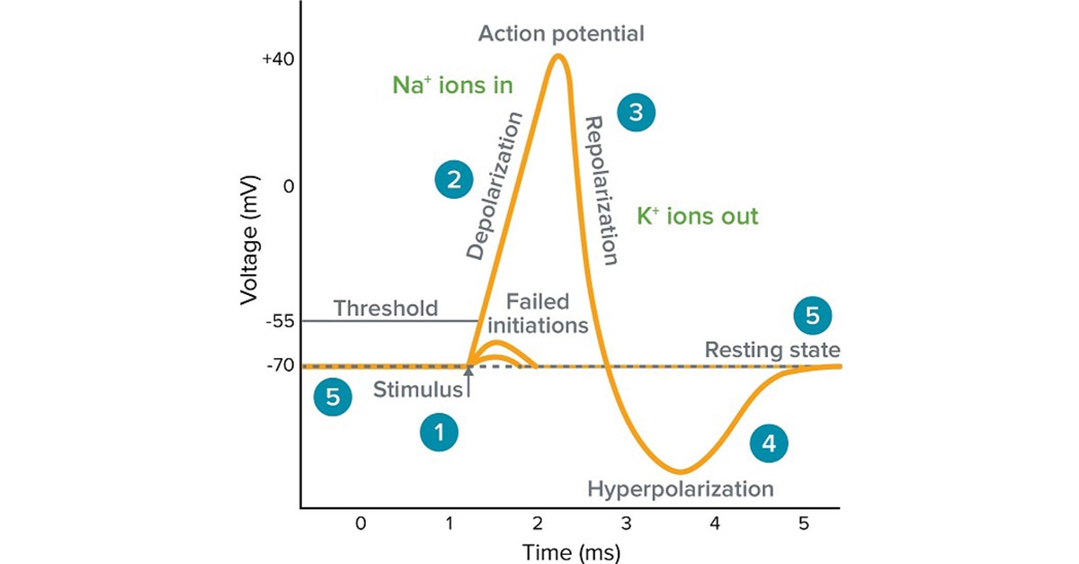
How does a voltage gated Na+ and K+ channels open/move during action potential
change in membrane potential/voltage opens these gates
Na+ comes into the cell
K+ leaves the cell
how is an action potential conducted along the axon?
Sodium channels depolarize neighboring regions by bringing these regions to threshold potential
inactive sodium channels during relative refactory period behind the zone prevent action potential from moving back
What is salatory conduction?
action potential jumps from node of ranvier. note: action potential is a domino effect where one action potential will start another action potential at another node
What is an absolute and relative refactory period?
absolute: cannot restimulate/start a second action potential bc Na+ channels are inactive (remember this is what starts the action potential)
relative: can be restimulated for a second one but needs a stronger stimulus. its bc Na+ channels are at rest and closed and can be reopened
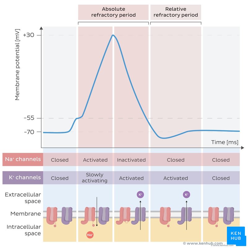
What happens after action potential? How does it get back to resting potential?
Sodium potassium pump to balance ions and charges
3 Na+ moves out of the cell
2 K+ moves inside the cell
req ATP
Synaptic communication, what happens at the axon terminal
action potential reaches axon terminal
Ca2+ channels open and get inside the cell
causes it to release neurotransmitters in vesicles
goes to synapse then receptors’
recycled or broken down
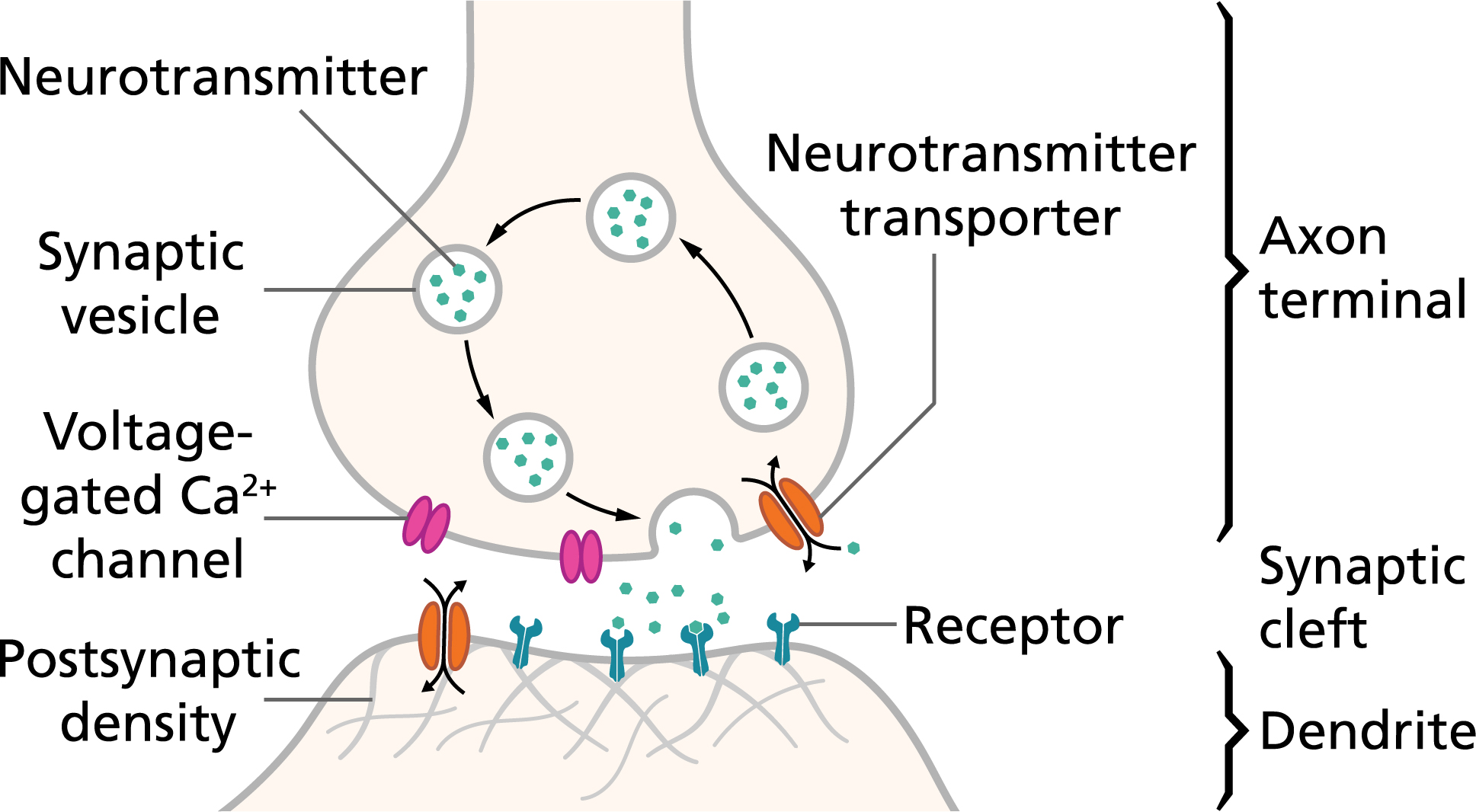
What are post synaptic potentials? What are the most effective ones
changes in the membrane potential of post synaptic neuron. graded potentials! influxation of ions creates these post-synpatic potentials, these converge at the hillock and neuron summates signals
Excitatory postsynaptic potential (EPSPs): depolarization that brings membrane potential towards threshold
Inhibitory postsynaptic potentials (IPSPs): hyperpolarization that brings potential away from threshold
Temporal Summation: two rapid ESP in succession
Spatial Summation: two diff ESP produced simultaneously
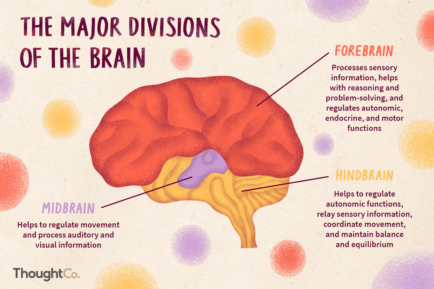
What does the brain do? what parts are in the forebrain, midbrain, and hindbrain
regulate homestasis, awareness, movement, cog, emotions
forebrain: cerebrum (cerebral cortext. white and grey matter)
midbrain: connects fore and hind
hindbrain: pons, cerebellum, medulla oblongata
What does the cerebrum do? what is cerebral cortext, grey and white matter
language, cog, memory, conciousness
cerebral cortext: receives input from sensory organs and somatosensory organs
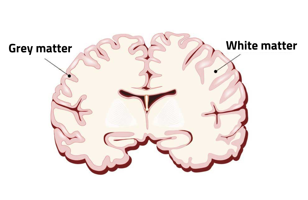
grey matter: neuron cell bodies, process data, memory thoughts
white: axon, commuication network ofrom grey and rest of body
what is lateralization
left side dominant for lang, math, logical
right dominat for pattern, recognition
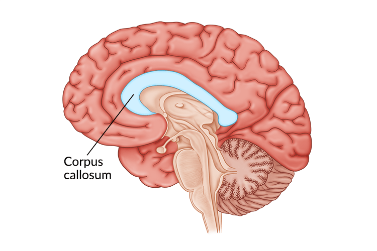
exchange info thorugh corpus callosum
What are the four lobes
Occipital lobe: visual
temporal lobe: auditory
parietal: reception and perception of somatosensory
frontal: volunatry movemnt, thinking
What does the thalamus, hypothalamus, brain stem, and cerebellum do?
thalamus: senses stimuli, synpatic integration center
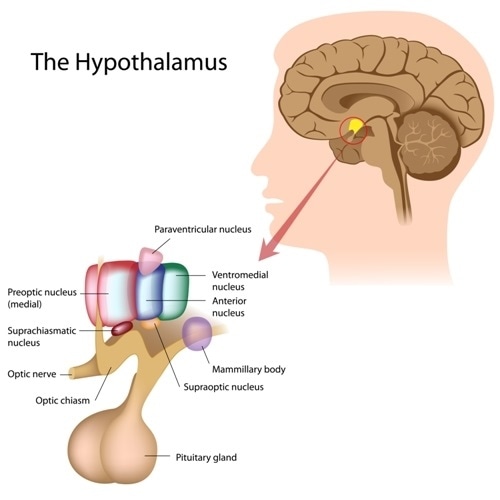
hypothalamus: homeostatsis, internal environment
brain stem: cardiovasular, digestive, and respiratory
cerebellum: balance and coordination
What is the biological clock regulation?
circadian rhythm is regulated in hypothalamus by suprachiasmatic nucleus
What is arousal and sleep and memory/emotions regualted
arousal and sleep: controlled by midbrain and pons
memory and emotion: amygdala, hippocammpus, thalamus
What does the spinal cord do?
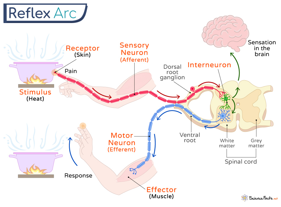
link between CNS and PNS, integrating center for spinal reflexes (withdrawl reflex)
What protects the CNS
mengines: three layers
Cerebrospinal fluid: cushioning fluid
blood-brain barrier: limits access of blood-borne material into brain tissue
What is neural plasticity
nervous system can be modified after birth
changes happen at synapses and can strenghten or weaken signaling
What are the different neurotransmitter pathways in the brain
Norepinephrine: sleep, learning, memory
Serotonin: emotion
Dopamine: reward center
What is the brains reward center and drugs
collection of structures and pathways that are responsible for desire, motivation, etc
cocaine/amphatamine: block dopamine from removal in clef
Opium/heronin: stops inhibitor
Nicotine: stimualte dopamine
Define the somatic and visceral components of the PNS
Somatic: brings sensory info from skeletal muscle, joints, and skin
Visceral: monitors other internal tissues like smooth muscles, cardiac muscle, glands
What is the first step in the sensory pathway?
Sensory reception: detects stimuli
What is the second step in the sensory pathway?
Sensory transduction
Receptor potential: stimulus causes a change in membrane potential
Graded potential!
Neuronal Receptor: sends signal directly to the CNS, larger change in receptor potential = more frequent
Non-neuronal Potential: uses neurotransmitters, larger change in receptor potential = more neurotransmitters released
What is the third step in the sensory pathway?
Perception
the brains construction of stimuli
the path of the action potential is how the brain distinguishes stimulli
cerebral cortex receives input from sensory organs
What is the fourth step in the sensory pathway?
Modification
Amplification: strengthening of stimulus energy by cells in sensory pathways
Adaptation: decrease in responsiveness to continued stimulation
What are the 6 types of sensory receptors
Mechanoreceptors: non-neuronal, sound, touch, pressure, motion
Electromagnetic receptors/ photo receptors: light, electricity, magnetism
Thermoreceptors: heat and cold; skin receptors and ant. hypothalamus sends info to post hypothalamus
Pain receptors (Nociceptors): trigger defense mechanisms; neuronal
Osmoreceptors: changes in solute conc; hypothalamus
Chemoreceptors
Smell (Olfaction): detection of order in the air; neuronal
Taste (Gustation): dectant of tastants in solution; non-neuronal
How is hearing detected by mechanoreceptors
Sound (moving particles in air) enters the outer ear into the canal
Vibrates the tympanic membrane
transmits vibration to the middle ear: Malleus, Incus, Stapes
transmits vibration to the oval window of the cochlea
pressure waves move through cochlea fluid
fluid movement bends the hair cells in the organ of Corti
Depending on the direction of the hair, the bending causes ion channels to open and close and release or release less of neurotransmitters
generates an action potential to the auditory nerve to the CNS
goes through thalamus
goes to auditory cortex in temporal lobe
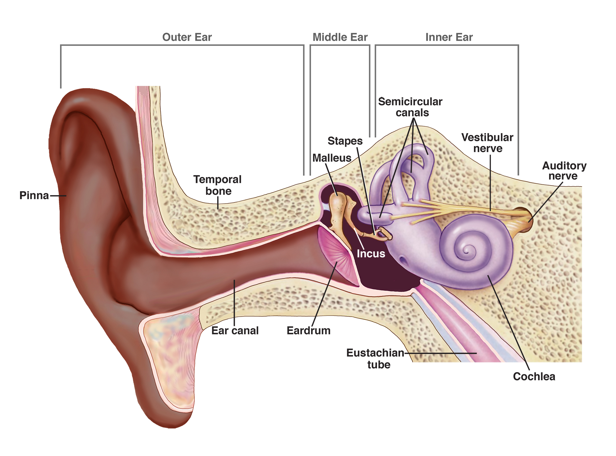
How is body equilibrium maintained? (mechanoreceptors)
three semicircular canals on cochela detect angular movements in any direction
Utricle and saccule detect linear motion and position relative to gravity
fluid movement in vesibular sense organs
hair cells bend and produces an action potential
signals carries to vestibulocochelar nerve to brainstem and cerebral regions
What are the parts of the eye
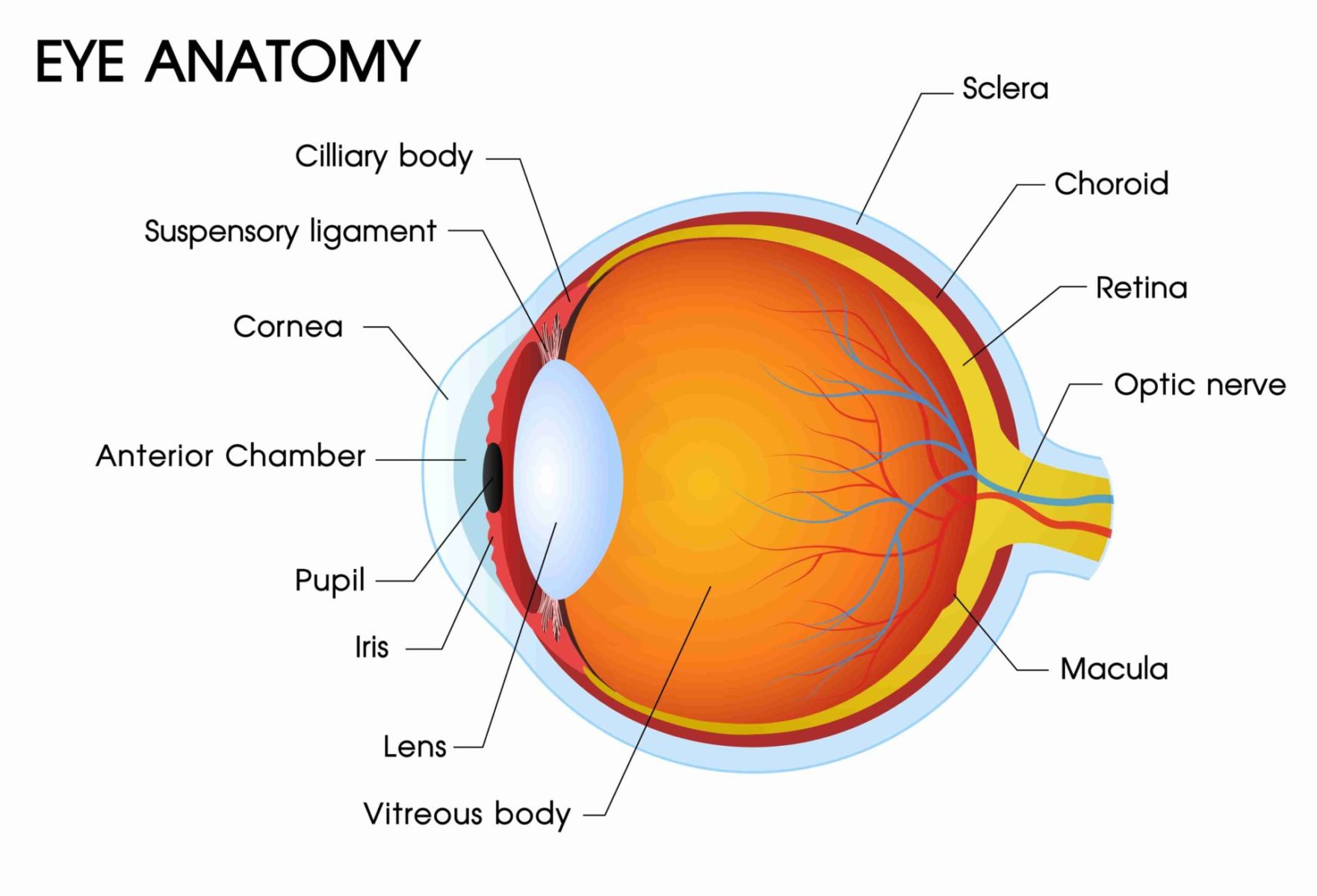
Pupil: light enters
Iris: controls how much light enters the pupil by constricting or dilating the pupil
Scalera: protection of the eye
Lens: focuses the light to send to the retina
Ciliary body: can contract and alter the shape of the lens to focus light on near or far objects
How is light travel in the eye
Light enters pupil
focuses through the lens
sent to the retina
What are the three components of the retina
Rods and cones
Bipolar cells: has synpases between the two
ganglion cells: bundles of axon form optic nerve
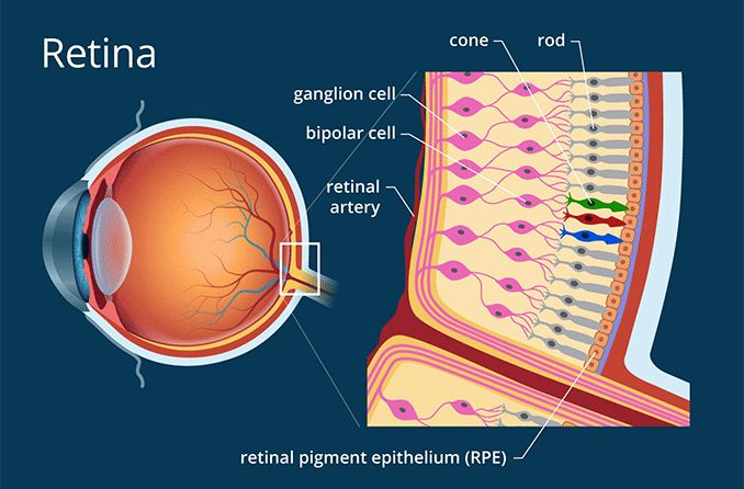
What is the structure of rods and what do they do?
Rods is where the light enters bc it has photoreceptors
outer structure: detects light stimulus, has photopigment molecules, faltten membranous disc, contains rhodopsin (opsin and retinal)
Inner structure: metabolic structure
Synaptic terminal: releases neurotransmitters to bipolar cells; depends if it is light or dark
What do cones do?
Three types of cones have three diff visual pigments: red, green, blue
Red and green color blindness is sex linked on X chromosomes
What happens in the eye when it is dark?
Photoreceptor ion channels are opened
dark photoreceptors are depolarized and releases inhibitory neurotransmitters onto the bipolar cells
bipolar cells cannot fire anymore
brain detects this as dark
cis retinal structure
What happens in the eye when it is light?
Recall: Rods have photoreceptor pigments
light travels to retina
activates photopigments (rhodopsin changes to a trans position)
photoreceptors hyperpolarized
does not send the inhibitory neurotransmitter
bipolar cell can activate and release neurotransmitters
signal sent to ganglion cells and into the optic nerve
What is the efferent division of the PNS and two parts?
Carries motor commands from the CNS to target muscles and glands
Autonomic Nervous System and Somatic Nervous System (motor)
What is the autonomic nervous system?
controls smooth and cardiac muscles, glands
involuntary movement
sympathetic and parasympathetic nervous system; dual innervation, antagonistic control
works with endocrine and behavioral state systems to maintain homeostasis
What is the autonomic nervous system pathway? What neurons?
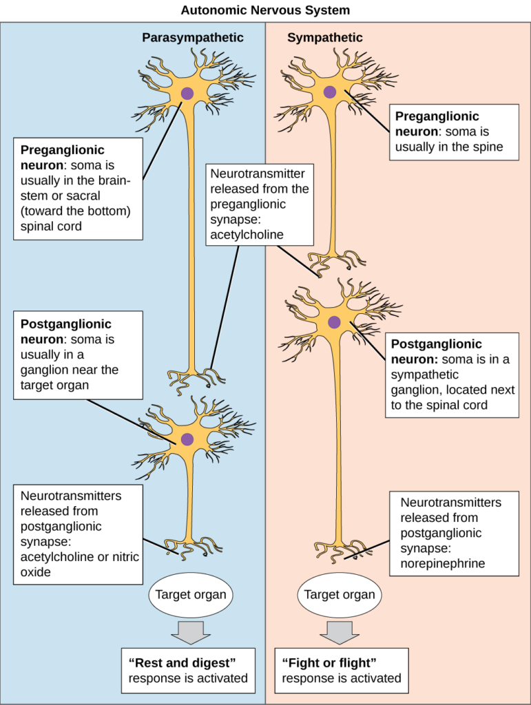
Two neuron chain: preganglionic axon and postganglionic axon
Preganglionic axon extends from CNS
Postgangolionic axon extends to target tissue
SNS: short preganglionic axons w/ long ganglionic axons, preganglionic axon physically extends from CNS and leaves through spinal nerves (T1-L2), ganglia are usually near spinal cords
PNS: long preganglionic axons w/ short ganglionic axons, pre axons have their cell bodies in brain stem and physically extends from CNS and leaves through crainal nervers and sacral spinal cord, ganglia are near or within target
what is the SNS and PNS
SNS
- flight or flight
inc heartrate, inc breathing, decreases motility of digestion
PNS
rest and digest
dec heartrate, bdecbreathing, inc motility of digestion
What is the difference btwn the Sympathetic and Parasympathetic neurotransmitters?
Both use acetylcholine for their preganglionic axons to ganglionic axon with nicotinic cholinergic receptor
SNS: uses norepinephrine for their postganglionic axon to target tissue with an adrenergic receptors
PNS: uses Ach for their postganglionic axon to target tissue with a muscarinic receptor
What is the basic anatomy of a skeletal muscle?
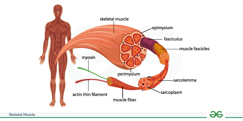
Thick filaments (myosin) and thin filaments (actin) make up the unit of a sacromere
Sacromere makes up a myofibril
Bunch of myofibril make up muscle fiber
bundle of muscle fiber makes up muscle
What is the pathway to a target muscle?
CNS axon extends to the target muscle and relases Ach to nicotinic receptors
What does the neuromuscular junction consist of
axon terminal (somatic neuron branches), motor end plate (receptors), schwann cells
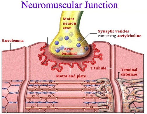
What is the muscle contraction process?
Action potential arrives at the end of the neuron
Releases Ach (from Ca2+ channels) into the synpase
Ach binds to the ligated nicotinic channels
causes sodium voltage gated channels to open and starts action potential
action potential travels down T-tubules which signals the sacroplasmic reticulum to relase Ca2+ into the cytosol
Ca2+ binds with troponin and allows myosin to bind with actin
filaments pull close together, causing a contraction
ATP binds with myosin head to stop and lower its position
What is excitation-contraction coupling
events that link muscle excitation and contraction, Ca2+ is the link
What are chemical agents and diseases that affect neuromusclar junctions?
Botulinum toxin blocks release of Ach
Curae blcoks Ach receptor
Myasthenia gravis, autoimmune disease that destorys Ach receptors
black window venom causes excess release
organophosphate inhibits AchE
How does skelton support muscles
muscles attach to skeleton
provide support, movement
antagonistc pairs
What is a behavior
an action carried out by muscles under the control of the CNS
behavior can affect survivial and reproduction, thus under natural selection
What is the endocrine system composed of?
Ductless endocrine glans
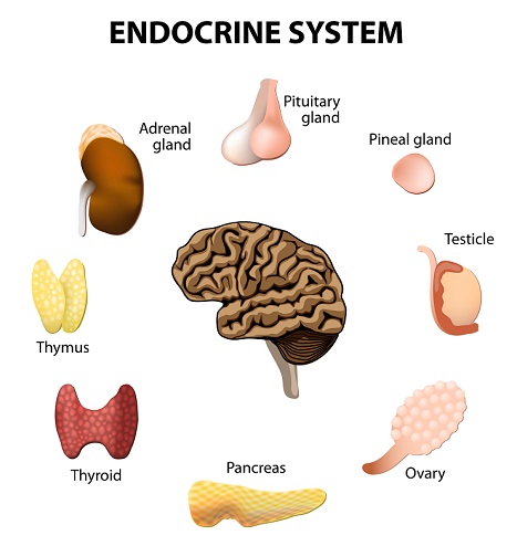
Central Endocrine glands: pineal, hypothalamus, pituitary
Peripheral Nervous glands
Pituitary, thyroid, and adrenal glands are solely endocrine functions

What are the endocrine system’s functions?
mediated by hormones
Control and integrate both the digestion and absorption of food and circulation
regulates metabolism (insulin and glucagon)
regulates red blood cell production
promotes sequential growth and development
controls reproduction
regulates H2O and electrolyte balance
Help body cope with stressful situations
What are hormones? Why do the same hormones have diff responses? Give an example using epinephrine
Chemical messengers secreted by endocrine glands into the blood stream to transport signals to distant target cells
Cells with certain receptors can respond to certain hormones
Same hormone can have different responses due to different receptors types or in the molecules that produce the response in target cells
Epinephrine on Adenergic Beta receptor on liver cell
Causes glycogen to break down glucose and release it
Epinephrine on Adnergic Beta receptor on skeletal muscle blood vessel
vessel dilates
Epinephrine on alpha receptor on intestinal blood vessel
constricts vessel


What are the different types of hormones?
Hydrophilic hormones
binds to membrane proteins and uses receptors, can manipulate protein expression. Ex: peptides (Insulin), catecholamines (norepinephrine)
Lipophilic
can penetrate through plasma membrane and travel directly to nucleus by a receptor complex moving inside the nucleus, alter gene expression. Ex: steroid hormones (sex hormones like testerone), thyroid hormones
Tropic hormones: a hormone can regulate secretion of another hormone from another endocrine gland

What is a simple endocrine pathway using pH control and digestive enzyme secretion in duodenum? Where is the exocrine portion of the pancreas and where is the endocrine?
Local stimulus → endocrine cell —> release hormone —> blood vessel —> targeting cell —> response
pH control
Stimulus: low pH (acidic) —> In the duodenum mucosa, S cells secrete secretin —> travel on blood vessel —> pancreatic duct cells (target cell) —> releases bicarbonate in duodenum to neutralize acid
Digestive Enzymes
Stimulus: proteins and fats in duodenum —> In the duodenum mucosa, I cells secrete CCK —> travel on blood vessel —> Pancreatic acinar cells (target cell) —> release enzyme
Exocrine = ducts — duct and acinar cells
Endocrine = blood — secrete insullin and glucagon

What is a simple endocrine pathway using blood glucose and energy metabolism regulation
Local stimulus → endocrine cell —> release hormone —> blood vessel —> targeting cell —> response
Insulin
Stimulus: Inc blood glucose —> Pancreatic Beta cells located in the Islets of Langerhans release Insulin —> travel through blood stream —> act on target cell/tissues —> dec blood glucose levels
Glucagon
Stimulus: Low blood glucose —> Pancreatic Alpha cells in the Islets of Langerhans release glucagon —> travel through blood stream —> act on target cell/tissues —> inc blood glucose lvls

What is a neuroendocrine pathway? Use thyroid hormones and infant suckling as examples
responds to stimuli from the external/internal environment and relay on a sensory in the nervous system
Neurosecretory cells: receive neuron impulses but releases hormones into the blood stream (hypothalamus)
Regulation of thyroid hormones
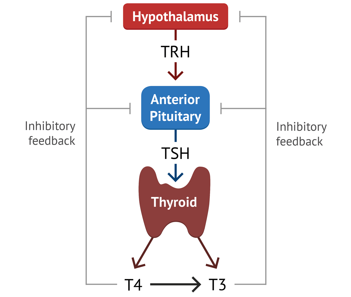
Stimulus: low thyroid levels, cold, etc —> sensory neuron detects —> information sent to hypothalamus —> action potential from this causes hypothalamus to release hormones: Thyrotropin-releasing hormone (TRH) —> sent to portal system (blood stream) —> Anterior pituitary —> causes the Ant. pituitary to release Thyroid-stimulating hormone —> acts on thyroid gland —> thyroid gland releases thyroid hormone (T3, T4) —> target body cells inc metabolism and heat production
*Negative feedback
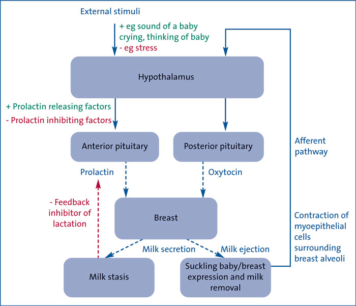
Infant Suckling
Stimulus: infant sucking —> sensory nerves in nipples detect —> hypothalamus and their axons extend all the way down to the post. pituitary gland —> release oxytocin technically from the post. pituitary gland —>travel in blood stream —> acts on smooth muscle in mammary gland —> milk release
*Positive feedback
*Prolactin makes milk, oxytocin secretes it

How does the hypothalamus and pituitary glands work together?
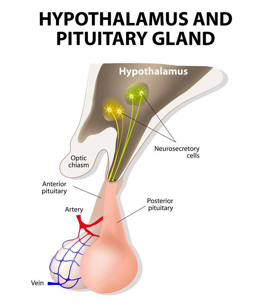
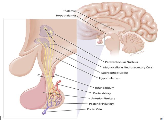
Hypothalamus integrates both the nervous system and endocrine system
Signals from the hypothalamus travel to the pituitary gland located at its base
hypo controls the release of hormones of the ant. and post. pituitary glans
Hypo secretes hypophysiotrophic hormones (releasing or inhibitory hormones) into portal systems (a specialized network of blood vessels that directly connects the hypothalamus to the anterior pituitary gland)
What does the anterior pituitary gland produce and secrete?
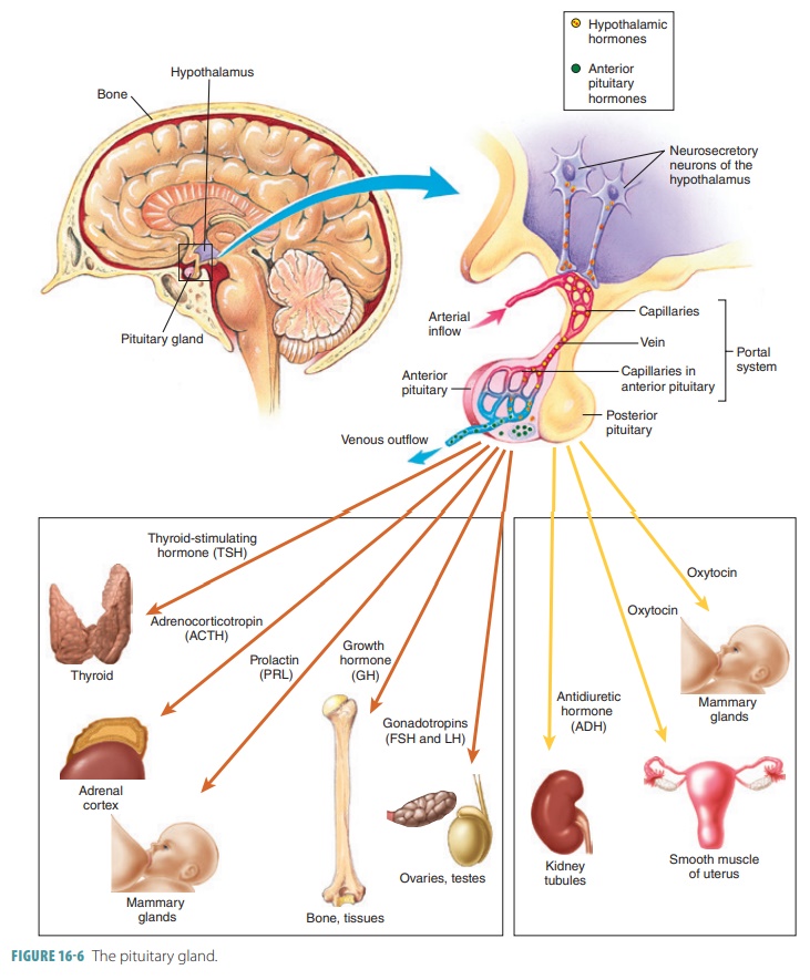
The hypothalamus hormones target the ant. pituitary gland to release their hormones
Thyroid-stimulating hormone (TSH): tropic hormone, tells thyroid gland to release thyroid hormone, T3, T4
Adrenocorticotropic Hormone (ACTH): tropic hormone, tells adrenal gland to release cortisol
Follicle-stimulating hormone (FSH) and Luteinzing Hormone (LH): tropic hormone, tells gonads (ovaries or testes) to secrete sex hormones (estrogen and progesterone or testosterone, and also ova or sperm)
Growth Hormone (GH): tropic hormone, tells liver to release IGF1, which affects bone and soft tissue and causes growth
Prolactin (PRL): non tropic, tells mammary gland to produce milk
What does the posterior pituitary gland do?
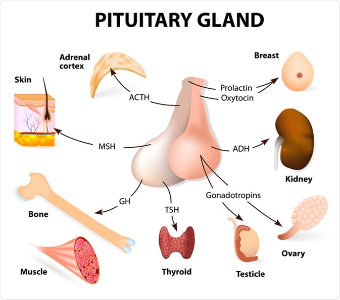
Stores two small peptide hormones (Vasopressin and Oxytocin) which are made in the Hypothalamus
The Hypothalamus axons extend to the neuronal terminals in post pituitary — release Vasopressin and Oxytocin
Vasopressin: act on nephrons in kidney and increase permeability of distal and collecting tubules to H2O (conserve water during urine), or acts on arterioles and causes vasoconstriction (narrowing)
Oxytocin: acts on uterus to stimulate uterine contractions during childbirth or mammary glands for ejecting milk

What do the thyroid gland and hormones do?
Thyroid gland contains two types of endocrine secretory cells
Follicular cells produce thyroid hormones T3 and T4
C cells produce calcitonin which secretes in response to an increase plasma Ca2+, and lwrs it by inhibiting the activity of bone osteoclats
Thyroid hormones
main determinant of basal metabolic rate
plays crucial role in normal development of nervous system
essential for normal growth
These hormones tell nearly every cell in your body how fast to turn food and oxygen into usable energy, dictating how many calories you burn at rest, how energetic you feel, and how your body stores or uses weigh


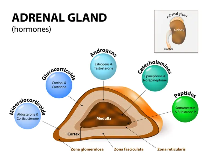
What does the adrenal gland do and secrete?
It is involved in nutrient metabolism and stress adaptation
Adrenal Cortex: secretes steroid hormones (lipohilic)
Adrenal Medulla: secretes catecholamines (hydophilic), Epinephrine 80%, Norepinephrine 20%
Steroid hormones
Aldosterone: helps maintain salt balance and blood pressure
Cortisol: involved in stress resistance and anti-inflammatory effects
Sex hormones: regulate sex drive
Catecholamine (Epinephrine)
reinforces sympathetic effects
maintain arterial blood pressure
inc blood glucose and fatty acids
What is the neuroendocrine reflex? Use stress response as an example (short vs long)
A bodily response that links the nervous system and the hormone system
EX: Stress response: A generalized nonspecific response of the body to any factory that overwhelms or threatens to overwhelm the body’s ability to maintain homeostasis
Short term stress: sends nerves impulse down spinal cord through preganglionic fibers—> adrenal medulla —> secrete catecholamines (epinephrine and norepinephrine)
Long term stress: sends signal to hypothalamus —> releases Corticotropin hormone (CRH) onto ant pituitary —> releases ACTH onto adrenal cortex —> secretes steroid hormones (cortisol)
* Note Vasopressin can also work with CRH to ramp up production of ACTH
What regulates hormone secreation?
Hierarchic chain of command: chain of hormonal communcation
Feedback control
negative: targets gland hormones and inhibits hypothalamic and ant. pituitary secretion, in simple pathway it stops the stimulus
positive: enhances response
Circadian rhythm: regulated by suprachiasmatic nucleus, reguaates concentration during light and dark
What are some examples of endocrine dysfunction?
Hypothyroidism
Causes: failure of thyroid gland, secondary cause due to deficient TRH and TSH, inadequate supply of Iodine (produces T3 and T4)
Energy and Weight: Constant fatigue, sluggishness, and unexpected weight gain.
Treatment: replacement thyroid hormone, dietary Iodine
Hyperthyroidism
Causes: Grave’s Disease (body produces thyroid stimulating immunoglobins which bind and continue secretion), secondary cause is excess TRH and TSH
Weight changes: Losing weight fast even with a big appetite. — break down storage
Digestion: More frequent bowel movements or loose stools. — contract faster
Treatment: removal of over-secreting thyroid lobe, radioactive iodine/anti-thyroid drugs

What is the evolution of the hormone function?
Thyroid hormone in frogs stimulate the reabsorption of tadpole tails during metamorphosis
Prolactin in birds regulate fat and metabolism, freshwater fish regulate salt and water balance
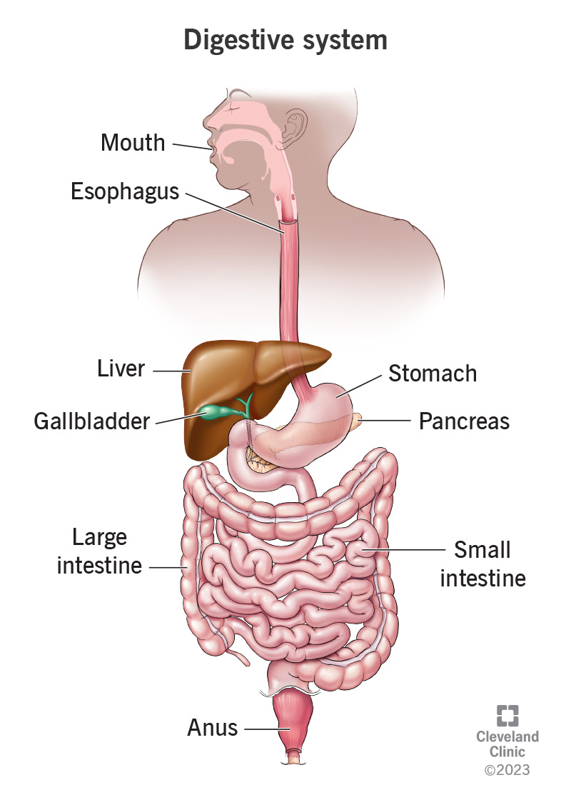
What are the functions of the digestive system? Which parts are regulated and why
Transfer nutrients, water, and electrolytes from ingested food into the bodys internal enviroment
Digestion: Biochemical (enzygmatic) breakdown of complex foodstuffs into absorbable units
Absorption: The end-products of digestion that is transferred from the digestive tract lumen into blood stream to specific regions
Secretion: the release of water, electrolytes, and specific organic constituents (enzyme) by cells and organs, released onto digestive tract through neuronal or hormonal stimulation, reabsorbed back into blood after digestion complete
Motility: muscular contraction that mix and move food forward
propulsion: pushing content forward
mixing: mixing food with digestive fluid
mass movement: happens in large intestine, intense and prolonged contractions, clear the large intestine
Secretion and motility regulated bc nothing stops digestion and absorption (passive) also bc of acidic enzymes

What are the digestive compartments?
Specialized compartments where food breaksdown
Intracellular digestion: Lysosomes breaking down food, waste, pathogens in cell
Extracellular digestion: breakdown of food outside of cells in cavity or tube
Gastrovascular cavity: not in humans, same hole for digesting and excretion, ex: flatworm
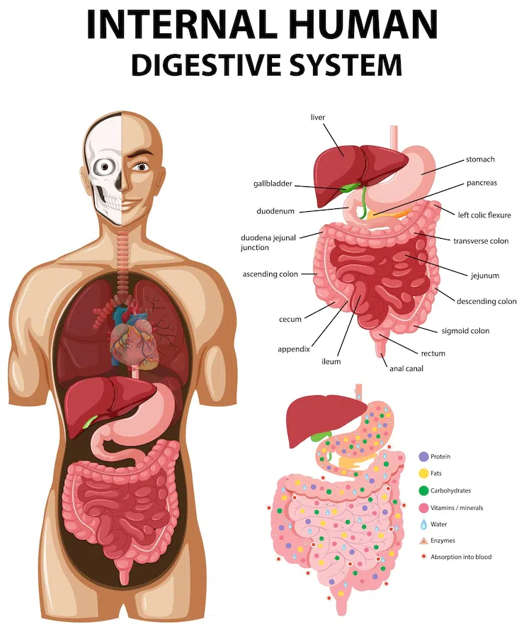
Digestive tract: food travels from mouth to anus
Accessory organs: help with digestion but food does not travel here, Ex: pancreas, gall-bladder, salivary glands, liver
Oral cavity with salivary glands (amylase)
Esophagus
Stomach
Small intestine
Large intestine
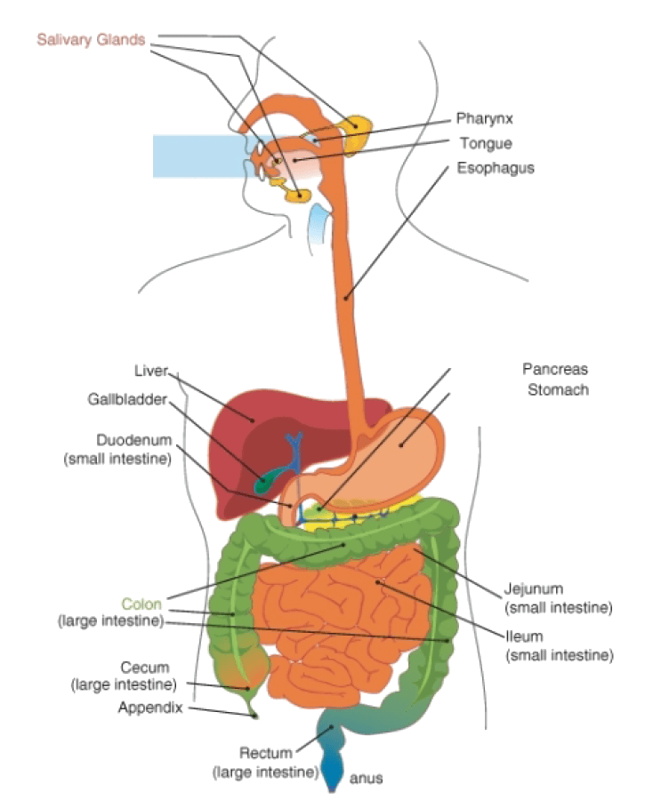

What is the anatomy of the digestive tract
Lumen: hallow space in digestive tract
Serosa: Outer connective tissue, secretes serosa fluid that lubricates prevents friction, continous w/ mesentary
Muscularis externa: major smooth muscle in the wall
Outer longitudinal muscle: can shorten the length of the GI Tube through contraction
Inner circular muscle: can shorten/dec diameter of the lumen through contraction
Myenteric plexus: network of nerves in between these two layers
Submucosa: thick connective tissue, provides distensibililty (stretch), and elasticity, has large blood and lymph vessels
Submucosal plexus: nerve network
Mucosa: lines luminal surface of digestive tract
Muscularis mucosa: outer, smooth muscle
Lamina propria: thin middle layer connective tissue
Mucous membrane: inner endothelial layer
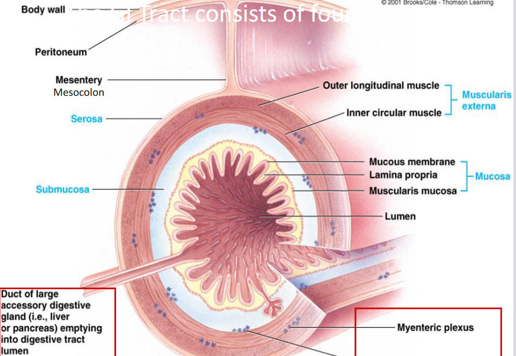
How is digestion regulated? talk abt the two systems and hormones
neural and hormonal inputs
Enteric Nervous System
Nerve network in GI tract — submucosal plexus and myenteric plexus
autonomous from CNS, regulates content, movement of smooth muscle,
controls muscle and secretion of glands
does communicate with CNS
CNS influences digestive tract motility and secretion, can alter the lvl of hormone
ENS and CNS communicate through nerve fibers of the ANS (sympathetic and parasympathetic branches)
GI hormones: produced by specialized endocrine cells within mucosa
Ex: Peptides and amino acids causes the Stomach to release Gastrin from G Cells —> acts on ECL Cells and and parietal cells —> secretes gastric acid
Ex: Small Intestine releasing secretin and CCK (duodenum)
Ex: Intestine can release motilin which stimulates fasting —> acts on gastric and intestinal smooth muscle —> causes migrating motor complex to be stimulated (facilitates transportation of indigestible substances to large intestine)
What is the first step of the digestive pathway? What are special enzymes
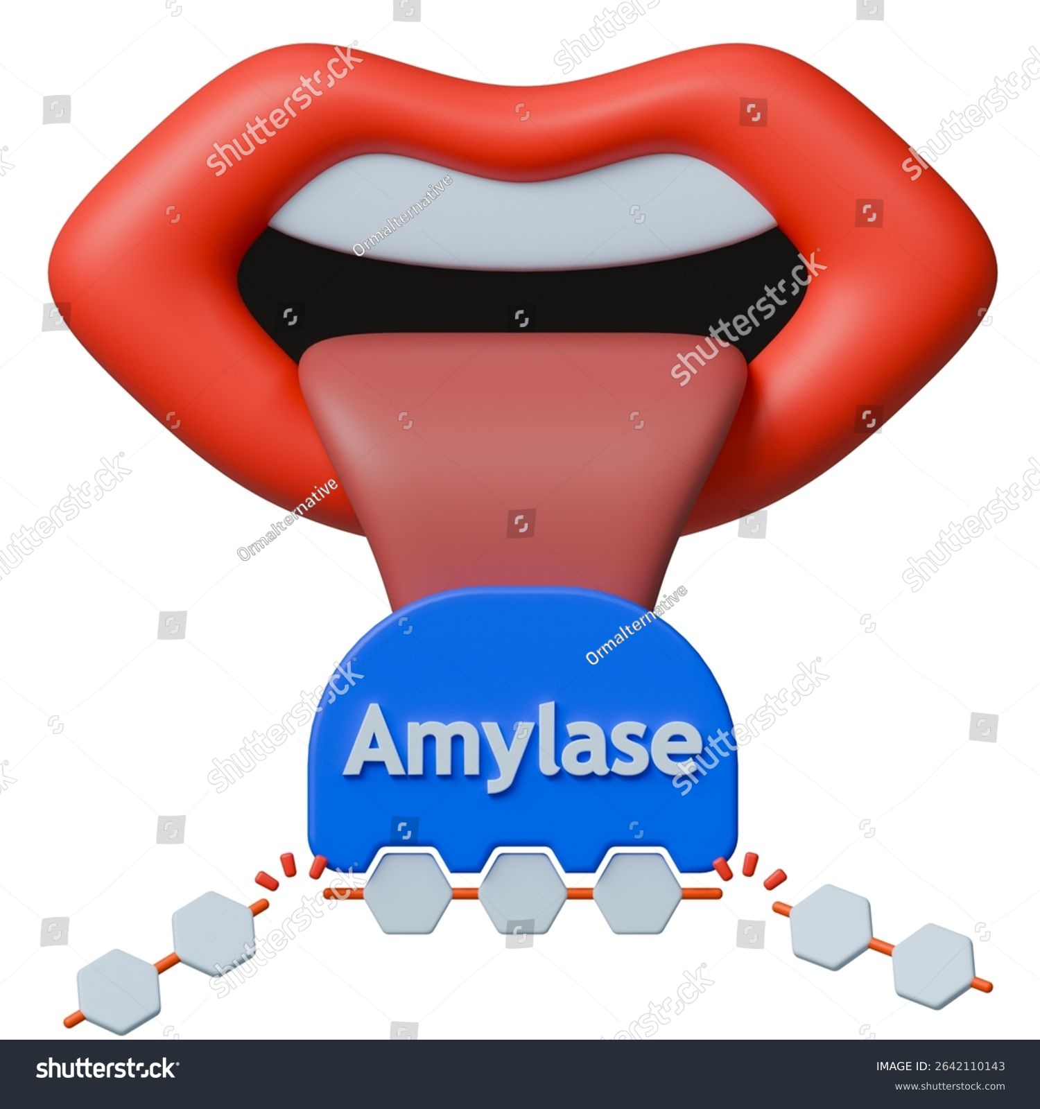
Mouth: mechanically chewing and breaking down food, amylase (carbohydrate enzyme)
What is the second step of the digestive pathway?
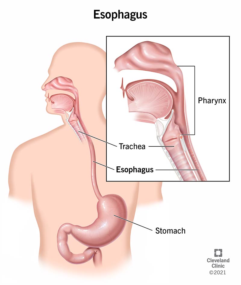
Mouth —> Food travels to esophagus

What is the third step of the digestive pathway? Describe the three specific cells, gastric juices, and feedback
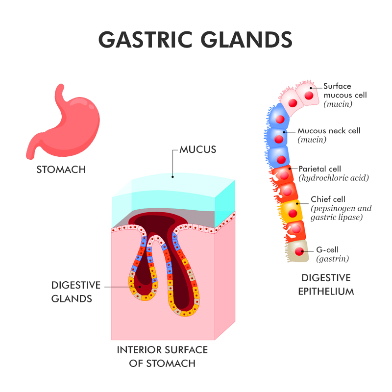
Stomach: store food, mixes food mechanically, secrete gastric juices which help break down food into chyme (soft food mix with digestive juices)
has gastric gland which makes gastric jucies
Parietal Cells (bottom): secrete CI, cleaves (breakdown) pepsinogen from chief cells into active pepsin
Chief cells (middle): make and secrete pepsinogen (inactive form to stop body from digesting its own body protein)
Mucus cell (top): make and secrete mucus as a physical barrier from acidic gastric juices
Gastric juices
HCI — low pH 2, denatures proteins
Pepsin — formed from HCI and pepsinogen mixing, breaks down the denatured protein into smaller pieces
Secretion of HCI from the parietal cells is controlled by Gastrin and can also be stimulated by acetycholine
Pathway
Stimulus: proteins/food enter stomach —> G cells secrete Gastrin —> travels through blood stream —> act on ECL cell —> releases histamine —→ travel through blood stream —> act on parietal cell —> release HCI *enteric neurons can release acetylcholine on parietal cell —> Chief cells release pepsinogen —> HCI and pepsinogen mix —> active pepsin —> HCI and pepsin break down food —> chyme
Feedback loop
Somatostatin-releasing cell can inhibit production of HCI
Releases somatostatin which works to inhibit G cell, ECL Cell, and parietal cell
What is the fourth step of the digestive pathway? describe the pH and digestive enzyme control. talk about transport and villi
Chyme enters Small intestine (major organ of digestion and absorption)
most digestion occurs at duodenum (first portion of small intestine)
Main absorption of nutrients and water happens in jujunum (middle) and ileum (terminal)
pH control in duodenum — hormonal control
chyme is acidic and we need to neutralize it
Stimulus (low pH) —> S cells release secretin —> travels on bloodstream → pancreatic duct cell —> release bicarbonate —> neutralize chyme
Digestive enzyme control in duodenum — hormal control
Stimulus (proteins and fats) —> I cells release CCK —> travel to bloodstream —> Pancreatic acinar cells —> release digestive enzyme —> food breakdown
Chyme (after neutralization) will mix with other digestive fluids in pancreas, liver, gallbladder and intestinal wall
Pancreas: trypsin and chymotrypsin (break down proteins), amylase, lipase
Liver: makes bile (stored in gallbladder)
Intestinal wall: digestive enzymes that complete the digestion of carbs, fats, proteins
Villi and microvilli in the intestinal lumen that create a brush border and inc the rate of nutrient absorption
Small particles that are broken down enough will leave to the blood stream and to other regions using transport in jejunum and ileum (absorption)
Passive transport: simple and facilitated diffusion (req a facilitator or protein)
Active transport: primary and secondary (relay on atpase gradient, does not directly use ATP)
Ex: Glucose transport
ATPase pump, pumps out 3 Na+ and pumps in 2K+
Na+ wants to go back in the cell naturally and glucose also wants to enter
uses Sodium glucose cotransporter which brings them both in the cell
Glucose travels through the cell and moves out of the cell through facilitated diffusion of GLUT-2
*Fructose uses GLUT-5
Pathway'
Chyme enters small intestine (duodenum) —> S cells release secretin —> travels on bloodstream → pancreatic duct cell —> release bicarbonate —> neutralize chyme —> Duodenum breaks down proteins—> I cells release CCK —> travel to bloodstream —> Pancreatic acinar cells —> release digestive enzyme —> food breakdown —> Chyme mixes with digestive fluids —>absorption in jejunum and ileum —> transporters take small particles into blood capillaries —> rest is sent to large intestine

What is the fifth step of the digestive pathway
Large intestine: four structural parts but we will focus on colon — recover water, vitamin K, salt that has entered the digestive tract, will turn into solid stool
no secretion of digestive enzymes, no absorption of nutrients
food enters through ileocecal valve
Motility
Mass movements move the food forward and trigger the defacation reflex (stool moves into rectum
What is the sixth and seventh step of the digestive pathway
Rectum (stores pee and poop) —> anus
What are some disorders of the digestive system?
Gastroparesis: delayed gastric emptying (food stays in stomach for too long), can be caused by glyconic control, or extrinisic or intrinisic neuropathy
Constipation: infrequent bowel movement or difficult passage of stool, can be caused by inadequate fiber diets, depression, diabetes, meds,etc
Diarrhea: loose, watery bowel movement, caused by viral parasitic infections, meds
Gluten-senstive enteropathy (Celiac disease): autoimmune inflammatory disease that causes destruction enterocytes (epithelial cells of intestines), resulting in shortening of villi, gluten destroys lining
Inflammatory bowel disease (Crohns, Ulcerative colitis): chronic inflammatory intestinal condition
Define nutrients
Chemical substances ini food that provide the energy for various body processes
chemical energy is converted into ATP
organic building blocks such as organic carbon and organic nitrogen synthesizes a variety of complex organic molecules in the body
Essential nutrients: must be obtained from dietary sources, cant be synthesized in the body
essential amino acids: meat, egg, cheese,
essential fatty acids: lionelic acid (Omega-6 found in vegetable oils) and alpha-lionelic acid (Omega-3 found in plant seeds and nuts)
6 Classifications: carbs, fats, proteins, minerals, vitamin, water
What is malnutrition and undernutrition
Malnutrition: long term absence of one or more essential nutrients from diet
Undernutrition: results when diet does not provide enough chemical energy
used by stored fat and carbs
breaks down its own protein, loss of muscle mass
suffer protein deficiency of brain
die or suffer irreversible damage
What hormones regulate appetite and consumption
Ghrelin secreted by the stomach wall increase appetite
Leptin produced by adipose tissue suppresses appetite
GLP-1 produced by intestines promotes satiety/suppresses appetite
What is metabolism? what is catabolic and anabolic pathways
Sum of chemical reactions in the body
extract energy from nutrients
uses energy for work
store excess energy
Catabolic pathway: break large molecules into smaller ones, release energy
Anabolic pathway: synthesize larger molecules from smaller one, uses energy
What is bioenergetics
the flow and transformation of energy
carbohydrates —> glucose or stored as glucogen
Proteins —> amino acids or excerted through urine
Fats/lipids —> fatty acids or stored in adipose tissue
Describe cellular respiration
Involves nutrient molecules and O2 to convert into ATP and CO2
Oxygen from breathing provides the O2 for input
Glycolysis: Glucose —> Pyrvate, produces ATP and NADH
Citric Acid Cycle (Kreb): purvate —> Ach-CoA, enter mitochondria, oxidizes to O2, forms ATP, NADH, and FADH
Oxidative phosphorylation/ETC: strip electrons away to a lower energy, energy release creates hydrogen gradient —> movement of protons makes enerfy for ATP synthesize
Regulation of energy storage
Glucose is stored as glucagon in liver and muscle. Excess —> fatty acids in adipose tissue
Fatty acids are stored in adipose tissue
Amino acids are stored as body proteins in muscle. Excess —> Glucose
Hormonal regulation of metabolism. How does insulin cause uptake? How does it benefit cellular respiration?
relies on endocrine system
Cell does not have a lot of glucose transporters naturally. Insulin causes activates intercellular pathways that inc the inseration of glucose transporters. Glucose can enter cell and be either stored as glycogen, broken down into pyurvate for ATP, or stored long term as fatty acid. BECAUSE GLUCOSE IS OUT OF THE BLOODSTREAM, THE BLOOD HAS DEC BLOOD GLUCOSE
Insulin
Stimulus: inc blood glucose —> Beta cells in pancreas release insulin —> travel in blood —> target cells or cellular uptake —> dec blood glucose
Insulin delivers glucose to the cell using glucose transporter. This is the ingredient for cells to use to start cellular respiration
Glucagon
stimulus: low blood glucose —> alpha cells in pancreas release glucagon—> breakdown glycogen and releases glucose —> bloodstream —> target cells —> inc blood glucose
Cellular respiration cannot happen without a constant supply of fuel (like glucose or fatty acids) to break down into ATP energy. Glucagon keeps cellular respiration supplied with fuel between meals and during sleep.
Type 1 and Type 2 diabetes
caused by deficiency of insulin or dec response to insulin
Type 1 diabetes: an autoimmune disorder (produce antibodies), immune system destorys pancreatic Beta cells, genetic predisposition
insulin shots
Type 2: failure of target cells to respond to insulin (something wrong with insulin receptor or glucose insertion receptor), unable to uptake glucose, fat becomes main substrate
metaformin: reduces glucose production in liver, encourages uptake
Acarbose: inhibits enzymes that break carb into glucose, slow absorption
GLP-1: promote insulin release, promote satiety

What is the difference between an open and closed circulatory system? What is single and double circulation for closed circulatory? What is the advantages of a double circulation? Why do we oump blood twice through the heart?
Open
circulatory fluid (hemolymph) bathes the organs directly
no distinction btwn circulatory fluid and intersitial fluid
Closed
blood is confined to vessels and is distinct from interstitual fluid
chemical exchange happens btwn blood and intersititual fluid and fluid and cell
Single circulation: One way flow, blood passes through the heart once per circuit
Double circulation: blood passes through heart twice, separate gas exchange and systemic circuit
Advantages to double
inc the pressure and flow rate
important for larger bodies, high metabolic rate, high body temp (thermal homeostasis)
Why two pumps
Capillaries create substantial resistance and pressure loss
returning to the heart can restore pressure

What are arteries, veins, and capillaries?
Arteries: carry blood away from the heart, thicker wall to tolerate higher pressure
Veins: carry blood towards heart, valves prevent backflow
Capillaries: site of chemical exchange between blood and interstial fluid, one cell thick wall

What are the components are the heart - Atrium, Ventricle, valves, arteries
Right Atrium: oxygen-poor blood enters from the vena cava
Left atrium: oxygen-rich blood enters from pulmonary veins
Right ventricle: oxygen-poor blood moves from R atrium to R Ventricle with valves
Left ventricle: oxygen-rich blood moves from L atrium to L ventricle with valves
Septum: thick muscular wall that divides the R and L sides
Atrioventricular valve (AV) : separate A & V — tricuspid valve and bicuspid valve
Semilunar valve: separate R &L ventricle from arteries
Sup and Inf vena cava: carries oxygen-poor blood from tissues and pumps it into the R Atricum
Pulmonary artery: carries oxygen -poor blood to lungs
Aorta: carries oxygen rich blood from L ventricle to rest of body
*left ventricle is the thickest bc it needs to pump blood to all organs so it contracts with more force BUT both ventricles have the same amount of blood vol
Describe blood flow
Deoxygenated blood arrives from vena cava to R atrium —> tricuspid valve —> R ventricle —> Pulmonary semilunar valve —> pulmonary artery —> lung capillaries
Oxygenated blood arrives from pulmonary veins into L atrium —> bicuspid valve —> Left ventricle —> aortic semilunar valve —> aorta → arteries and capillaries —> rest of body

Why are valves important in blood flow?
prevent back flow and ensures blood is moving in the right direction
Valves open due to pressure differences
pressure > behind valve = open
pressure > infront of valve = close
Valves: AV and semilunar valves
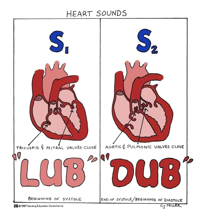
What are the heart sounds you hear. what is a heart murmur
“lub” (S1): caused by the closing of the AV valves (tri and bi), occurs at the oneset of ventricular systole, ventricular pressure > atrial pressure
“dub” (S2): caused by closing of semilunar valves (aortic and pulmonary)
heart murmur: defective valve function produces turbulent flow, can have back flow
What are the heart walls made out of
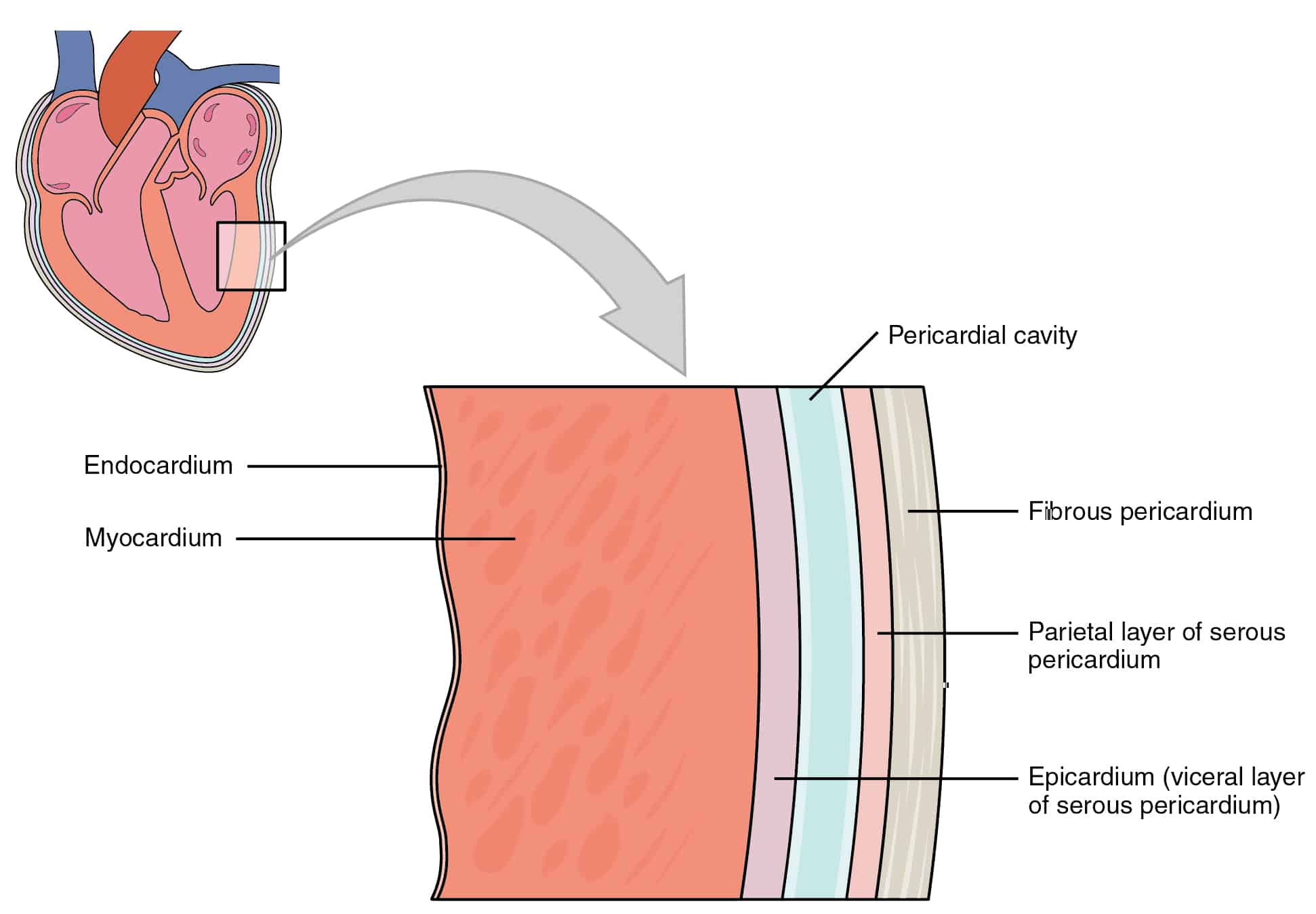
3 layers
endothelium: inner layers, lines the heart
Myocardium: cardiac muscle, middle layer
Epicardium: external layer, covers the heart