Pelvic US, Embryology, Pelvic Muscles + cavity, correlative imaging
 Call Kai
 Learn
Learn Practice Test
 Spaced Repetition
 Match
Match Flashcards
 Knowt Play
1/100

There's no tags or description
Looks like no tags are added yet.
Name | Mastery | Learn | Test | Matching | Spaced | Call with Kai | Chat |
|---|
No analytics yet
Send a link to your students to track their progress
101 Terms
most common used tdx for pelvic US
curvilinear/convex probe
2nd most common probe for pelvic US
transvaginal (endovaginal)
common reasons for pelvic US
pain, abnormal bleeding, palpable mass, procedure guidance, etc
sagittal plane notch is at
12 o’clock
sagittal plane labeled top/bottom and sides
anterior, posterior, superior/cephalic, inferior/caudal
transverse plan notch is at
9 o’clock
transverse plane labeled top/bottom and sides
anterior, posterior, right, left
transabdominal usually requires a — — and shows a — view
full bladder, global
transvaginal US usually requires a — — and a more — view
empty bladder, narrow
transperineal US is most similar to a — view
transvaginal (TV)
MRI uses a
very strong magnet and radiofrequency waves
CT uses
ionizing radiation and images in 3 planes
in MRI T1 shows fluid as — (low signal intensity)
dark
in MRI T2 shows liquid as —
bright
genitourinary tracts develop from
mesoderm (embryonic tissue)
embryology is dating from the
day of conception
in obstetrics (gestational age), dating is from the
LMP (2 weeks before conception)
embryo stage is
10 weeks from LMP
fetal stage is
10 weeks until delivery from LMP
ovum contributes — chromosomes + — chromosome, —
22, sex, X
sperm contributes — chromosomes + — chromosome, either - or -
22, sex, X or Y
fertilization yields a —, which is — chromosomes
diploid, 46
How many chromosomes (including sex chromosome) are in the non-fertilized ovum?
23
How many chromosomes (including sex chromosome) are in the sperm?
23
How many TOTAL chromosomes (including sex chromosome) are in the fertilized ovum?
46
gametes are either — or —
sperm or oocyte
gametes are created by —- migrating to the wall of the yolk sac in the 2nd week after conception
primordial germ cells
gonads are either
testicles or ovaries
gonads are formed by — developing on — part of —
urogenital ridges, anteromedial, mesonephros
paramesonephric duct also known as
Mullerian duct
mesonephric duct also known as
Wolffian duct
without testosterone, — ducts disappear and — ducts develop in both directions along gonads
mesonephric, paramesonephric
proximal paramesonephric ducts remain unfused and become —
fallopian tubes
distal paramesonephric ducts create —
uterus, cervix, upper 4/5 vagina
in genetic males, growth of — ducts occur and — ducts regress
mesonephric, paramesonephric
the vagina has dual origin, which are from the — and the —
paramesonephric duct, urogenital sinus
the paramesonephric duct forms the — part of the vagina
upper
the urogenital sinus forms the — part of the vagina
lower
hymen
thin tissue separating vagina from urogenital sinus
What is the embryologic origin of the lower part of the vagina?
urogenital sinus
What duct regresses in the biologic female?
mesonephric duct
What duct forms the fallopian tubes, uterus, and upper 4/5 of the vagina?
paramesonephric duct
Primordial germ cells migrate to the ________ _______ and form the gametes (sperm/oocytes)
yolk sac
external genitalia is the same in both genders until
week 7
genital tubercle elongates to become the — —-
primordial phallus
primordial phallus becomes the — or —
penis, clitoris
day 1 of embryology
conception/fertilization 46X or 46XY, genetic gender determined
week 2 embryology
gametes are created by primordial germ cells migrating to yolk sac wall
week 5 embryology
primordial germ cells enter embryo from yolk sac
by the end of the 5th week of embryology, PGCs are on the — — of the mesonephros
urogenital ridge
week 6 embryology
inducer germ cells implant on the genital ridges, eventually forming gonads
before week 7 embryology
male and female embryos have indifferent gonads
after week 7 embryology
SRY on Y chromosome causes testosterone production forming male structures
4th month oogenesis
primitive germ cells become oogonia
primary oocytes
surrounded by epithelial cells to become primordial follicles
primordial follicles
precursor to follicle that releases oocytes with menstrual cycle
primordial germ cells become —, which become primary —
oogonia, oocytes
genital tubercle
primordial (early) phallus
urogenital sinus forms the
lower vagina
rectus abdominis muscles
anterior muscles just lateral to midline surrounded by RECTUS SHEATH
arcuate line
invisible line that marks the lower limit of the posterior rectus sheath
Inferior to arcuate line, the — —- lies directly on the TRANSVERSALIS FASCIA
rectus abdominis
linia alba
fascia between 2 rectus abdominis muscles (the midline)
pelvic cavity is the — portion of the abdominopelvic cavity
lower
organs in the pelvic cavity
part of the large intestine, rectum, urinary bladder, reproductive organs
muscles of the pelvic diaphragm include
iliopsoas, piriformis, obturator internus, levator ani, coccygeus, rectus abdominis muscle

iliopsoas
combined iliacus (pelvic side wall) + psoas major muscles (distally) + psoas minor

piriformis
posterolateral wall muscle forming most of posterior pelvic floor
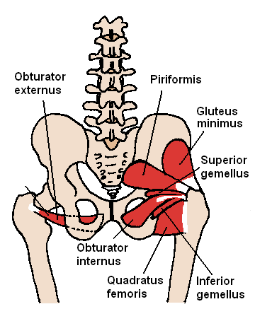
obturator internus
anterolateral pelvic sidewall, lateral to ovaries l
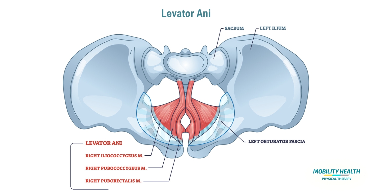
levator ani
pelvic floor, keeps organs from falling through the hole in the bottom of the boney pelvis

coccygeus
posterior muscle part of posterior pelvic floor

rectus abdominis muscle
anterior midline abdominal wall
inferior pelvic muscles include
piriformis, coccygeus, levator ani
what are the muscles of the pelvic floor collectively called?
pelvic diaphragm
what muscle lies on the lateral pelvic wall and is the pelvic sidewall
obturator internus
the pelvic skeleton includes what 5 bones?
ilium, ischium, pubis, sacrum, coccyx
what are the 4 innominate bones?
ilium, pubis, ischium, sacrum
male vs. female pelvis
female pelvis is wider
male vs female peritoneal cavity
male is a closed cavity, female has communication due to fallopian tubes
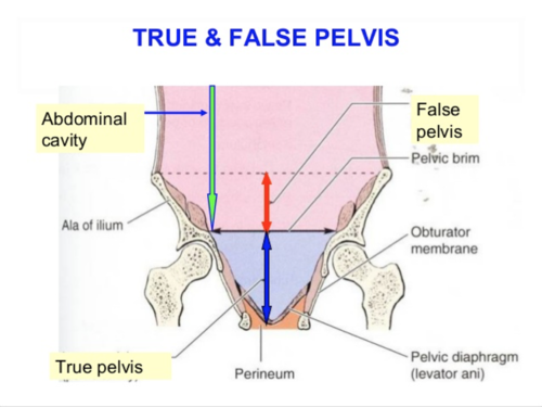
true vs false pelvis separated by —
linea terminalis
false pelvis is — and includes the —
above, bowel
true pelvis is — and includes — and —
below, pelvic organs and urinary bladder
true pelvis anterior compartment contains the — and —
bladder and reproductive organs
the true pelvis posterior compartment contains the (4)
posterior cul-de-sac, rectosigmoid muscle, perirectal fat, and presacral space
the cartilage that separates the 2 anteroinferior pelvic bones is called the
symphysis pubis
potential spaces is a
location where fluid could accumulate, normal or abnormal
the uterus is — to the rectum and — to the bladder
anterior, posterior
vesicouterine pouch also known as the
anterior pouch
rectouterine pouch also known as
posterior pouch, pouch of douglas, posterior cul de sac
space of Retzius
retropubis between bladder + pubic bone
Space of Retzius located between
symphysis pubis + bladder
space of Retzius is also know as
retropubic pouch
anterior cul de sac located between the
bladder and uterus
posterior cul de sac located between
uterus and rectum
vesicouterine pouch also known as
retropubic pouch
innominate bone is fused
ischium, ilium, and pubic bones
iliospoas
iliacus m + psoas major m (lateral)
psoas major is the large —- from — abdomen to —
bilateral muscle, upper, pelvis
linea alba is the the
midline fascia between 2 rectus abdominis muscles
rectus sheath is the
covering of rectus abdominis muscles