Exotics
1/207
There's no tags or description
Looks like no tags are added yet.
Name | Mastery | Learn | Test | Matching | Spaced |
|---|
No study sessions yet.
208 Terms
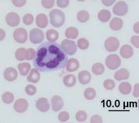
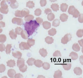
African hedgehog
Toxic neutrophil
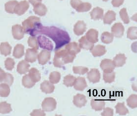
Mouse and rat, what cell type?
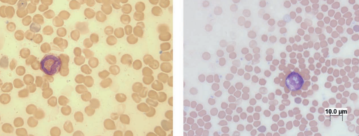
Eosinophil
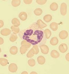
Mouse
Monocyte
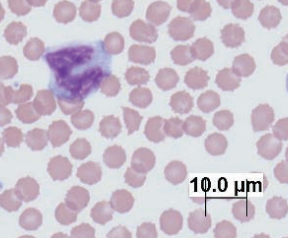
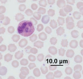
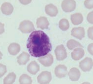
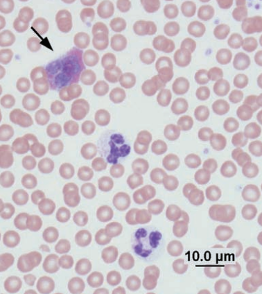
Guinea pig
Monocytes
Guinea pig
Large granular lymphocyte
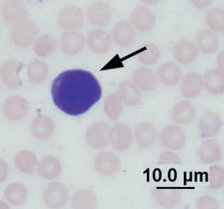
Guinea pig
Lymphocyte with Kurloff body
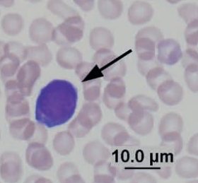
Chinchilla
Heterophil
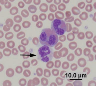
Rabbit
Heterophil
Rabbit
Basophil
Rabbit
Eosinophil arrow, heterophil below
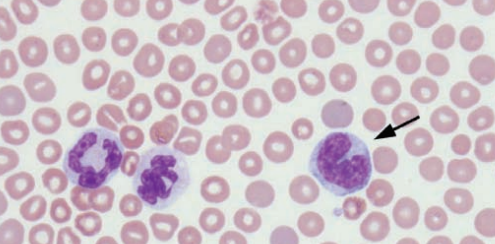
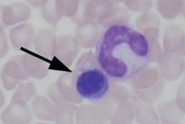
Rabbit
Heterophil and lymphocyte
Rabbit
Monocyte
Ferret
Eosinophil and neutrophils
Hedgehog
Neutrophil and eosinophils
Hedgehog
Neutrophils and monocytes
Guinea pig bone marrow, identify each
Rubriblast, prorubricyte, rubricyte and band
Guinea pig bone marrow
Myeloblast
Guinea pig bone marrow
Promyelocytes
Guinea pig bone marrow

Basophil myelocyte
Guinea pig bone marrow
Promegakaryocyte next to megakaryocyte
Guinea pig bone marrow
Promegakaryocyte
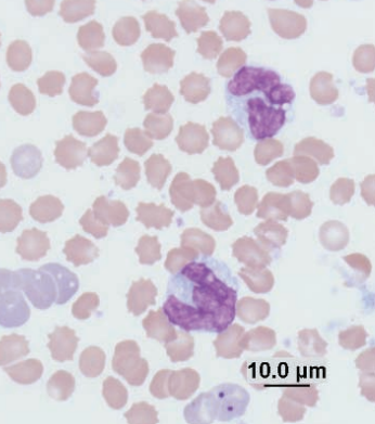
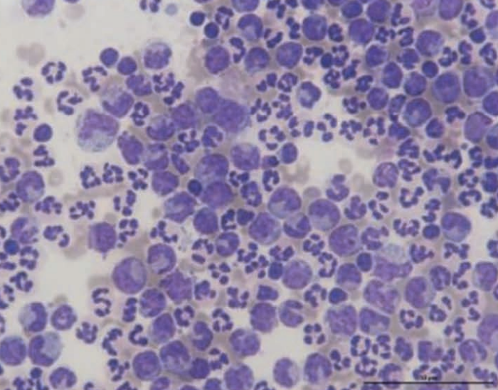
Chinchilla
Toxic seg and band
Chinchilla
Toxic band
What infectious agent causes diarrhea in ferrets, often with green mucus, and in chronic conditions a bird-seed-like appearance?
Two main ddx?
Ferret enteric coronavirus (epizootic catarrhal enteritis, ECE)
Ddx: foreign body and Helicobacter mustelae gastritis
Cytospin from tracheal wash of a guinea pig
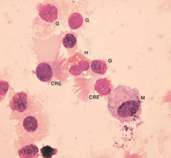
Normal. Ciliated respiratory epithelial cells (CRE), goblet cells (G), heterophil (H), macrophage (M)
Conjunctival scraping from a guinea pig
Normal melanocytes
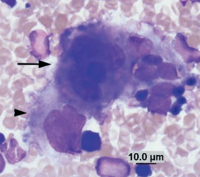
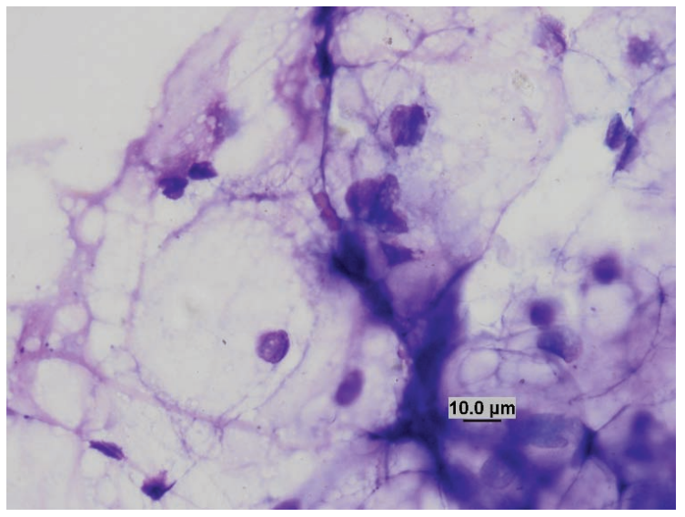
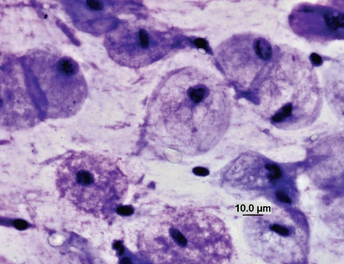
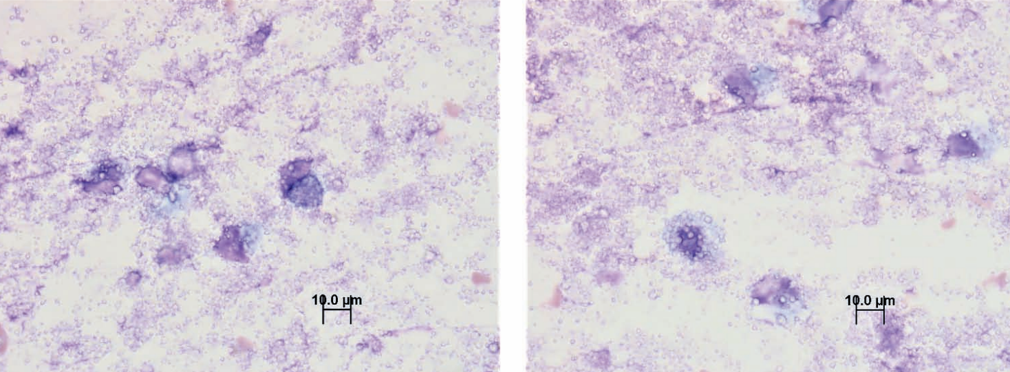
Choroid plexus imprint (from CNS)
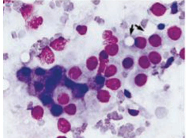
Alpha cells have dark blue cytoplasm and densely stained nuclei, and beta cells have paler nuclei with a cribriform chromatin distribution. Two gamma cells with intracellular vesicles are seen (arrows).
Guinea pig conjunctival scraping
Normal epithelium
Ferret lymph node
Normal, two mast cells in centre
Ferret, liver
Intracytoplasmic bile pigment
Rabbit, nasal flush
Heterophilic rhinitis
Ferret, subcutaneous mass
Mixed-cell inflammation
Mass from a hamster
Fibroblasts from an abscess
Ferret abdominal fluid
Macrophagic inflammation in peritonitis
Hedgehog, skin
Eosinophilic inflammation
Tiger, tongue
Neutrophils, eosinophils, and a mast cell
Sugar glider, skin
Calcinosis circumscripta
Ferret, liver
Macrophagic inflammation with Mycobacterium (goodii)
Rabbit, conjunctival swab
Heterophilic inflammation
Rat, skin
Lipoma
Ferret, skin
Histiocytoma
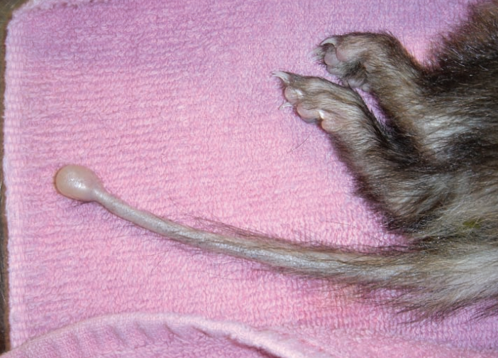
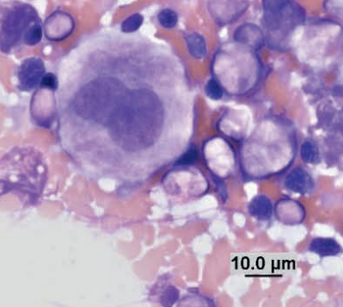
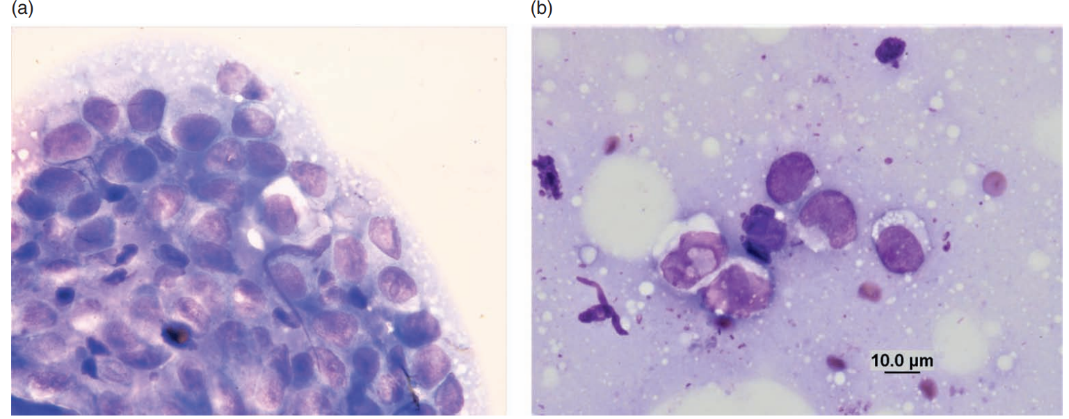
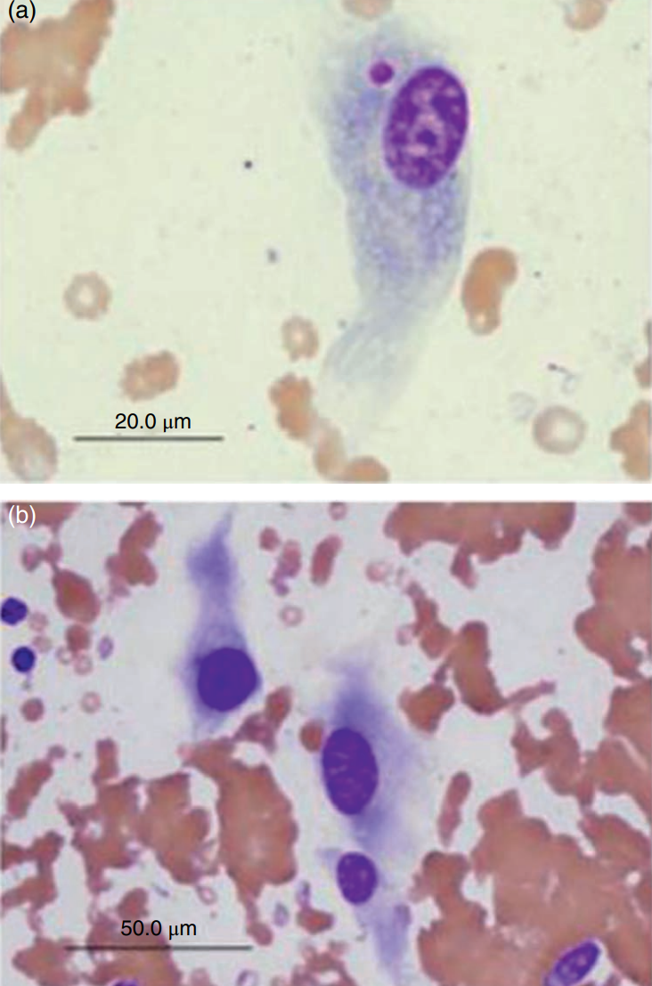
Ferret, chordoma
Ferret, tail
Chordoma
Large foamy physaliphorous cells with a background of mucinous pinkish material
Ferret, tail
Chordoma
Large foamy physaliphorous cells with a background of mucinous pinkish material
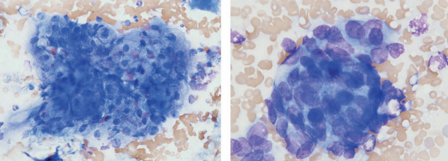
Rat, ear
Zymbal gland adenoma
Rat, ear
Zymbal gland adenoma
Caudal abdominal mass in a hedgehog
Uterine leiomyoma
Ferret, prostate
Squamous metaplasia
Ferret, lymph node
Reactive
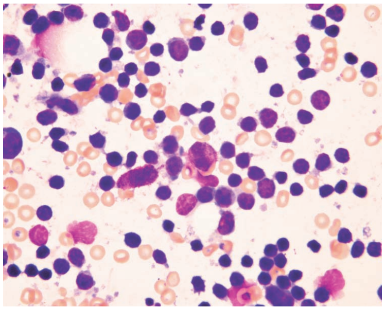
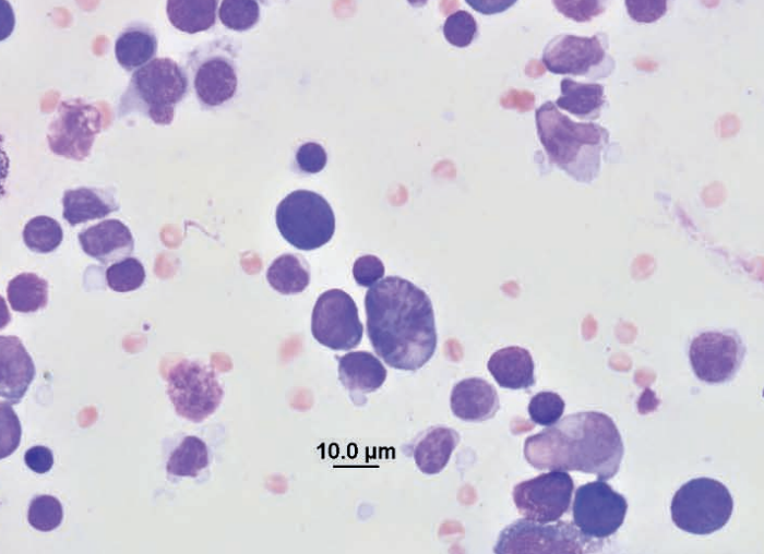
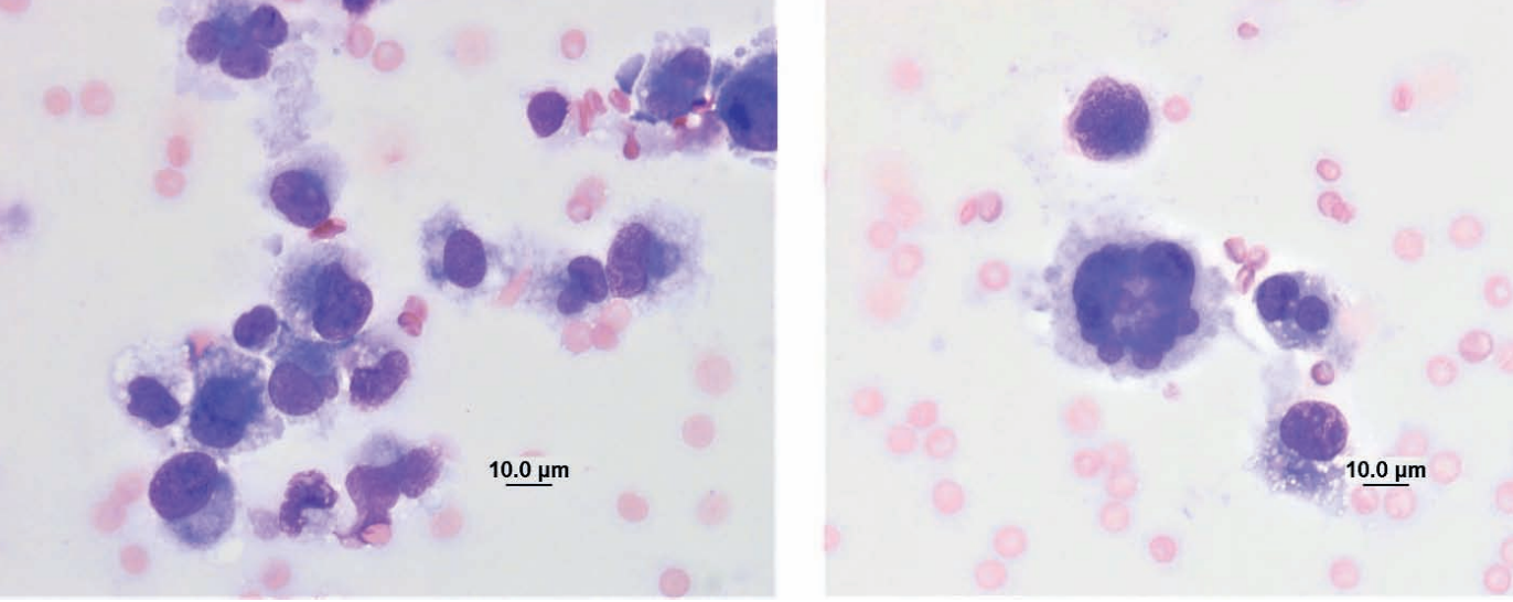
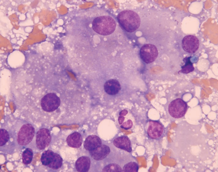
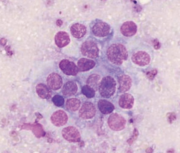
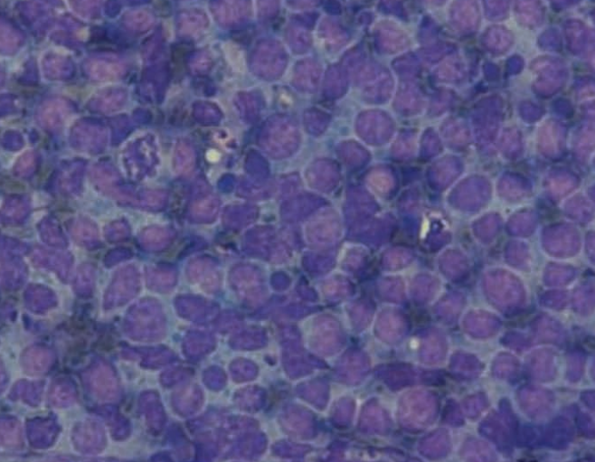
Ferret, spleen
Multiple myeloma
Guinea pig, neck mass
Thyroid adenoma
Tiger, skin
MCT
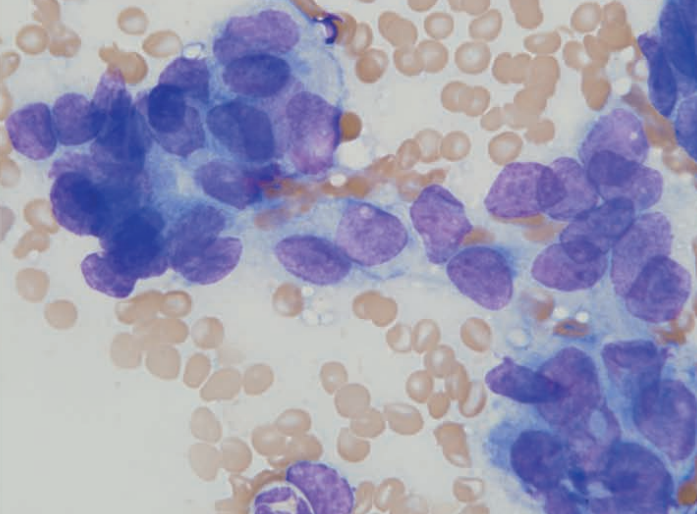
Rat
Histiocytic sarcoma
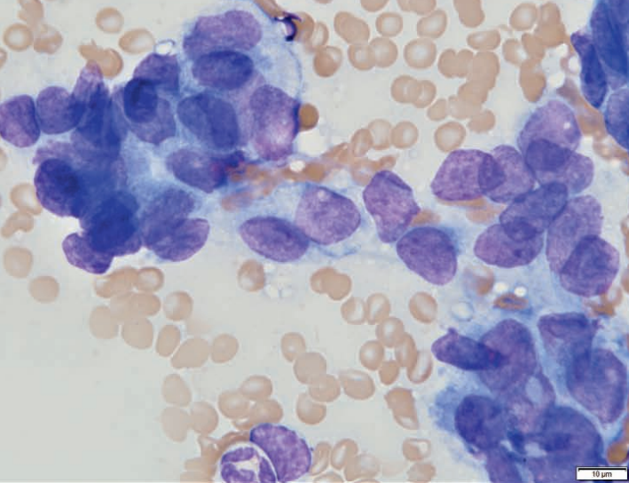
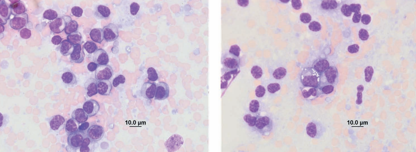
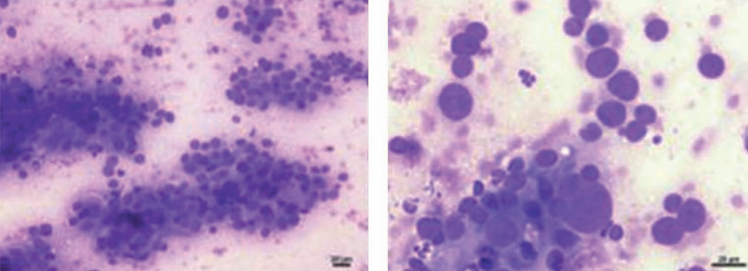
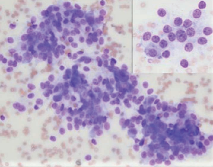
Ferret, lymph node
Lymphoma
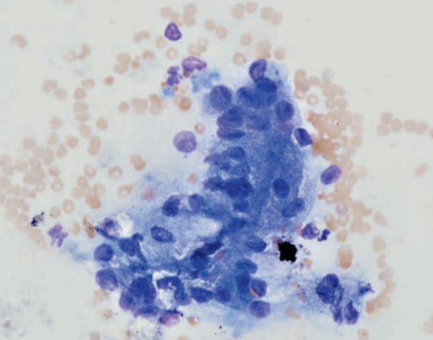
Ferret, lymph node
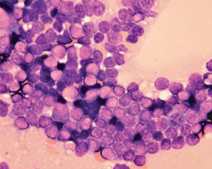
Lymphoma
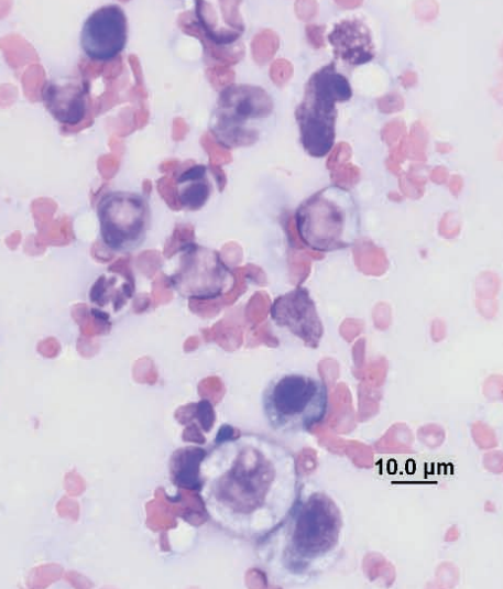
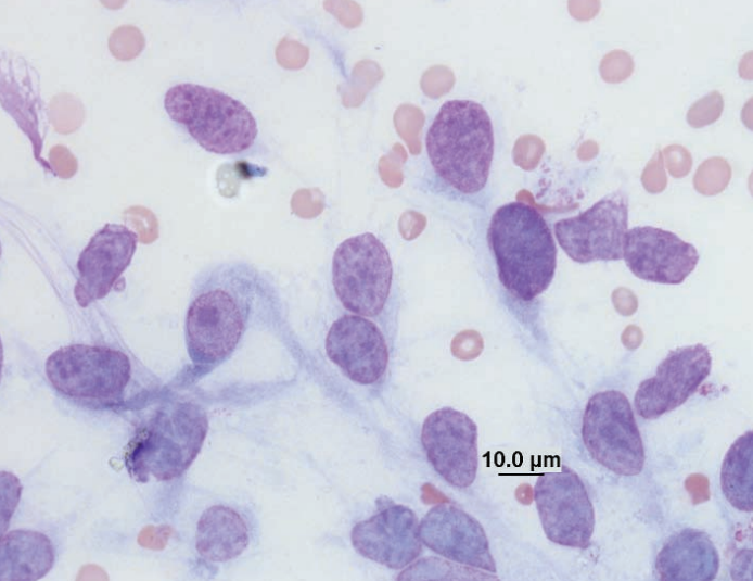
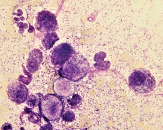
Ferret, abdominal mass
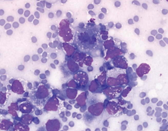
T-cell lymphoma with pleomorphism
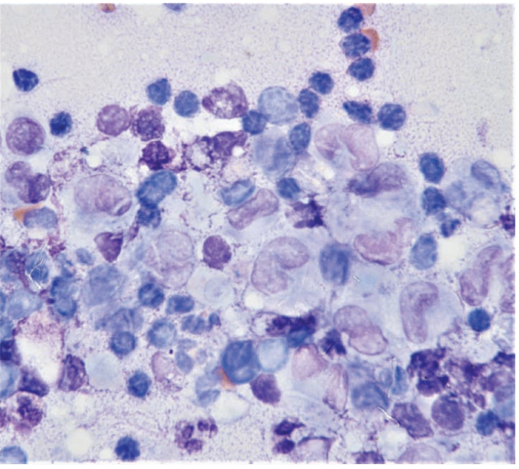
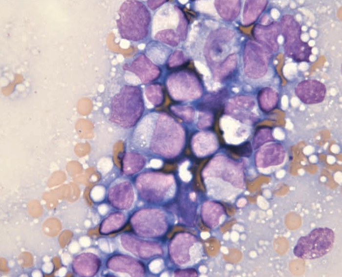
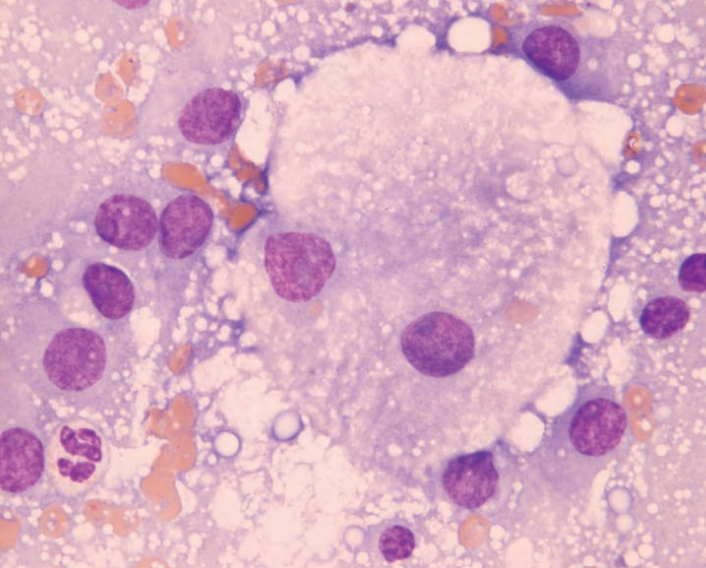
Ferret, spleen
Multiple myeloma
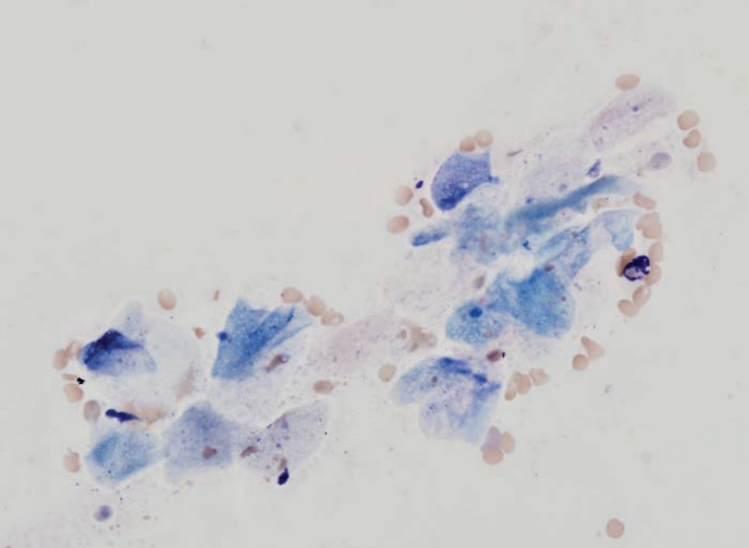
Ferret
Carcinoma
Guinea pig, uterine impression
Endometrial adenoma
Hamster, ventral skin
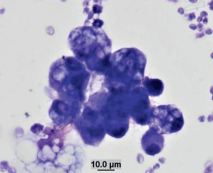
Mammary adenocarcinoma
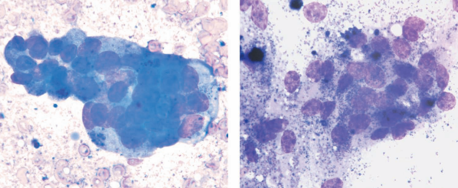
Ferret, abdominal mass
Adrenal adenocarcinoma
Ferret, abdominal mass
Adrenal adenocarcinoma
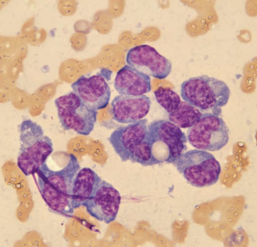
Hedgehog, oral FNA
SCC
Gerbil, abdominal mass
Poorly-differentiated sarcoma
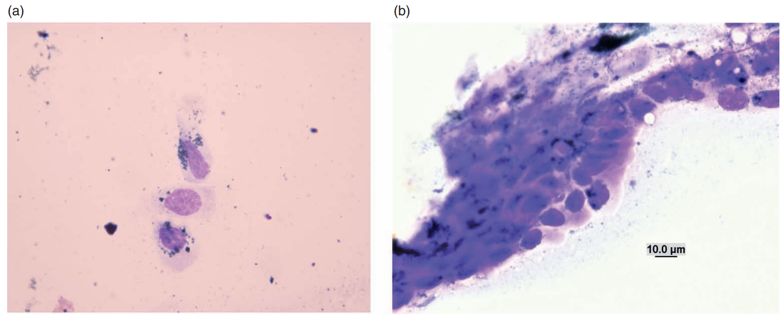
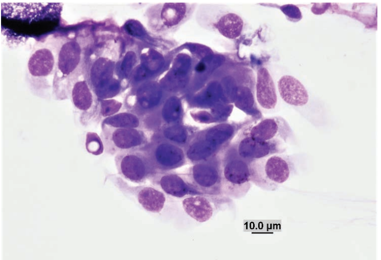
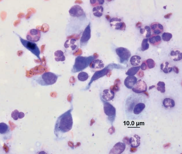
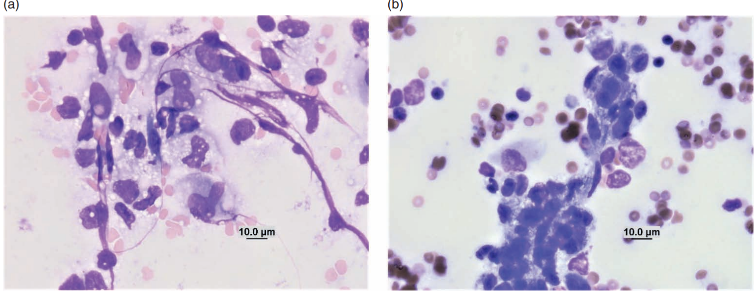
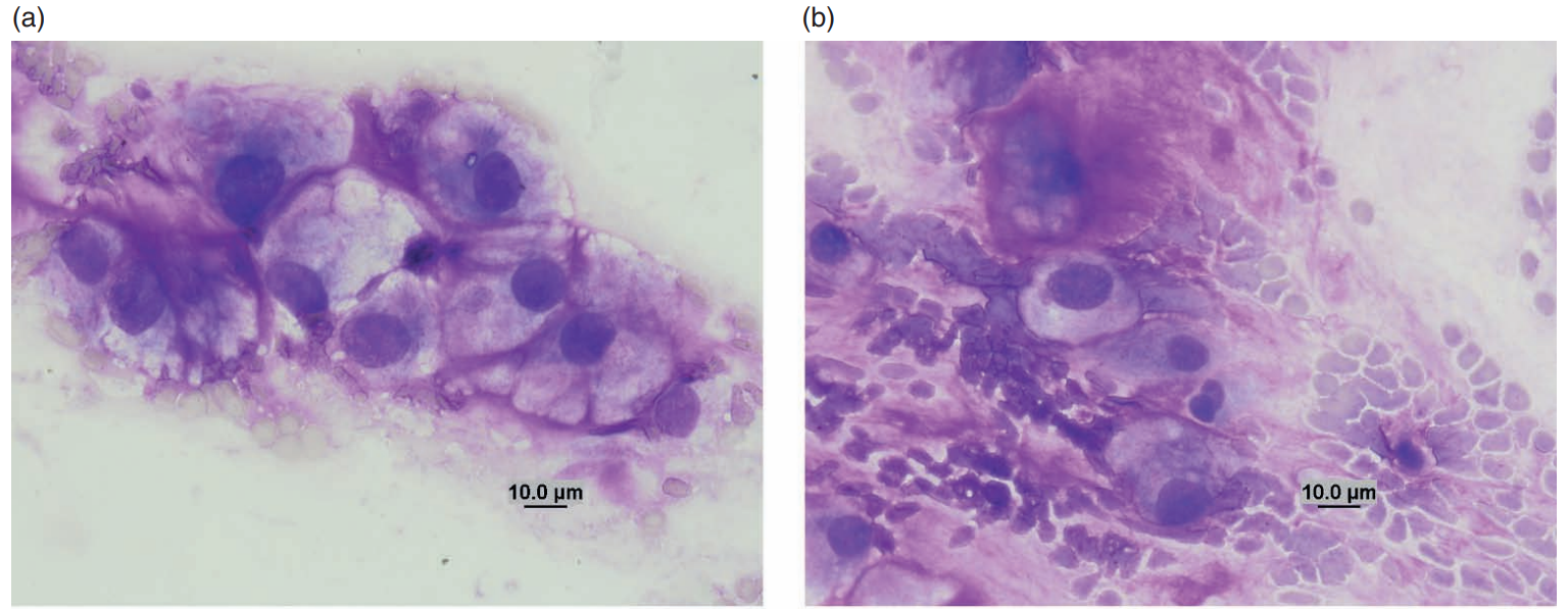
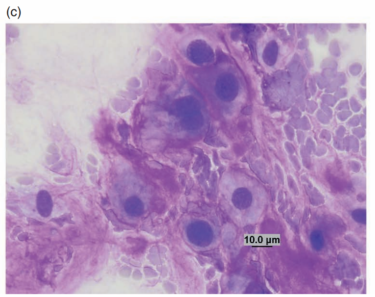
Rabbit, nodule on muzzle
Shope fibroma
Rabbit (Shope) fibroma virus (Poxviridae)
The virus ordinarily produces localized fibromas where cytologic preparations consist of spindle-shaped cells exhibiting moderate anisocytosis and anisokaryosis (Figures 7.86a and 7.86b). The nuclei are large (1–3 times the size of erythrocytes), round to oval, eccentric, and exhibit finely stippled chromatin with multiple nucleoli. The basophilic cytoplasm occasionally contains round to oval eosinophilic inclusions that vary in size (3–5 μm) and shape
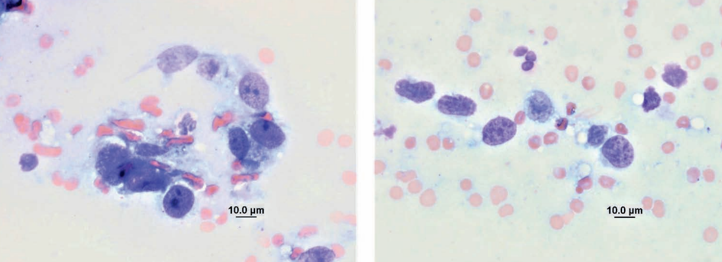
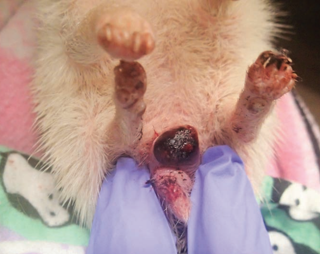
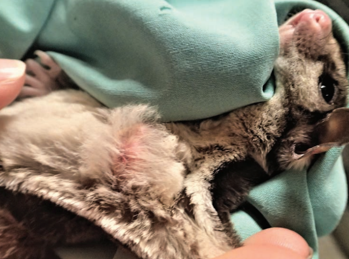
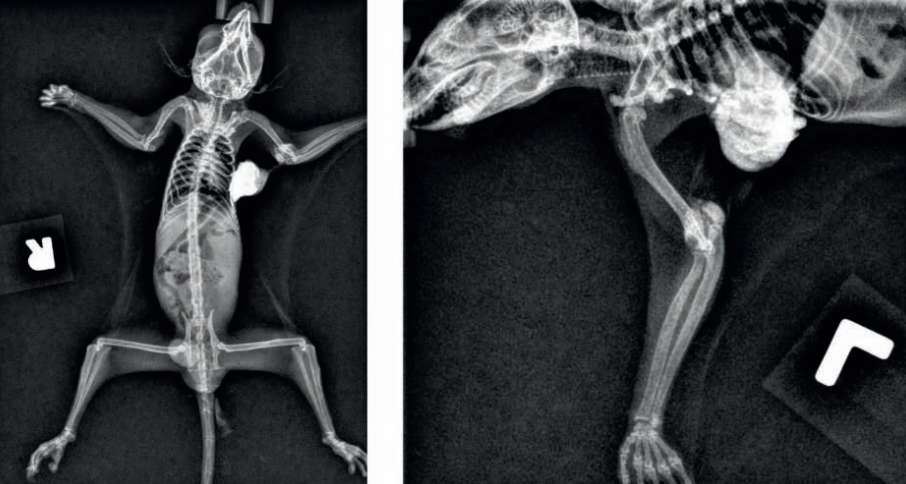
Sugar glider, mass projecting from right aspect of marsupium
Carcinoma or ACA, ddx amelanotic melanoma
Ferret, perianal mass
Anal sac adenocarcinoma
Guinea pig, skin

Melanoma
Ferret, abdominal mass
Ferret, abdominal mass
Term
Adrenal gland adenoma
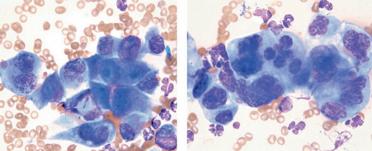
Hedgehog, right cervical mass
Thyroid carcinoma
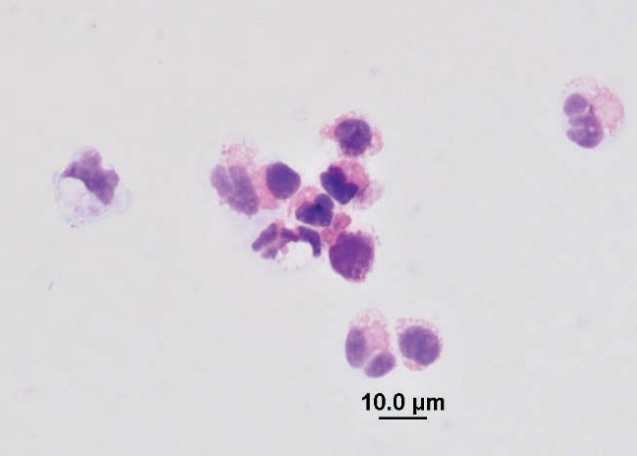
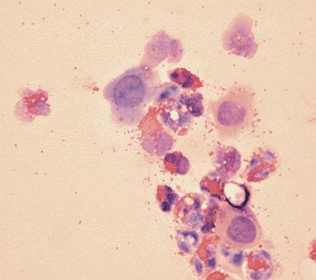
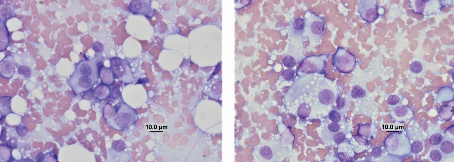
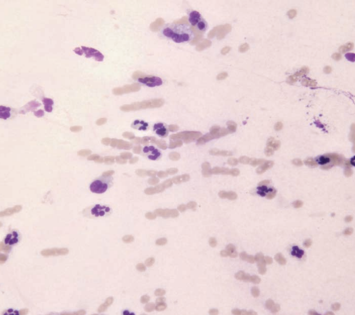
Ferret, abdominal fluid cytospin
Chylous effusion, but see mixed population of nondegenerate neutrophils and macrophages that show intermittent foamy cytoplasm. Small lymphocytes are in low numbers.
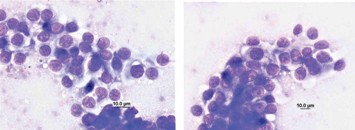
Ferret, abdominal mass
Lymphoma
Ferret, fluctuant mass on the head
Mucocele
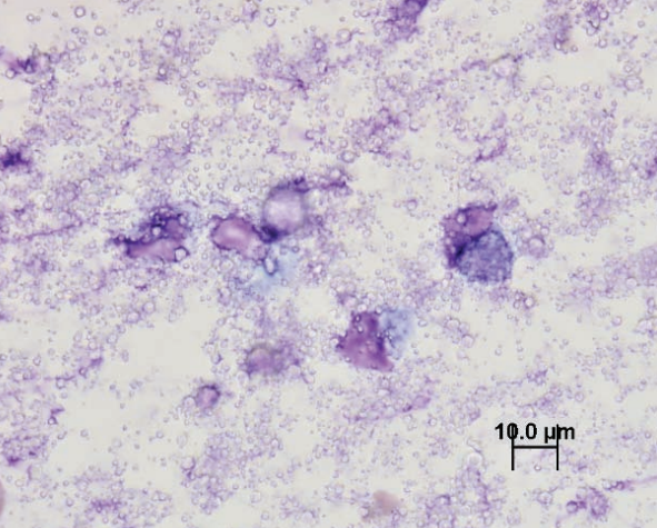
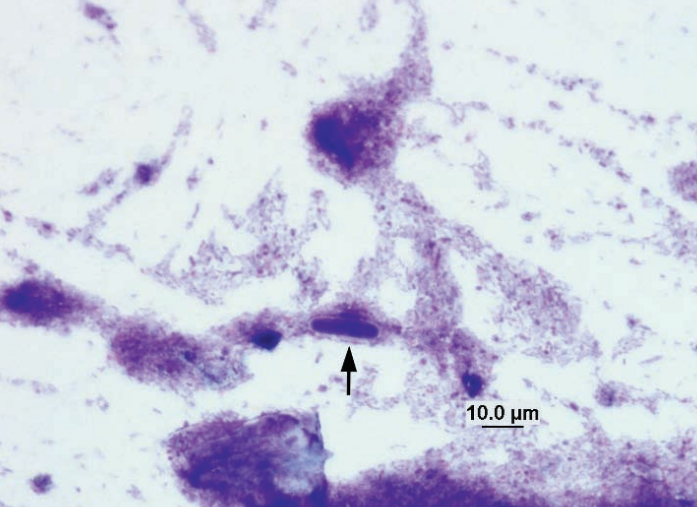
Hamster, fecal smear
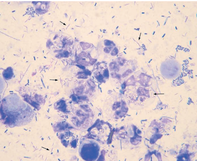
Campylobacter-like organisms in case of septic colitis
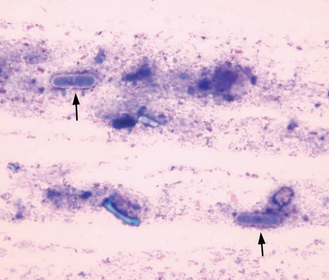
Hamster, rectal swab
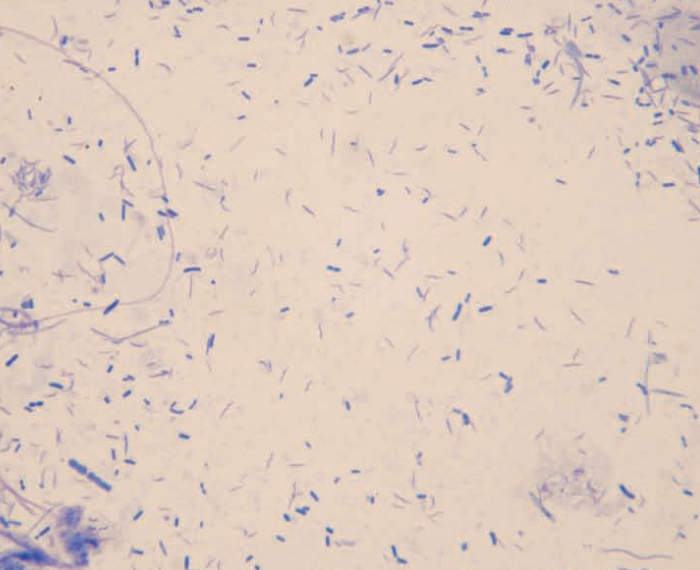
Campylobacter-like organisms in a case with colitis
Hamster, fecal smear
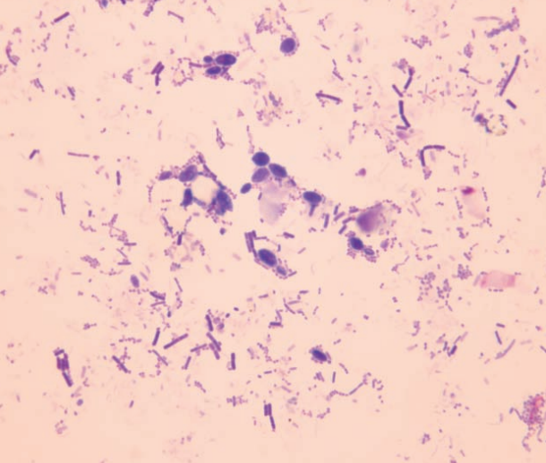
Budding yeast in a case with diarrhea
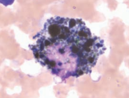
Ferret, lung
Blastomyces dermatitidis
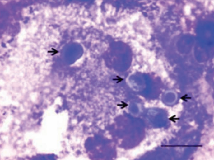
Rabbit, fecal smear
Saccharomyces spp. yeast, part of normal flora of the cecum
Rabbit, fecal smear
Saccharomyces spp. yeast, part of normal flora of the cecum
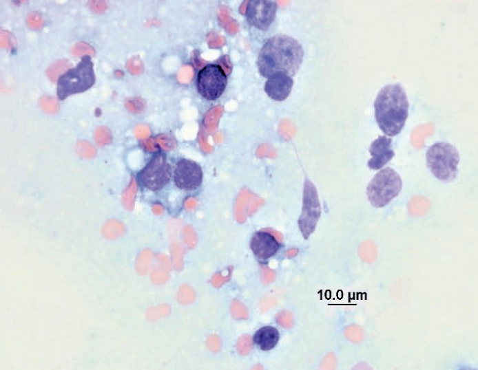
Rat, fecal smear
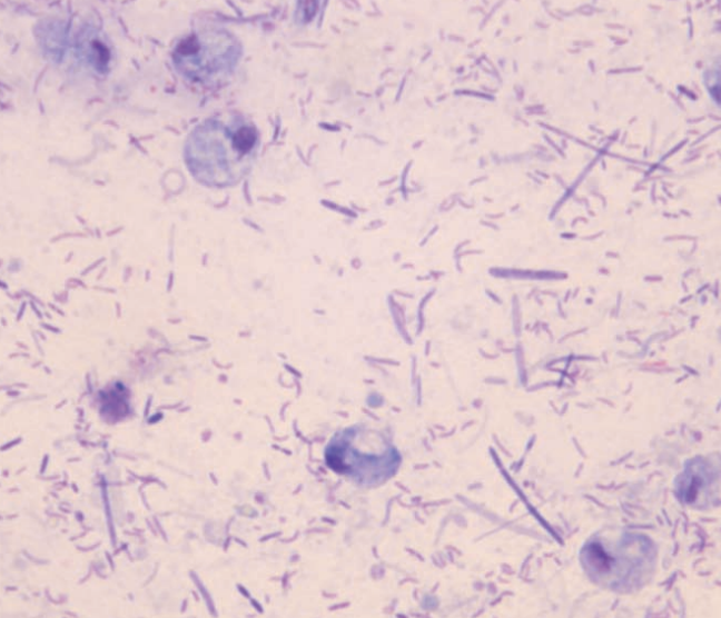
Trichomonas in a case with diarrhea
Hedgehog, swelling under right forelimb
Mammary adenocarcinoma
Rat, rapidly growing mass on face
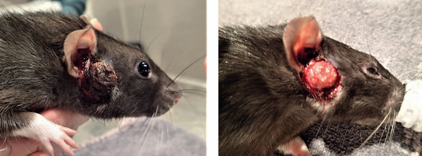
Ddx sebaceous carcinoma, Zymbal gland adenocarcinoma based on location
Hedgehog, vaginal mass
Leiomyosarcoma, was also within the uterine horn
Sugar glider, mass in left axilla
Calcinosis circumscripta
Blood smear from a turkey
Clumping, vacuolation, and pyknotic nuclei from 48h in EDTA
Avian blood smears
Clumping due to heparin
Blood smear from eclectus parrot
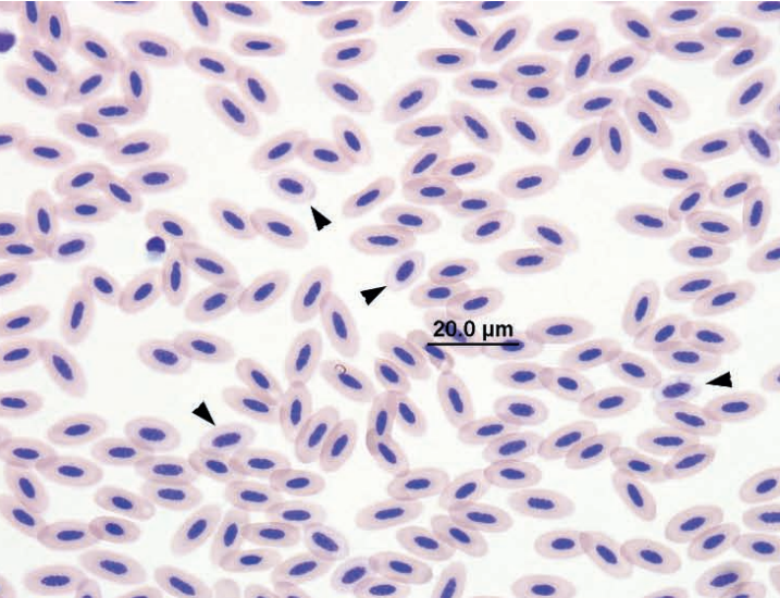
Hypochromic RBCs
Blood smear from a turkey vulture
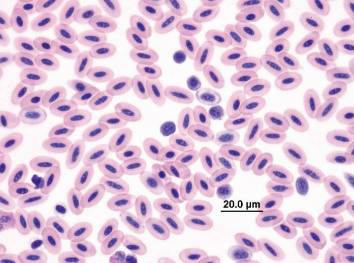
Increased numbers of immature erythrocytes (early and mid-polychromatic rubricytes) without an appropriate increase in polychromasia due to lead poisoning
Avian blood smear
Artifact from slow drying
Blood smear from an eclectus parrot
Agglutination and erythrophagia
Blood smear from a hawk
Basophilic rubricyte and increased polychromatophils
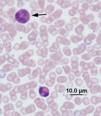
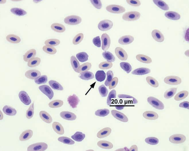
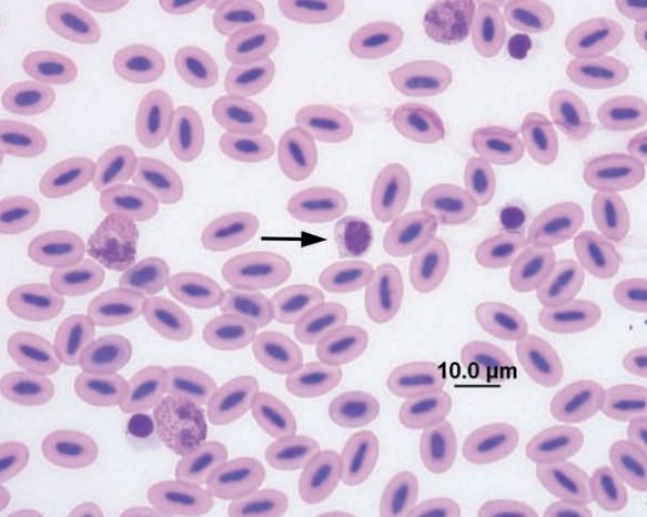
Amazon parrot blood
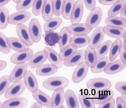
Lymphocyte
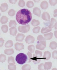
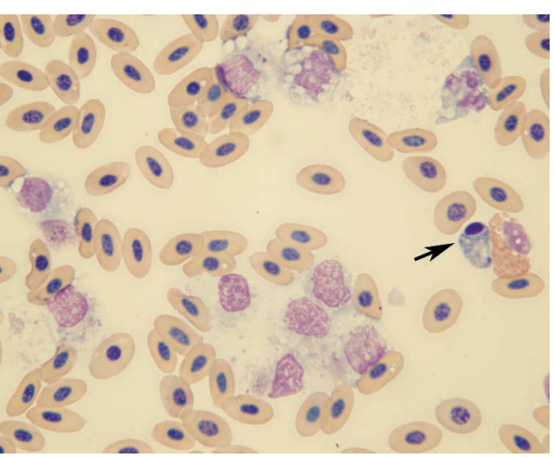
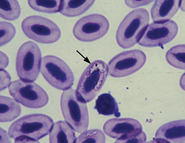
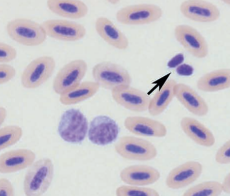
Conure blood
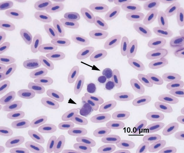
Immature erythrocyte—early polychromatic rubricyte (arrow), lymphocyte (arrowhead), and thrombocytes
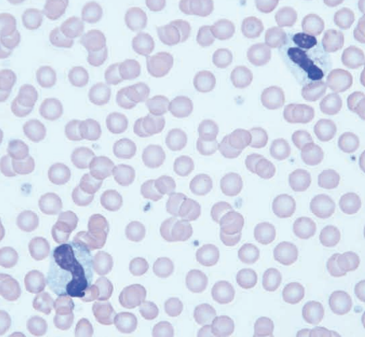
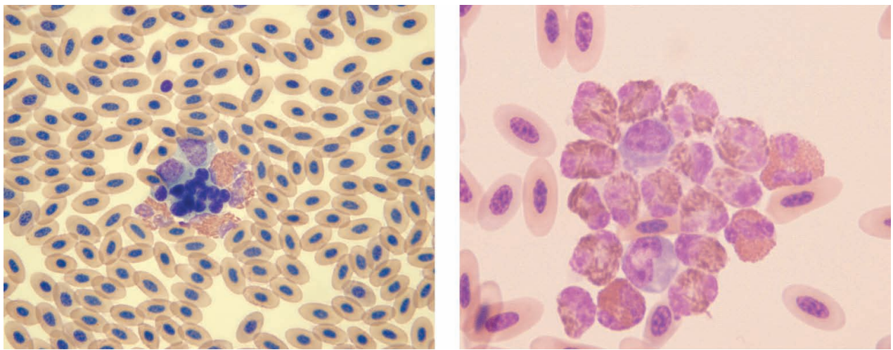
Domestic chicken blood
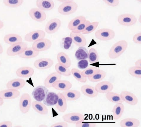
Large lymphocytes (arrowheads), medium lymphocyte (arrow), and thrombocyte
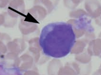
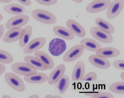
Amazon parrot
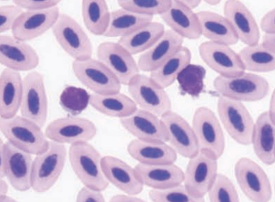
Lymphocytes
Barred owl
Lymphocyte with azurophilic cytoplasmic granules (arrow) and thrombocyte
Domestic duck
Two large lymphocytes (center) and two thrombocytes (arrow)
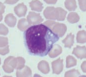
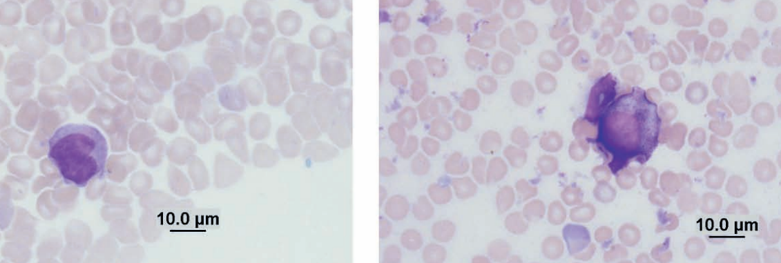
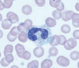
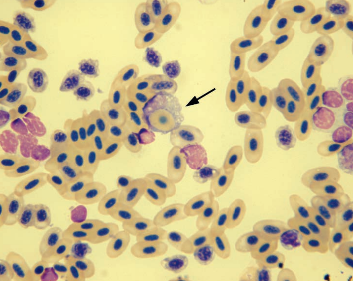
Conure
Large reactive lymphocyte
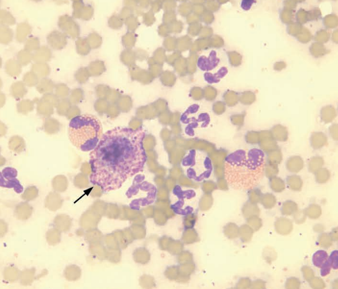
Gannet (a sea bird) blood smear
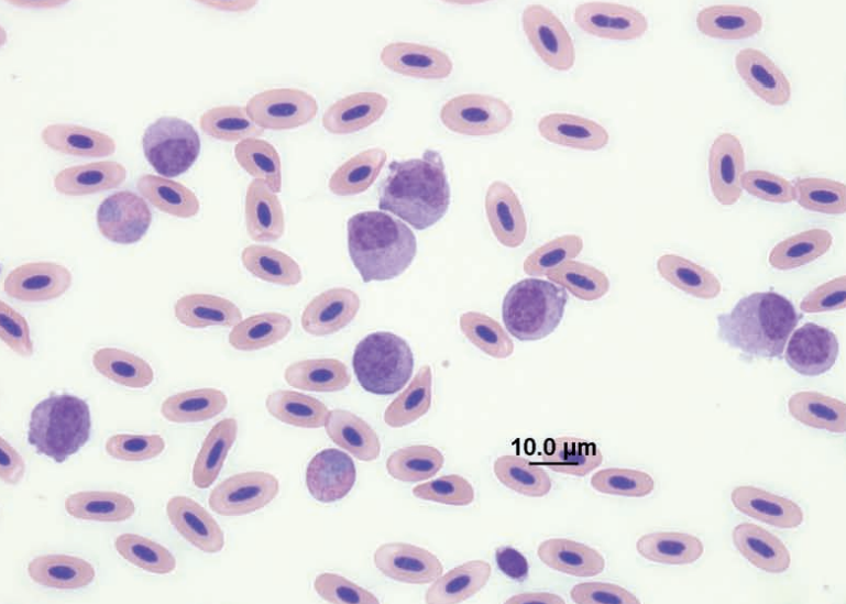
Monocytosis