BSCI422 FINAL

0.0(0)
Studied by 0 people
call kaiCall Kai
Locked
learnLearn
examPractice Test
spaced repetitionSpaced Repetition
heart puzzleMatch
flashcardsFlashcards
GameKnowt Play
Card Sorting

1/47

encourage image

There's no tags or description

Looks like no tags are added yet.

Last updated 4:12 AM on 5/15/26
Name
Mastery
Learn
Test
Matching
Spaced
Call with Kai
Chat

No analytics yet

Send a link to your students to track their progress

48 Terms

1
New cards

Anti-Viral Immunity

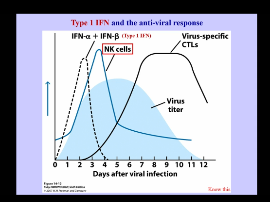
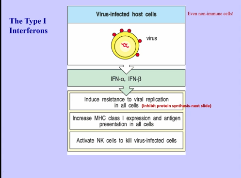
  • IFN-alpha and IFN-beta (Type 1 IFN)

    • innate IFN production occurs quickly in response to viral infection

    • NK cell production follows shortly after

    • Virus-specific CTLs appear later (antigen specific, require clonal expansion, etc → all of which take time, leading to their presence later in the process)
<ul><li><p>IFN-alpha and IFN-beta (Type 1 IFN) </p><ul><li><p>innate IFN production occurs quickly in response to viral infection</p></li><li><p>NK cell production follows shortly after</p></li><li><p>Virus-specific CTLs appear later (antigen specific, require clonal expansion, etc → all of which take time, leading to their presence later in the process)</p></li></ul></li></ul><p></p>
2
New cards
<p>Type 1 IFN</p>

Type 1 IFN

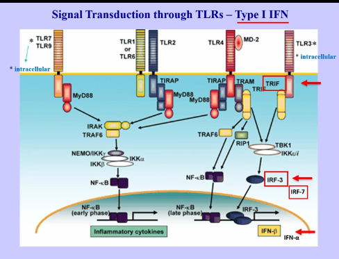
  • can be made during innate immune responses

  • image outdate: TLR3 would be inside the cell on endosomes

  • TLR3 signals on adaptor protein, TRIF, which activates IRF-3 and IRF-7 (regulatory factors), turning on production of type 1 interferon production (IFN-beta and alpha)

  • TLR → on plasma membranes in endosomes

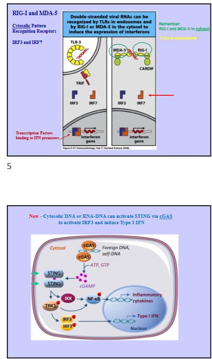
  • Cytosolic Pattern Recognition Receptors: RIG-I and MDA-5, which can recognize viral RNAs, also activating IRF-3 and IRF-7, producing transcription of interferon genes

  • cGAS

    • recognizes double stranded cytosolic DNA or RNA-DNA, which activates STING (stimulator of interferon signaling), which activates IRF3 and other interferon regulatory factors to provide type 1 interferons
<ul><li><p>can be made during innate immune responses </p></li><li><p>image outdate: TLR3 would be inside the cell on endosomes </p></li><li><p>TLR3 signals on adaptor protein, TRIF, which activates IRF-3 and IRF-7 (regulatory factors), turning on production of type 1 interferon production (IFN-beta and alpha) </p></li><li><p>TLR → on plasma membranes in endosomes</p></li><li><p>Cytosolic Pattern Recognition Receptors: RIG-I and MDA-5, which can recognize viral RNAs, also activating IRF-3 and IRF-7, producing transcription of interferon genes </p></li><li><p>cGAS </p><ul><li><p>recognizes double stranded cytosolic DNA or RNA-DNA, which activates STING (stimulator of interferon signaling), which activates IRF3 and other interferon regulatory factors to provide type 1 interferons </p></li></ul></li></ul><p></p>
3
New cards
<p>Type 1 Interferons (cont) </p>

Type 1 Interferons (cont)

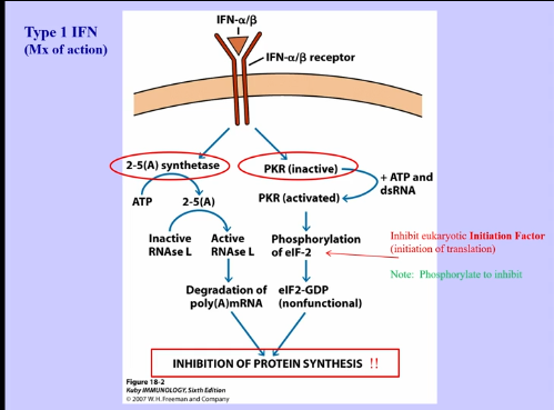
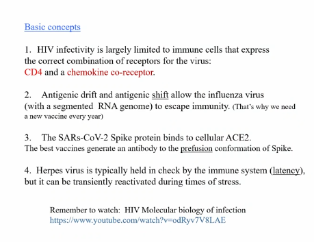
  • present on all cells, because all cells have the potential to be infected by viruses

  • potent inhibitor of protein synthesis and can activate NK cells

    • inhibition of protein synthesis:

      • activates 2-5(A) synthetase, which leads to degradation of mRNA, meaning proteins are unable to be made

      • also activates PKR, leading to phosphorylation of initiation factors (elF-2), preventing initiation of transcripts, lading to inhibited protein synthesis

      • inhibits protein production of both viruses and host cells (can be a problem)

<ul><li><p>present on all cells, because all cells have the potential to be infected by viruses</p></li><li><p>potent inhibitor of protein synthesis and can activate NK cells</p><ul><li><p>inhibition of protein synthesis:</p><ul><li><p>activates 2-5(A) synthetase, which leads to degradation of mRNA, meaning proteins are unable to be made</p></li><li><p>also activates PKR, leading to phosphorylation of initiation factors (elF-2), preventing initiation of transcripts, lading to inhibited protein synthesis </p></li><li><p>inhibits protein production of both viruses and host cells (can be a problem) </p></li></ul></li></ul></li></ul><p></p>
4
New cards
<p>NK Cells</p>

NK Cells

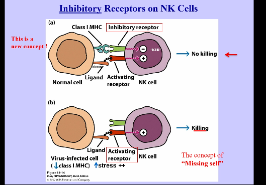
  • similar to CTLs, however, they recognize targets in very different ways

  • Recognition occurs from combination of activating and inhibitory receptors:

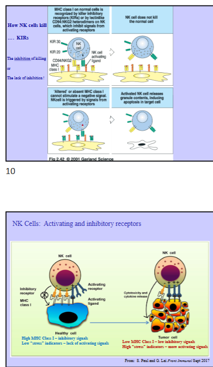
    • NK cells have receptors that recognize MHC class 1 (all healthy, nucleated cells are expressing MHC class 1, meaning if NK cell sees MHC class 1, it transmits a negative signal into the NK cell, telling it to not kill the cell)

      • receptors that recognize healthy cells with MHC class 1 = Killer Inhibitory Receptors (KIR)

    • NK cells also contain activating receptors, which recognize stress (when cell infected, they begin expressing stress-induced ligands, which get pushed out from the cytosol to outside the plasma membrane when cell becomes stressed, and are recognized by NK activating receptors)

      • stress:

        • oxidative stress during inflammation

        • protein synthetic stress (virus steals machinery)

    • killing activity mediated by balance of activating and inhibiting signals

      • inhibiting signals are slightly more potent

    • Missing Self → concept that missing MHC class 1 or lower expression of MHC class 1 is recognized by NK cells

      • if host cell is virally infected, its protein machinery will be taken over, leading to decreased host protein production, meaning less MHC class 1 is being generated and cell begins expressing stress signals (combination of low MHC class 1 and high stress signals → results in NK cell killing)


NK Cell Killing:

  • Killer Inhibitory Receptors (KIRs, CD94, NKG2, etc) transduce negative signals, which turn off NK cells and preventing signaling and killing

  • If activating signals are received (stressed) = induces killing

  • low stress levels will not result in killing if MHC class 1 is recognized, as inhibitory signal will over power activating signal

  • tumor cells = growing fast = no time to synthesize MHC class 1 = causes expression of stress induced ligands on cells = NK cell activating signals and lower MHC class 1 = lower inhibitory signals = NK cell killing occurs
<ul><li><p>similar to CTLs, however, they recognize targets in very different ways</p></li><li><p>Recognition occurs from combination of activating and inhibitory receptors:</p><ul><li><p>NK cells have receptors that recognize MHC class 1 (all healthy, nucleated cells are expressing MHC class 1, meaning if NK cell sees MHC class 1, it transmits a negative signal into the NK cell, telling it to not kill the cell)</p><ul><li><p>receptors that recognize healthy cells with MHC class 1 = Killer Inhibitory Receptors (KIR) </p></li></ul></li><li><p>NK cells also contain activating receptors, which recognize stress (when cell infected, they begin expressing stress-induced ligands, which get pushed out from the cytosol to outside the plasma membrane when cell becomes stressed, and are recognized by NK activating receptors)</p><ul><li><p>stress:</p><ul><li><p>oxidative stress during inflammation</p></li><li><p>protein synthetic stress (virus steals machinery)  </p></li></ul></li></ul></li><li><p>killing activity mediated by balance of activating and inhibiting signals</p><ul><li><p>inhibiting signals are slightly more potent</p></li></ul></li><li><p>Missing Self → concept that missing MHC class 1 or lower expression of MHC class 1 is recognized by NK cells</p><ul><li><p>if host cell is virally infected, its protein machinery will be taken over, leading to decreased host protein production, meaning less MHC class 1 is being generated and cell begins expressing stress signals (combination of low MHC class 1 and high stress signals → results in NK cell killing)  </p></li></ul></li></ul></li></ul><p></p><p>NK Cell Killing:</p><ul><li><p>Killer Inhibitory Receptors (KIRs, CD94, NKG2, etc) transduce negative signals, which turn off NK cells and preventing signaling and killing</p></li><li><p>If activating signals are received (stressed) = induces killing </p></li><li><p>low stress levels will not result in killing if MHC class 1 is recognized, as inhibitory signal will over power activating signal </p></li><li><p>tumor cells = growing fast = no time to synthesize MHC class 1 = causes expression of stress induced ligands on cells = NK cell activating signals and lower MHC class 1 = lower inhibitory signals = NK cell killing occurs</p></li></ul><p></p>
5
New cards
<p>NK Cell Receptors Structures</p>

NK Cell Receptors Structures

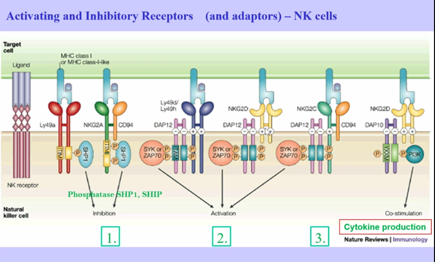
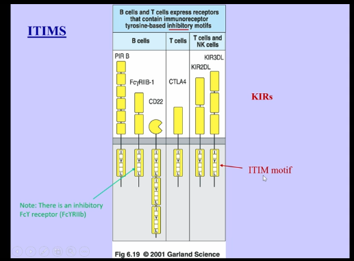
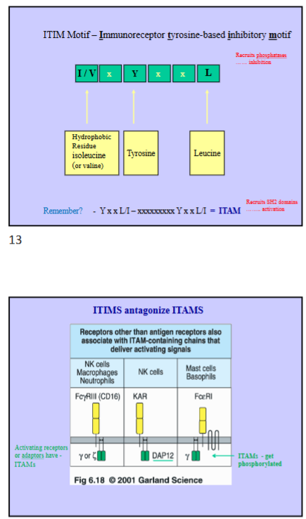
  • Killer Inhibitory Receptors signal through ITIMS (immuno-based tyrosine inhibition motif)

    • similar to single ITAM

    • I _Y _ L

    • tyrosines become phosphorylated (just single tyrosine)

    • ITIMS require phosphatases (remove phosphates), which inhibit activating signals associated with ITAMs

    • vs ITAMs (phosphorylated in 2 sequentially spaced tyrosines, which activate SH2 domains)

    • ITIMs dominate ITAMs, preventing phosphorylation and activation of ITAMs

    • receptors and adaptors have ITIMs or ITAMs (receptors associated with adaptors)

<ul><li><p>Killer Inhibitory Receptors signal through ITIMS (immuno-based tyrosine inhibition motif) </p><ul><li><p>similar to single ITAM</p></li><li><p>I _Y _ L </p></li><li><p>tyrosines become phosphorylated (just single tyrosine)</p></li><li><p>ITIMS require phosphatases (remove phosphates), which inhibit activating signals associated with ITAMs </p></li><li><p>vs ITAMs (phosphorylated in 2 sequentially spaced tyrosines, which activate SH2 domains) </p></li><li><p>ITIMs dominate ITAMs, preventing phosphorylation and activation of ITAMs</p></li><li><p>receptors and adaptors have ITIMs or ITAMs (receptors associated with adaptors) </p></li></ul></li></ul><p></p>
6
New cards
<p>NK Cell Receptor Process</p>

NK Cell Receptor Process

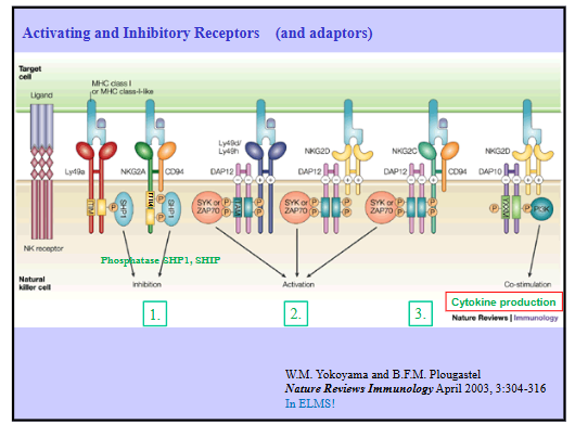
  1. Inhibitory receptors that recognize MHC class 1 on healthy cells

    • contain ITIMs, which allows for the recruitment of phosphatases (SHP1 or SHIP), which then inhibit tyrosine phosphorylation (especially associated with ITAMs)

  2. Activating Receptors recruit adaptor molecules (DAP12) as ITAMs, which can then be phosphorylated, recruiting T and B cell master kinases (SYK or ZAP70), leading to downstream activation

    • recognize stress-induced ligands on virally infected cells = provides activating signals

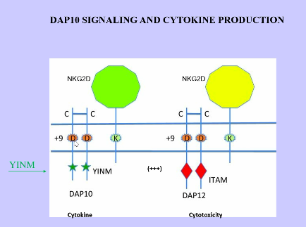
  3. NK Cells - Cytokine Production

    • different receptor (not inhibitory or activating receptor), which associates with a slightly different adaptor (DAP10), which contains a different cytosolic domain (not ITAM or ITIM), which activates cytokines by recruiting PI3K (PI3 kinase)

    • similar receptors (not same), slightly different adaptors, and entirely different cytosolic domains = produces very different responses (cytotoxicity vs cytokine production)
<ol><li><p>Inhibitory receptors that recognize MHC class 1 on healthy cells </p><ul><li><p>contain ITIMs, which allows for the recruitment of phosphatases (SHP1 or SHIP), which then inhibit tyrosine phosphorylation (especially associated with ITAMs) </p></li></ul></li><li><p>Activating Receptors recruit adaptor molecules (DAP12) as ITAMs, which can then be phosphorylated, recruiting T and B cell master kinases (SYK or ZAP70), leading to downstream activation </p><ul><li><p>recognize stress-induced ligands on virally infected cells = provides activating signals </p></li></ul></li><li><p>NK Cells - Cytokine Production</p><ul><li><p>different receptor (not inhibitory or activating receptor), which associates with a slightly different adaptor (DAP10), which contains a different cytosolic domain (not ITAM or ITIM), which activates cytokines by recruiting PI3K (PI3 kinase) </p></li><li><p>similar receptors (not same), slightly different adaptors, and entirely different cytosolic domains = produces very different responses (cytotoxicity vs cytokine production) </p></li></ul></li></ol><p></p>
7
New cards
<p>Activating and Inhibitory Receptors</p>

Activating and Inhibitory Receptors

  • chronic immune response → leads to stress

  • hypoxia during inflammation → leads to collection of cells, which leads to a lack of blood vessels in a region, and leads to stress

  • protein synthetic stress


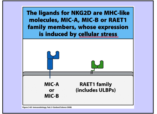
NKG2D → important activating receptor that binds MIC-A, MIC-B (and other stress-induced ligands include RAET1 family)

  • cellular ligands for activating receptors are induced by stress (made in the cytosol and then go to cell surface)

  • LY 49a and NKG2A → inhibitory receptors (ITIMS)


Adaptors + Receptors Determines Receptor Status:

  • CD94:NKG2A → inhibitory signal

  • CD94:NKG2D → activating signal


CTLs → recognize antigen in antigen-specific manner within MHC class 1, producing/activating CTLs for cytotoxicity, however, in some cases, viruses inhibit MHC class 1 production, thereby decreasing CTL cytotoxicity, however, it instead activates NK Cell cytotoxicity


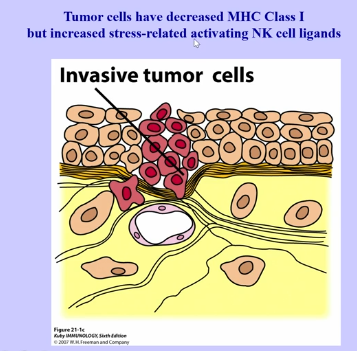
Tumor Cells

  • induce neovascularization

  • collection of cancer cells (all cells require oxidation), but as tumor grows, cells inside are unable to get required oxygen, leading to blood vessel formation

  • have decreased MHC class 1 and increased stress-related activating NK cell ligands

<ul><li><p>chronic immune response → leads to stress</p></li><li><p>hypoxia during inflammation → leads to collection of cells, which leads to a lack of blood vessels in a region, and leads to stress</p></li><li><p>protein synthetic stress </p></li></ul><p></p><p>NKG2D → important activating receptor that binds MIC-A, MIC-B (and other stress-induced ligands include RAET1 family)</p><ul><li><p>cellular ligands for activating receptors are induced by stress (made in the cytosol and then go to cell surface) </p></li><li><p>LY 49a and NKG2A → inhibitory receptors (ITIMS)</p></li></ul><p></p><p>Adaptors + Receptors Determines Receptor Status:</p><ul><li><p>CD94:NKG2A → inhibitory signal </p></li><li><p>CD94:NKG2D → activating signal </p></li></ul><p></p><p>CTLs → recognize antigen in antigen-specific manner within MHC class 1, producing/activating CTLs for cytotoxicity, however, in some cases, viruses inhibit MHC class 1 production, thereby decreasing CTL cytotoxicity, however, it instead activates NK Cell cytotoxicity </p><p></p><p>Tumor Cells</p><ul><li><p>induce neovascularization</p></li><li><p>collection of cancer cells (all cells require oxidation), but as tumor grows, cells inside are unable to get required oxygen, leading to blood vessel formation </p></li><li><p>have decreased MHC class 1 and increased stress-related activating NK cell ligands </p></li></ul><p></p>
8
New cards
<p>Antibody Dependent Cellular Toxicity (ADCC) </p>

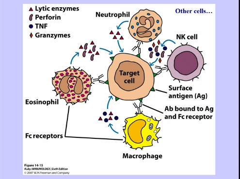
Antibody Dependent Cellular Toxicity (ADCC)

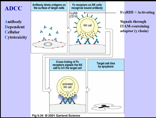
  • NK cells are a part of innate immune response, because they can lyse cells

    • can also be a part of the adaptive immune response

  • NK cells also have Fc receptors (Fc gamma receptor 3), which leads to antibody dependent cellular cytotoxicity

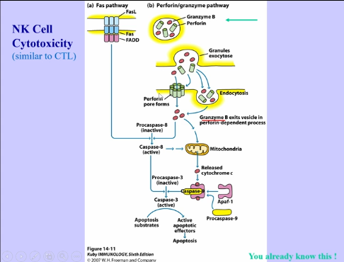
  • cytotoxicity is similar to that of CTLs (perforin, granzymes, apoptosis, etc)

<ul><li><p>NK cells are a part of innate immune response, because they can lyse cells</p><ul><li><p>can also be a part of the adaptive immune response </p></li></ul></li><li><p>NK cells also have Fc receptors (Fc gamma receptor 3), which leads to antibody dependent cellular cytotoxicity </p></li><li><p>cytotoxicity is similar to that of CTLs (perforin, granzymes, apoptosis, etc) </p></li></ul><p></p>
9
New cards
<p>Cytotoxicity + Negative Signaling</p>

Cytotoxicity + Negative Signaling

  • many cells express stress-induced ligands

  • many can also express cytotoxicity


Negative Signaling

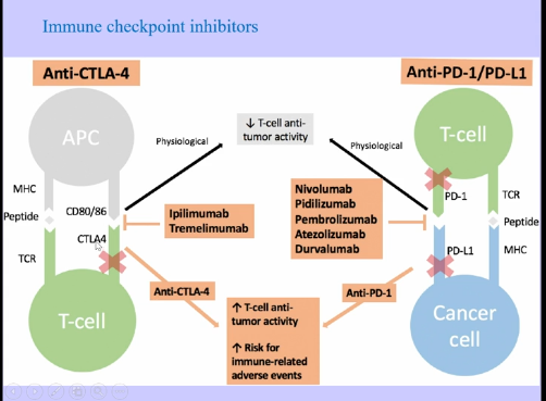
  • CTLA-4 → can be coinhibitory, binding to CD80/CD86, turning T cells off

  • Tumor cells can express PD-L1, which binds to PD-1, also turning T cells off

<ul><li><p>many cells express stress-induced ligands</p></li><li><p>many can also express cytotoxicity</p></li></ul><p></p><p>Negative Signaling</p><ul><li><p>CTLA-4 → can be coinhibitory, binding to CD80/CD86, turning T cells off</p></li><li><p>Tumor cells can express PD-L1, which binds to PD-1, also turning T cells off </p></li></ul><p></p>
10
New cards

Autoimmunity + Self-Tolerance (Overview)

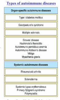
  • autoimmunity → state in which immune system attacks self-tissues

    • normally prevented by multiple self-tolerance mechanisms

    • can occur when infection or tissue damage overwhelms tolerance, but most initiating autoimmune disease events unknown

    • can involved all components of immune system (systemic or tissue-specific)

    • susceptibility can be genetic or environmentally related

    • animal models (spontaneous and induced) for number of autoimmune diseases, but few good treatments


General

  • autoimmunity = breakdown of tolerance

    • immune system attacking body’s own tissues, rather than a pathogen

    • under normal conditions, autoimmunity is prevented by multiple self-tolerance mechanisms (receptors that are self reactive can’t be selected against → VDJ recombination is random, so instead, mechanisms are put in place to destroy self-reactive ones)

      • negative selection can’t catch every self-reactive lymphocyte

    • genetic and environmental factors lead to breakdown of self-tolerance, allowing activation of self-reactive lymphocytes

11
New cards
<p>Mechanisms of Self-Tolerance</p>

Mechanisms of Self-Tolerance

  1. Clonal Deletion → within bone marrow/thymus, during lymphocyte development

    • negative selection of B cells in bone marrow and T cells in the thymus

    • if rearranged receptor (BCR or TCR) recognizes self-antigen, if it finds the antigen during development, is deleted

    • problem: specific antigens that might be present only in some regions (tissues, brain, etc) are not found during development or during puberty and after different proteins are made that could not have been selected against

    • solution:

      • mice with autoimmunity, mice when homozygous deficient developed T cells that attack all organs within the body

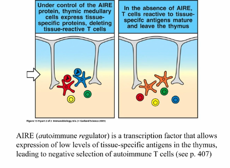
      • point mutation in AIRE (autoimmune regulator), which is a transcription factor expressed in hematopoietic cells within the thymus, allowing for expression of low levels of tissue-specific antigens in the thymus, leading to negative selection of autoimmune T cells

        • binds to lots of genes = low level expression of many genes, even in regions where they’re not normally found

        • antigens get presented, therefore allowing for negative selection against those that react to antigens

        • insulin

        • increased range of antigens that could be negatively selected against, however, mutation prevented this

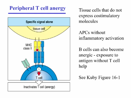
  2. Clonal Inactivation by tissue-specific antigens presented in absence of costimulatory signals → at periphery

    • if lymphocyte interacts with specific antigen in periphery for first time, without inflammatory/costimulatory signals, becomes deactivated instead of activated

    • T cell anergy

      • in absence of inflammatory cytokines (TNF-alpha, IFN-gamma, etc)

      • in absence of costimulatory molecules

      • in absence of licensing of antigen presenting cells

      • absence of TLR ligands

    • B cell Anergy

      • in absence of T cell help


<ol><li><p>Clonal Deletion → within bone marrow/thymus, during lymphocyte development</p><ul><li><p>negative selection of B cells in bone marrow and T cells in the thymus </p></li><li><p>if rearranged receptor (BCR or TCR) recognizes self-antigen, if it finds  the antigen during development, is deleted </p></li><li><p>problem: specific antigens that might be present only in some regions (tissues, brain, etc) are not found during development or during puberty and after different proteins are made that could not have been selected against </p></li><li><p>solution:</p><ul><li><p>mice with autoimmunity, mice when homozygous deficient developed T cells that attack all organs within the body</p></li><li><p>point mutation in AIRE (autoimmune regulator), which is a transcription factor expressed in hematopoietic cells within the thymus, allowing for expression of low levels of tissue-specific antigens in the thymus, leading to negative selection of autoimmune T cells</p><ul><li><p>binds to lots of genes = low level expression of many genes, even in regions where they’re not normally found </p></li><li><p>antigens get presented, therefore allowing for negative selection against those that react to antigens </p></li><li><p>insulin </p></li><li><p>increased range of antigens that could be negatively selected against, however, mutation prevented this </p></li></ul></li></ul></li></ul></li><li><p>Clonal Inactivation by tissue-specific antigens presented in absence of costimulatory signals → at periphery</p><ul><li><p>if lymphocyte interacts with specific antigen in periphery for first time, without inflammatory/costimulatory signals, becomes deactivated instead of activated </p></li><li><p>T cell anergy</p><ul><li><p>in absence of inflammatory cytokines (TNF-alpha, IFN-gamma, etc)</p></li><li><p>in absence of costimulatory molecules </p></li><li><p>in absence of licensing of antigen presenting cells </p></li><li><p>absence of TLR ligands </p></li></ul></li><li><p>B cell Anergy</p><ul><li><p>in absence of T cell help </p></li></ul></li></ul></li></ol><p></p>
12
New cards

Mechanisms of Self-Tolerance (cont)

  1. Immunological Ignorance

    • antigen that immune system is unable to detect, doesn’t have access to it, or at low levels that receptors are unable to detect it

    • problem: if upregulated due to stimulus or released from unavailable tissue following tissue damage, leading to immune system lacking tools to deal with it

  2. Immunologically Privileged Sites

    • sites where antigen is available, but presented in a way to deliberately induce tolerance

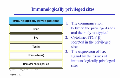
    • places where you wouldn’t want inflammation, because it likely leads to organismal death (brain, eye, testes, etc)

    • antigens and antigen presented cells within these tissues communicate differently compared to other cells

      • presenting antigen in the eye induces general tolerance throughout the rest of the body

      • anti-inflammatory cytokines (TGF-beta) present in fluid of these regions, inducing tolerance in lymphocytes

      • tissues also express Fas ligand, which binds to Fas receptors, inducing apoptosis

  3. Immune Suppression by Special T Cells

    • regulatory T cells

    • 3-day-old mice had their thymus removed, leading to multiorgan autoimmune disease

    • Regulatory T cells were missing

      • CD4+ T cells that constitutively express CD25+ (alpha subunit of interleukin 2 receptor, which increases affinity for receptor)

      • upon stimulation, regulatory T cells do not produce IL-2 or typical T helper cytokines (IFN-gamma, IL-4, IL-17, etc), but instead secrete inhibitory cytokines IL-10 and TGF-beta; can also inhibit APC function (through cell-cell contact, convert APC from activating immune response to tolerizing them)

        • without IL-2 → lose cells = generates autoimmunity

        • CD4+CD25+ T cells can block or even cure autoimmune disease in several animal models

  4. Activation-induced Cell Death (Fas/FasL)

    • chronic stimulation of T cells results in upregulation of Fas receptor, leading to apoptosis

<ol><li><p>Immunological Ignorance</p><ul><li><p>antigen that immune system is unable to detect, doesn’t have access to it, or at low levels that receptors are unable to detect it</p></li><li><p>problem: if upregulated due to stimulus or released from unavailable tissue following tissue damage, leading to immune system lacking tools to deal with it</p></li></ul></li><li><p>Immunologically Privileged Sites</p><ul><li><p>sites where antigen is available, but presented in a way to deliberately induce tolerance</p></li><li><p>places where you wouldn’t want inflammation, because it likely leads to organismal death (brain, eye, testes, etc)</p></li><li><p>antigens and antigen presented cells within these tissues communicate differently compared to other cells</p><ul><li><p>presenting antigen in the eye induces general tolerance throughout the rest of the body</p></li><li><p>anti-inflammatory cytokines (TGF-beta) present in fluid of these regions, inducing tolerance in lymphocytes</p></li><li><p>tissues also express Fas ligand, which binds to Fas receptors, inducing apoptosis</p></li></ul></li></ul></li><li><p>Immune Suppression by Special T Cells</p><ul><li><p>regulatory T cells</p></li><li><p>3-day-old mice had their thymus removed, leading to multiorgan autoimmune disease</p></li><li><p>Regulatory T cells were missing</p><ul><li><p>CD4+ T cells that constitutively express CD25+ (alpha subunit of interleukin 2 receptor, which increases affinity for receptor)</p></li><li><p>upon stimulation, regulatory T cells do not produce IL-2 or typical T helper cytokines (IFN-gamma, IL-4, IL-17, etc), but instead secrete inhibitory cytokines IL-10 and TGF-beta; can also inhibit APC function (through cell-cell contact, convert APC from activating immune response to tolerizing them)</p><ul><li><p>without IL-2 → lose cells = generates autoimmunity</p></li><li><p>CD4+CD25+ T cells can block or even cure autoimmune disease in several animal models</p></li></ul></li></ul></li></ul></li><li><p>Activation-induced Cell Death (Fas/FasL)</p><ul><li><p>chronic stimulation of T cells results in upregulation of Fas receptor, leading to apoptosis</p></li></ul></li></ol><p></p>
13
New cards
<p>Regulatory T Cell Development </p>

Regulatory T Cell Development

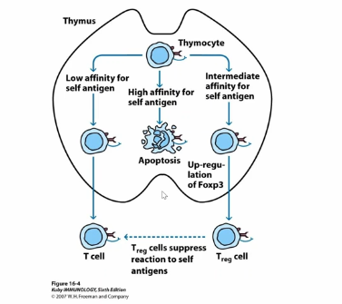
  • develop in thymus and go through normal negative and positive selection

  • TCR is somewhere between negative and positive selection (intermediate affinity)

    • high enough to get positively selected

    • low enough to avoid negative selection

    • still recognizes self-antigen, but not high enough to get negatively selected against

  • leads to upregulation of master transcription factor (Foxp3), which leads to regulatory T cell development, which suppress immune response


Previous Studies

  • scurfy mouse → autoimmune strain of mice

  • mutation in gene called scurfin, which encodes transcription factor Foxp3, leading to the failure in development of regulatory T cells

  • compared to this, overexpression of FoxP3 in normal CD4+ T cells → converts to CD4+CD25+ regulatory-like T cells
<ul><li><p>develop in thymus and go through normal negative and positive selection </p></li><li><p>TCR is somewhere between negative and positive selection (intermediate affinity) </p><ul><li><p>high enough to get positively selected</p></li><li><p>low enough to avoid negative selection </p></li><li><p>still recognizes self-antigen, but not high enough to get negatively selected against </p></li></ul></li><li><p>leads to upregulation of master transcription factor (Foxp3), which leads to regulatory T cell development, which suppress immune response </p></li></ul><p></p><p>Previous Studies</p><ul><li><p>scurfy mouse → autoimmune strain of mice </p></li><li><p>mutation in gene called scurfin, which encodes transcription factor Foxp3, leading to the failure in development of regulatory T cells</p></li><li><p>compared to this, overexpression of FoxP3 in normal CD4+ T cells → converts to CD4+CD25+ regulatory-like T cells </p></li></ul><p></p>
14
New cards
<p>Autoimmune Disease Causes </p>

Autoimmune Disease Causes

  1. Injury → trauma releases antigen from immunologically privileged site or increases antigen concentration past threshold of ignorance

  2. Infection → pathogen may have cross-reactive antigen, cause tissue damage, or induce costimulatory molecules via inflammatory response

    • Infection leads to production of PAMPs and inflammatory signals, which a naive T cell can recognize antigen for the very first time, meaning it doesn’t know that your antigen is not an infection and therefore reacts to it

    • foreign antigen could have similar structure to self-antigen, leading to memory response being generated for foreign antigen (memory response has lower threshold for activation = fewer antigen molecules or lower affinity)

  3. Spontaneous → underlying cause of most autoimmune diseases are unknown, but probably due to intrinsic defects in tolerance mechanisms


Process:

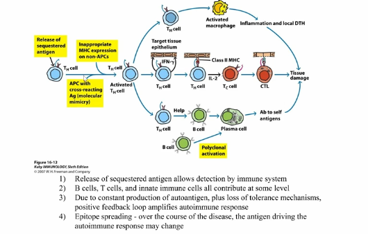
  • starts with some initiating event (traumatic event that leads to release of antigen, cross-reactivity with infectious agent, etc), which leads to inflammatory response that triggers autoimmune disease

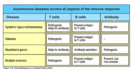
  • leads to activation of self-reactive T cell, which provide help to activate macrophages, which then lead to tissue damage, releasing more antigens and cytokines

  • T cells can also give help to B cells, producing autoreactive B cells, which release antibodies that activate complement, activate neutrophils through the Fc receptor, etc

  • all feedback to activating more T cells, leading to upregulation of MHC molecules on cells, leading to activating of CTLs, which lead to further tissue damage and more autoimmune response

  • positive feedback loop → constant production of autoantigen, loss of tolerance mechanisms, which cause amplified autoimmune response and release of new antigens (epitope spreading)

<ol><li><p>Injury → trauma releases antigen from immunologically privileged site or increases antigen concentration past threshold of ignorance </p></li><li><p>Infection → pathogen may have cross-reactive antigen, cause tissue damage, or induce costimulatory molecules via inflammatory response</p><ul><li><p>Infection leads to production of PAMPs and inflammatory signals, which a naive T cell can recognize antigen for the very first time, meaning it doesn’t know that your antigen is not an infection and therefore reacts to it</p></li><li><p>foreign antigen could have similar structure to self-antigen, leading to memory response being generated for foreign antigen (memory response has lower threshold for activation = fewer antigen molecules or lower affinity) </p></li></ul></li><li><p>Spontaneous → underlying cause of most autoimmune diseases are unknown, but probably due to intrinsic defects in tolerance mechanisms </p></li></ol><p></p><p></p><p>Process:</p><ul><li><p>starts with some initiating event (traumatic event that leads to release of antigen, cross-reactivity with infectious agent, etc), which leads to inflammatory response that triggers autoimmune disease</p></li><li><p>leads to activation of self-reactive T cell, which provide help to activate macrophages, which then lead to tissue damage, releasing more antigens and cytokines </p></li><li><p>T cells can also give help to B cells, producing autoreactive B cells, which release antibodies that activate complement, activate neutrophils through the Fc receptor, etc</p></li><li><p>all feedback to activating more T cells, leading to upregulation of MHC molecules on cells, leading to activating of CTLs, which lead to further tissue damage and more autoimmune response </p></li><li><p>positive feedback loop → constant production of autoantigen, loss of tolerance mechanisms, which cause amplified autoimmune response and release of new antigens (epitope spreading) </p></li></ul><p></p>
15
New cards

Mechanisms for Autoimmune Pathogenesis

Pathogenic Antibodies:

  1. Complement-mediate lysis (autoimmune hemolytic anemia)

    • especially for red blood cells (no energy investment into cell defense, because they rapidly turnover) → leads to susceptibility

    • type 2 hypersensitivity reaction → antibody binds to the surface, activates complement, and leads to lysis

    • transfusion reaction

  2. antibody and complement mediated opsonization

    • taken up by phagocytosis

  3. complement-induced inflammation (tissue damage)

    • type 2 hypersensitivity → antibody binds to surface, activating to complement, leading to inflammation (anaphylatoxin production)

  4. antibody-antigen complex deposition (kidney damage)

    • type 3 hypersensitivity → antibody-antigen complexes deposited in tissues, leading to complement activation and tissue

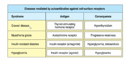
  5. blockage or stimulation of cell-surface receptors (Graves’ disease, myasthenia gravis)

    • antibody binds to receptor, blocking receptor and preventing ligand from binding, blocking function

    • can artificially crosslink receptors (overactivation of receptors)


Pathogenic T Cells:

  1. Help for B cells (autoantibody production)

  2. Activation and recruitment of innate immune cells (macrophages, neutrophils, etc)

  3. Cytokine-mediated inflammation and cytotoxicity

    • TNF-alpha is toxic at high concentrations

  4. Direct killing of target cells

    • CTLs


<p>Pathogenic Antibodies: </p><ol><li><p>Complement-mediate lysis (autoimmune hemolytic anemia)</p><ul><li><p>especially for red blood cells (no energy investment into cell defense, because they rapidly turnover) → leads to susceptibility </p></li><li><p>type 2 hypersensitivity reaction → antibody binds to the surface, activates complement, and leads to lysis </p></li><li><p>transfusion reaction </p></li></ul></li><li><p>antibody and complement mediated opsonization </p><ul><li><p>taken up by phagocytosis </p></li></ul></li><li><p>complement-induced inflammation (tissue damage)</p><ul><li><p>type 2 hypersensitivity → antibody binds to surface, activating to complement, leading to inflammation (anaphylatoxin production) </p></li></ul></li><li><p>antibody-antigen complex deposition (kidney damage)</p><ul><li><p>type 3 hypersensitivity → antibody-antigen complexes deposited in tissues, leading to complement activation and tissue </p></li></ul></li><li><p>blockage or stimulation of cell-surface receptors (Graves’ disease, myasthenia gravis) </p><ul><li><p>antibody binds to receptor, blocking receptor and preventing ligand from binding, blocking function </p></li><li><p>can artificially crosslink receptors (overactivation of receptors) </p></li></ul></li></ol><p></p><p>Pathogenic T Cells:</p><ol><li><p>Help for B cells (autoantibody production)</p></li><li><p>Activation and recruitment of innate immune cells (macrophages, neutrophils, etc)</p></li><li><p>Cytokine-mediated inflammation and cytotoxicity</p><ul><li><p>TNF-alpha is toxic at high concentrations </p></li></ul></li><li><p>Direct killing of target cells </p><ul><li><p>CTLs</p></li></ul></li></ol><p></p>
16
New cards

Animal Models for Autoimmune Diseases

Spontaneous:

  1. NOD (non-obese diabetic) mouse → type 1 diabetes

  2. NZB (New Zealand black) mouse → hemolytic anemia

  3. NZM * NZW (New Zealand white) F1 → systemic lupus erythematosus (SLE)


Induced:

  1. EAE (experimental autoimmune encephalomyelitis) → multiple sclerosis (induced by immunization with myeline proteins + adjuvants)

  2. Autoimmune arthritis → rheumatoid arthritis (induced by immunization with collagen + adjuvant, or injection of adjuvant into joints)

  3. transfer of pathogenic antibodies or T cells from autoimmune mouse into healthy mouse can transfer disease

  4. TCR Transgenic T cells → expression of TCR found in spontaneous or induced autoimmune disease can also cause disease


17
New cards

Alloreactivity and Transplant Rejection - Immunosuppression (overview)

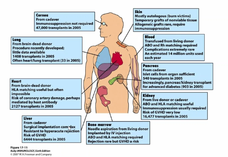
  • tissue/organ transplantation requires the prevention of immune responses against foreign tissue antigens

  • blood group antigens (ABO, Rh factor, etc) are important transplantation antigens → mostly antibody responses

  • MHC differences are responsible for rapid graft rejection due to alloreactivity of T cells

  • Minor histocompatibility antigens (polymorphisms in non-MHC proteins) are responsible for slower graft rejection

  • fetus is a kind of allograft, but generally not rejected, because of a variety of protective mechanisms

  • immunosuppression can be used to prevent graft rejection

18
New cards
<p>ABO Blood-Type Rejection</p>

ABO Blood-Type Rejection

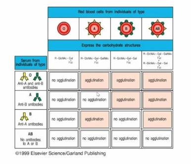
  1. Antibody dependent (IgM), but T-independent (carbohydrate antigens don’t generate a T cell response)

  2. Relies on pre-existing antibodies against surface carbohydrates (no activation of immune response)

  3. Donor RBC destroyed by complement-mediate lysis


Immune Response to ABO vs Rh Factor

  • ABO antibodies are pre-existing (induced by related antigens on gut bacteria); Rh antibodies require previous exposure to antigen (Rh- mother gives birth to Rh+ baby)

  • ABO antibodies are almost always IgM; Rh antibodies can be IgG (able to cross placenta)

  • Rh antibodies can therefore attack RBC of fetus and newborn, causing hemolytic disease of newborn (erythroblastosis fetalis)

    • only occurs during childbirth

    • no exposure of blood during first pregnancy, which means antigens are not exposed and no response developed

    • later pregnancies following exposure will result in this

    • Rhogam can be injected to control this

  • *transfusion reactions do not require MHC matching, because RBC don’t express MHC


<ol><li><p>Antibody dependent (IgM), but T-independent (carbohydrate antigens don’t generate a T cell response)</p></li><li><p>Relies on pre-existing antibodies against surface carbohydrates (no activation of immune response) </p></li><li><p>Donor RBC destroyed by complement-mediate lysis </p></li></ol><p></p><p></p><p>Immune Response to ABO vs Rh Factor </p><ul><li><p>ABO antibodies are pre-existing (induced by related antigens on gut bacteria); Rh antibodies require previous exposure to antigen (Rh- mother gives birth to Rh+ baby) </p></li><li><p>ABO antibodies are almost always IgM; Rh antibodies can be IgG (able to cross placenta) </p></li><li><p>Rh antibodies can therefore attack RBC of fetus and newborn, causing hemolytic disease of newborn (erythroblastosis fetalis) </p><ul><li><p>only occurs during childbirth</p></li><li><p>no exposure of blood during first pregnancy, which means antigens are not exposed and no response developed</p></li><li><p>later pregnancies following exposure will result in this </p></li><li><p>Rhogam can be injected to control this</p></li></ul></li><li><p>*transfusion reactions do not require MHC matching, because RBC don’t express MHC</p><p></p></li></ul><p></p>
19
New cards
<p>Transplantation</p>

Transplantation

Types:

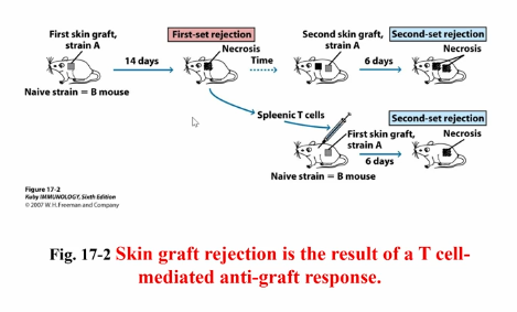
  • autograft (same individual)

    • no requirement for protection, because your own cells are being transplanted

  • syngeneic graft (genetically identical individuals)

    • identical siblings

    • immune systems would be different

    • little to no immunosuppression required

  • allograft (different MHC)

    • more than likely to have different MHC

  • Xenograft (cross-species)

    • pig organs and human organs are similar sizes

    • genetically engineering to prevent triggering of immune responses


Challenges to Organ Transplantation:

  1. Blood types

  2. MHC → major histocompatibility complexes

  3. Minor histocompatibility complexes


*mostly T cell mediated → CD4+ T cells most important for initiating graft rejection

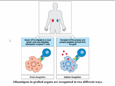
  • Direct Recognition → T cells recognize foreign MHC cells as alloreactivity; recognize resident APC within the graft

  • Indirect Recognition → own APC migrate into tissue and present foreign antigens on self-MHC molecules (MHC proteins presented themselves)

<p>Types:</p><ul><li><p>autograft (same individual)</p><ul><li><p>no requirement for protection, because your own cells are being transplanted</p></li></ul></li><li><p>syngeneic graft (genetically identical individuals)</p><ul><li><p>identical siblings</p></li><li><p>immune systems would be different</p></li><li><p>little to no immunosuppression required</p></li></ul></li><li><p>allograft (different MHC)</p><ul><li><p>more than likely to have different MHC</p></li></ul></li><li><p>Xenograft (cross-species)</p><ul><li><p>pig organs and human organs are similar sizes</p></li><li><p>genetically engineering to prevent triggering of immune responses</p></li></ul></li></ul><p></p><p>Challenges to Organ Transplantation:</p><ol><li><p>Blood types</p></li><li><p>MHC → major histocompatibility complexes</p></li><li><p>Minor histocompatibility complexes</p></li></ol><p></p><p>*mostly T cell mediated → CD4+ T cells most important for initiating graft rejection</p><ul><li><p>Direct Recognition → T cells recognize foreign MHC cells as alloreactivity; recognize resident APC within the graft</p></li><li><p>Indirect Recognition → own APC migrate into tissue and present foreign antigens on self-MHC molecules (MHC proteins presented themselves)</p></li></ul><p></p>
20
New cards

MHC Match

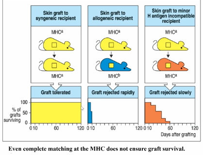
MHC match doesn’t guarantee graft acceptance → other polymorphic genes besides MHC molecules, which are recognized by the immune system

  • allelic differences can give rise to epitopes (minor histocompatibility complexes)

  • smaller number of T cells response to these epitope changes, meaning it takes more time to reject

<p>MHC match doesn’t guarantee graft acceptance → other polymorphic genes besides MHC molecules, which are recognized by the immune system </p><ul><li><p>allelic differences can give rise to epitopes (minor histocompatibility complexes) </p></li><li><p>smaller number of T cells response to these epitope changes, meaning it takes more time to reject</p></li></ul><p></p>
21
New cards
<p>Allogenic Bone Marrow Transplantation + Graft vs Host Disease</p>

Allogenic Bone Marrow Transplantation + Graft vs Host Disease

  • cause: mature donor T cells that contaminate the allogenic bone marrow recognize the tissues of the recipient as foreign

  • result: severe inflammatory response

  • symptom: rashes, diarrhea, and liver disease

  • prevention: before grafting bone marrow, remove mature T cells

    • can help in cases of leukemia, where bone marrow will help treat leukemia by rejecting them

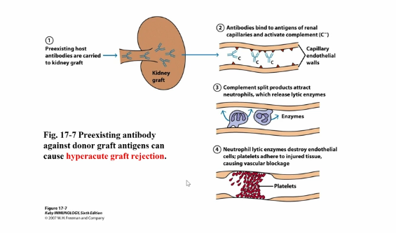
  • hyperacute graft rejection → occurs after transplant, rejection, and new transplant

    • allelic similarities

    • antibodies generated against first kidney, followed by rejection

    • second kidney that contains antigens that recognize those antibodies = very rapid and severe rejection

22
New cards
<p>Preventing Graft Rejection </p>

Preventing Graft Rejection

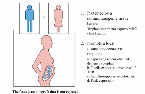
  • especially prevalent with pregnancy, since fetus is an allograft within mother


  1. protected by a nonimmunogenic tissue barrier

    • trophoblasts (fetal component of placenta) do not express MHC class 1 and 2

    • T cells entering placenta are not immediately exposed to foreign MHC molecules

    • potentially a target of NK cells, because they don’t express MHC class 1

      • doesn’t get rejected by them, because of nonclassical MHC molecules (HLA-E, HLA-G, etc) that interact with the inhibitory receptors of NK cells to prevent regulation

  2. Promote a local immunosuppressive response (privilege site)

    • expressing an enzyme that deplete tryptophan

      • in absence of tryptophan, T cells differentiate into regulatory T cells, which leads to immunosuppression

    • T cells express lower level of TCR

    • immunosuppressive cytokines

    • FasL expression


23
New cards
<p>Advances in Organ Transplantation </p>

Advances in Organ Transplantation

  • dependent on the development of immunosuppressive therapies

    • better immunosuppressive drugs

      • immunosuppression → intentional induction of immunodeficiency

      • used to control unwanted immune responses (autoimmune diseases, lymphoproliferation, and graft rejection)

      • classes (increasing order of specificity = decreasing order of negative side effects):

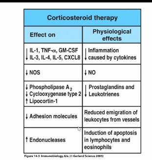
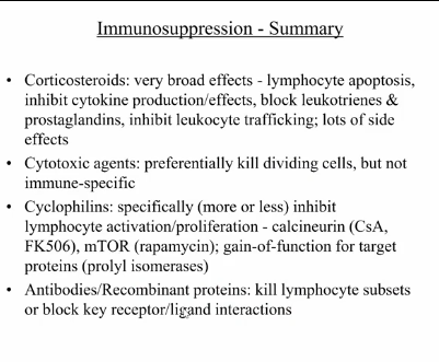
        • corticosteroids

          • very general

          • stress response hormones → conserve energy, decreasing immune response

        • cytotoxic agents

          • selectively kill dividing cells (activated, but not resting, lymphocytes)

          • drugs that inhibit de novo purine synthesis: azathioprine (AMP and GMP synthesis) and mycophenolate (GMP synthesis)

          • cyclophosphamide (nitrogen mustard) is DNA alkylating agent

          • lots of unpleasant side effects → kill all dividing cells, so must be used very carefully (often in combination with other drugs at lower dosages)

        • immunophilin-binding drugs

          • isolated from microorganisms: fungus (cyclosporin A), streptomyces spp. (FK506/tacrolimus and rapamycin)

          • bind cellular proteins that gain novel function upon binding drug (proteins have nothing to do with immunosuppression, but gain functions that inhibit signaling)

          • cyclosporin A and FK506 inhibit calcineurin - interrupts TCR calcium signaling pathway; can prevent long-term tolerance

            • would need to be on for life, however, it blocks all T cell activation, leading susceptible to other pathogens

          • rapamycin inhibits mTOR → interrupts distal signaling pathways, preventing proliferation without blocking T cell activation; can lead to tolerance induction

          • fewer side effects than corticosteroids or cytotoxic agents, but still broad immunosuppression

          • later down pathway: doesn’t prevent TCR signaling, but instead prevents secondary signal

        • antibodies/recombinant proteins

          • target lymphocyte subsets to give specific immunosuppression

          • depleting antibodies → target and induce death of lymphocytes (anti-CD4 and anti-CD8): useful in depleting mature T cells from bone marrow (prevents GVHD)

          • non-depleting antibodies → block receptor-ligand interactions to induce specific tolerance (anti-CD4, anti-40L, anti-integrins)

          • anti-cytokine/receptor antibodies: block effects of immune response (anti-TNF-alpha, anti-IL6R)

          • recombinant proteins: compete with normal receptor ligand interactions (CTLA4-Ig, LFG3-Ig, TNFR-Fc, IL-1Ra)

    • induction of specific tolerance


<ul><li><p>dependent on the development of immunosuppressive therapies</p><ul><li><p>better immunosuppressive drugs</p><ul><li><p>immunosuppression → intentional induction of immunodeficiency </p></li><li><p>used to control unwanted immune responses (autoimmune diseases, lymphoproliferation, and graft rejection) </p></li><li><p>classes (increasing order of specificity = decreasing order of negative side effects):</p><ul><li><p>corticosteroids </p><ul><li><p>very general </p></li><li><p>stress response hormones → conserve energy, decreasing immune response</p></li></ul></li><li><p>cytotoxic agents</p><ul><li><p>selectively kill dividing cells (activated, but not resting, lymphocytes) </p></li><li><p>drugs that inhibit de novo purine synthesis: azathioprine (AMP and GMP synthesis) and mycophenolate (GMP synthesis)</p></li><li><p>cyclophosphamide (nitrogen mustard) is DNA alkylating agent</p></li><li><p>lots of unpleasant side effects → kill all dividing cells, so must be used very carefully (often in combination with other drugs at lower dosages) </p></li></ul></li><li><p>immunophilin-binding drugs</p><ul><li><p>isolated from microorganisms: fungus (cyclosporin A), streptomyces spp. (FK506/tacrolimus and rapamycin)</p></li><li><p>bind cellular proteins that gain novel function upon binding drug (proteins have nothing to do with immunosuppression, but gain functions that inhibit signaling) </p></li><li><p>cyclosporin A and FK506 inhibit calcineurin - interrupts TCR calcium signaling pathway; can prevent long-term tolerance</p><ul><li><p>would need to be on for life, however, it blocks all T cell activation, leading susceptible to other pathogens</p></li></ul></li><li><p>rapamycin inhibits mTOR → interrupts distal signaling pathways, preventing proliferation without blocking T cell activation; can lead to tolerance induction </p></li><li><p>fewer side effects than corticosteroids or cytotoxic agents, but still broad immunosuppression</p></li><li><p>later down pathway: doesn’t prevent TCR signaling, but instead prevents secondary signal </p></li></ul></li><li><p>antibodies/recombinant proteins </p><ul><li><p>target lymphocyte subsets to give specific immunosuppression</p></li><li><p>depleting antibodies → target and induce death of lymphocytes (anti-CD4 and anti-CD8): useful in depleting mature T cells from bone marrow (prevents GVHD)</p></li><li><p>non-depleting antibodies → block receptor-ligand interactions to induce specific tolerance (anti-CD4, anti-40L, anti-integrins) </p></li><li><p>anti-cytokine/receptor antibodies: block effects of immune response (anti-TNF-alpha, anti-IL6R) </p></li><li><p>recombinant proteins: compete with normal receptor ligand interactions (CTLA4-Ig, LFG3-Ig, TNFR-Fc, IL-1Ra) </p></li></ul></li></ul></li></ul></li><li><p>induction of specific tolerance </p></li></ul></li></ul><p></p>
24
New cards
<p>Viruses</p>

Viruses

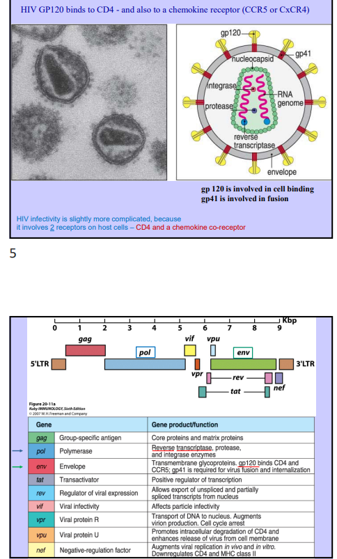
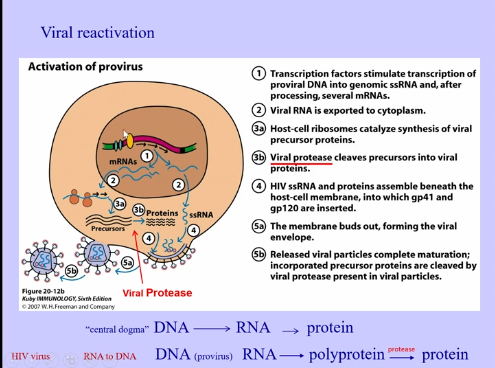
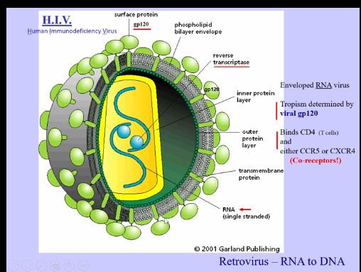
  • Human Immunodeficiency Virus (HIV)

    • kills CD4+ T cells, leading to patients overcome by opportunistic pathogens

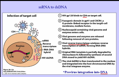
    • RNA virus (retrovirus), converts RNA back to DNA to establish itself into the genome

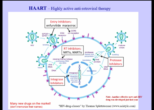
    • gp120 → envelope protein on surface, which binds to CD4 on T cells and chemokine coreceptor (CCR5 or CXCR4) to help virus get into cells

    • reverse transcriptase → takes single stranded RNA and convert it into DNA, allowing it to be integrated into our genome (DNA can just sit within the genome until virus is reactivated)

    • gp120 → binds to CD4, depleting body of T helper cells

<ul><li><p>Human Immunodeficiency Virus (HIV)</p><ul><li><p>kills CD4+ T cells, leading to patients overcome by opportunistic pathogens </p></li><li><p>RNA virus (retrovirus), converts RNA back to DNA to establish itself into the genome</p></li><li><p>gp120 → envelope protein on surface, which binds to CD4 on T cells and chemokine coreceptor (CCR5 or CXCR4) to help virus get into cells </p></li><li><p>reverse transcriptase → takes single stranded RNA and convert it into DNA, allowing it to be integrated into our genome (DNA can just sit within the genome until virus is reactivated) </p></li><li><p>gp120 → binds to CD4, depleting body of T helper cells </p></li></ul></li></ul><p></p>
25
New cards
<p>Viral Infection Process </p>

Viral Infection Process

knowt flashcard image
26
New cards
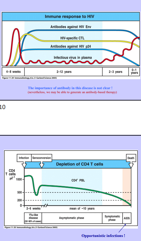
<p>Immune Response to HIV</p>

Immune Response to HIV

  • spike in virus, mount immune response, begin eliminating virus, virus stays in not quite latent stage (latent implies dormant, but the virus is actively replicating, but at very low levels)

  • antibodies against HIV made

  • HIV-specific CTLs

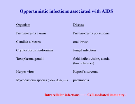
  • eventually, viral numbers increase, leading to susceptibility of opportunistic pathogens

  • all opportunistic pathogens are dealt with through cell-mediated immunity, which is what is deficient in HIV patients
<ul><li><p>spike in virus, mount immune response, begin eliminating virus, virus stays in not quite latent stage (latent implies dormant, but the virus is actively replicating, but at very low levels) </p></li><li><p>antibodies against HIV made </p></li><li><p>HIV-specific CTLs </p></li><li><p>eventually, viral numbers increase, leading to susceptibility of opportunistic pathogens </p></li><li><p>all opportunistic pathogens are dealt with through cell-mediated immunity, which is what is deficient in HIV patients </p></li></ul><p></p>
27
New cards
<p>Chemokine Co-Receptors + Medications</p>

Chemokine Co-Receptors + Medications

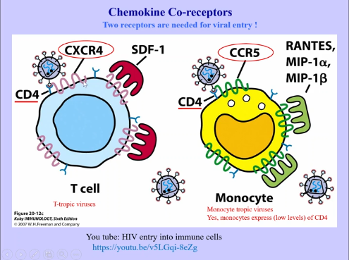
  • T-tropic viruses → have CD4 and chemokine receptors (CXCR4)

  • Monocyte Tropic Viruses → express low levels of CD4 and have their own chemokine co-receptor (CCR5)


Medications:

  • reverse transcriptase → humans don’t do this, however, virus would mutate to avoid these medications

  • drugs prevent HIV transmission

  • behavior modification can also decrease infections

  • new antibodies to block HIV attachment

<ul><li><p>T-tropic viruses → have CD4 and chemokine receptors (CXCR4)</p></li><li><p>Monocyte Tropic Viruses → express low levels of CD4 and have their own chemokine co-receptor (CCR5)</p></li></ul><p></p><p>Medications:</p><ul><li><p>reverse transcriptase → humans don’t do this, however, virus would mutate to avoid these medications </p></li><li><p>drugs prevent HIV transmission </p></li><li><p>behavior modification can also decrease infections </p></li><li><p>new antibodies to block HIV attachment </p></li></ul><p></p>
28
New cards
<p>Influenza Virus (flu)</p>

Influenza Virus (flu)

  • RNA virus, but it is not a retrovirus (doesn’t go from RNA to DNA)

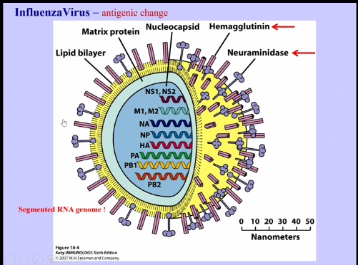
    • contains segmented RNA genome (similar to chromosomes)

    • when two viruses get together, they can swap segments, allowing rapid change in virus

    • contains Hemagglutinin and Neuraminidase, which are used to infect mammalian cells

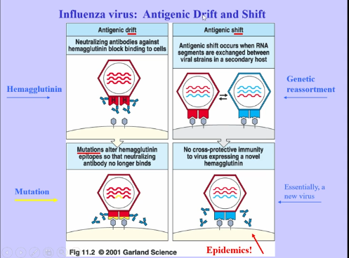
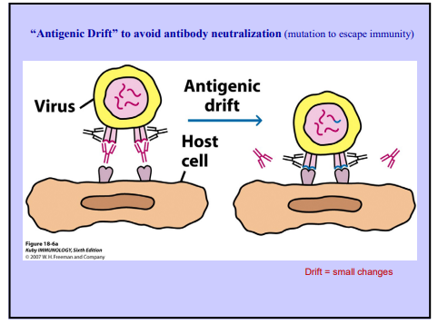
  • Antigenic Drift

    • hemagglutinin undergoes mutation, allowing hemagglutinin to no longer bind to antibodies

    • happens every year, allowing escape from immune response

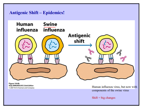
  • Antigenic Shift

    • two different forms of the virus exchange RNA segments, acquiring a different hemagglutinin, which is entirely different from the one that antibodies respond to

    • leads to epidemics


<ul><li><p>RNA virus, but it is not a retrovirus (doesn’t go from RNA to DNA)</p><ul><li><p>contains segmented RNA genome (similar to chromosomes) </p></li><li><p>when two viruses get together, they can swap segments, allowing rapid change in virus </p></li><li><p>contains Hemagglutinin and Neuraminidase, which are used to infect mammalian cells</p></li></ul></li><li><p>Antigenic Drift</p><ul><li><p>hemagglutinin undergoes mutation, allowing hemagglutinin to no longer bind to antibodies</p></li><li><p>happens every year, allowing escape from immune response  </p></li></ul></li><li><p>Antigenic Shift</p><ul><li><p>two different forms of the virus exchange RNA segments, acquiring a different hemagglutinin, which is entirely different from the one that antibodies respond to </p></li><li><p>leads to epidemics </p></li></ul></li></ul><p></p>
29
New cards
<p>Antigenic Shift + Drift </p>

Antigenic Shift + Drift

knowt flashcard image
30
New cards
<p>SARS-CoV-2</p>

SARS-CoV-2

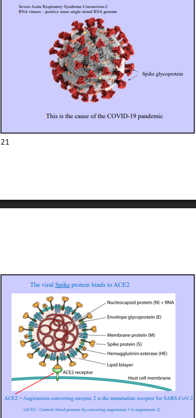
  • sever acute respiratory syndrome coronavirus 2

  • RNA viruses → positive sense single strand RNA genome

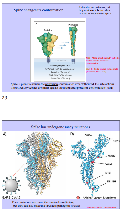
  • contain spike glycoproteins on their outside, allowing virus to enter and infect cells

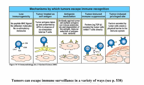
    • bind to ACE2 (angiotensin converting enzyme 2 → mammalian receptor for SARS-CoV-2), which controls blood pressure by converting angiotensin 1 to angiotensin 2

    • spike protein like spring and change conformation (goes from pre-fusion state to post-fusion state when binding to ACE2)

    • different antibodies are required for the different conformations

      • must stabilize prefusion conformation through mutations

    • mutations in virus can make vaccine and antibody response less effective, but they can also make the virus less pathogenic (or more)

      • could prevent binding to ACE2 if too many mutations

      • antibodies can still work, because changes are not massive, but they become less and less effective
<ul><li><p>sever acute respiratory syndrome coronavirus 2  </p></li><li><p>RNA viruses → positive sense single strand RNA genome</p></li><li><p>contain spike glycoproteins on their outside, allowing virus to enter and infect cells </p><ul><li><p>bind to ACE2 (angiotensin converting enzyme 2 → mammalian receptor for SARS-CoV-2), which controls blood pressure by converting angiotensin 1 to angiotensin 2</p></li><li><p>spike protein like spring and change conformation (goes from pre-fusion state to post-fusion state when binding to ACE2)</p></li><li><p>different antibodies are required for the different conformations </p><ul><li><p>must stabilize prefusion conformation through mutations</p></li></ul></li><li><p>mutations in virus can make vaccine and antibody response less effective, but they can also make the virus less pathogenic (or more) </p><ul><li><p>could prevent binding to ACE2 if too many mutations </p></li><li><p>antibodies can still work, because changes are not massive, but they become less and less effective </p></li></ul></li></ul></li></ul><p></p>
31
New cards
<p>Herpes Virus </p>

Herpes Virus

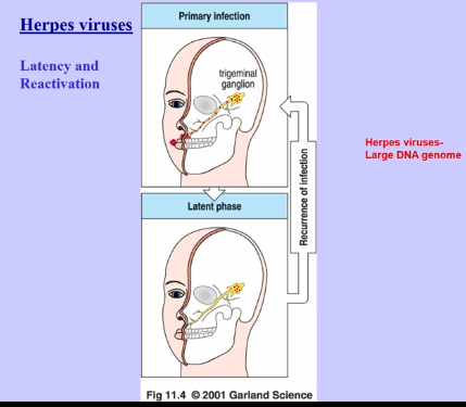
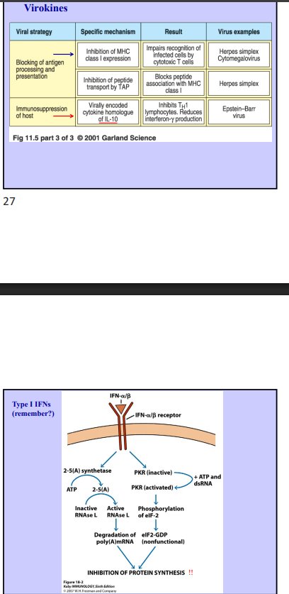
  • large DNA virus

  • latency and reactivation (virus goes dormant and then gets reactivated)

  • contain genes that appear like mammalian genes

    • viroceptors and virokines

      • can make homologs of things like IL-10, which is an immunosuppress cytokine, turning off immune response

      • can make a receptor that binds to own cytokines to decrease their effectiveness

<ul><li><p>large DNA virus </p></li><li><p>latency and reactivation (virus goes dormant and then gets reactivated) </p></li><li><p>contain genes that appear like mammalian genes</p><ul><li><p>viroceptors and virokines </p><ul><li><p>can make homologs of things like IL-10, which is an immunosuppress cytokine, turning off immune response </p></li><li><p>can make a receptor that binds to own cytokines to decrease their effectiveness </p></li></ul></li></ul></li></ul><p></p>
32
New cards
<p>Immunity to Viruses </p>

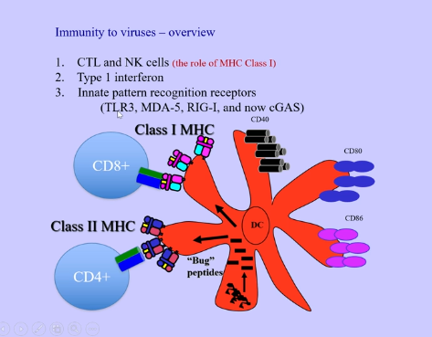
Immunity to Viruses

knowt flashcard image
33
New cards
<p>Tumor Immunity</p>

Tumor Immunity

  • tumor immunosurveillance

    • tumors generally express antigens that the immune system can recognize (mutated, overexpressed, or germ/embryonic proteins)

    • tumor-infiltrating lymphocytes (TILs) can often be found in solid tumors

    • some tumors spontaneously regress

    • bone marrow transplant after leukemia radiation therapy can lead to graft vs leukemia response

    • mice can be protected from some tumors by vaccination with killed tumor cells (lots of ways to prevent and cure cancer in mice)


Problem

  • immunodeficient mice and people don’t seme to have a much higher incidence of spontaneous tumors

    • there may be increased susceptibility to some carcinogens or virally-induced cancers, but that may reflect immune responses against causative agents, rather than tumors

    • also increased susceptibility to some tumors in NK cell deficiencies

  • TILs appear to be tolerant, rather than part of a productive response against tumors

  • tumor immunotherapies have generally been unsuccessful in human trials


cancer cells are not that different from the rest of your cells

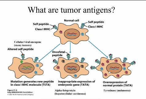
  • don’t express MHC, costimulatory molecules, etc and have normal antigens

  • antigens are taken up by normal APC and presented in absence of inflammatory signals (leads to no response)

  • can lead to removal of certain antigens that cause response from tumor cells

  • selective pressure → selects for those that secrete immunosuppressive cytokines

  • might have protective capsule, preventing phagocytosis

<ul><li><p>tumor immunosurveillance</p><ul><li><p>tumors generally express antigens that the immune system can recognize (mutated, overexpressed, or germ/embryonic proteins)</p></li><li><p>tumor-infiltrating lymphocytes (TILs) can often be found in solid tumors</p></li><li><p>some tumors spontaneously regress</p></li><li><p>bone marrow transplant after leukemia radiation therapy can lead to graft vs leukemia response</p></li><li><p>mice can be protected from some tumors by vaccination with killed tumor cells (lots of ways to prevent and cure cancer in mice)</p></li></ul></li></ul><p></p><p>Problem</p><ul><li><p>immunodeficient mice and people don’t seme to have a much higher incidence of spontaneous tumors</p><ul><li><p>there may be increased susceptibility to some carcinogens or virally-induced cancers, but that may reflect immune responses against causative agents, rather than tumors</p></li><li><p>also increased susceptibility to some tumors in NK cell deficiencies </p></li></ul></li><li><p>TILs appear to be tolerant, rather than part of a productive response against tumors</p></li><li><p>tumor immunotherapies have generally been unsuccessful in human trials </p></li></ul><p></p><p>cancer cells are not that different from the rest of your cells </p><ul><li><p>don’t express MHC, costimulatory molecules, etc and have normal antigens</p></li><li><p>antigens are taken up by normal APC and presented in absence of inflammatory signals (leads to no response) </p></li><li><p>can lead to removal of certain antigens that cause response from tumor cells</p></li><li><p>selective pressure → selects for those that secrete immunosuppressive cytokines</p></li><li><p>might have protective capsule, preventing phagocytosis </p></li></ul><p></p>
34
New cards
<p>Immune System + Cancer</p>

Immune System + Cancer

  • immune system normally plays a small role at most in cancer prevention

  • doesn’t mean immune response can’t be used against tumors

  • strategies:

    • tumor vaccines (peptide, killed cell, and enhanced immunogenicity cell vaccines)

      • find common antigens in tumors (tumor specific tumor antigens), where antigen is required in tumors

      • tumor associated tumor antigens → antigens that aren’t essential, but result from normal dysfunction of tumors (express oncofetal proteins, which are only found in tumors or overexpression of normal proteins)

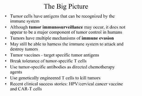
    • break tolerance of TILs (or other tumor specific lymphocytes)

    • tumor-specific antibodies

      • most effective

      • take antibodies or engineered T cells that are specific for tumor antigens

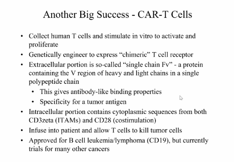
      • activates complement, recruits NK cells via ADCC to then kill tumor, antibody bound to tumor then becomes tag, or attach toxins/radioactive to kill the tumor
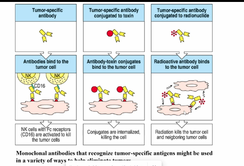

<ul><li><p>immune system normally plays a small role at most in cancer prevention</p></li><li><p>doesn’t mean immune response can’t be used against tumors</p></li><li><p>strategies:</p><ul><li><p>tumor vaccines (peptide, killed cell, and enhanced immunogenicity cell vaccines)</p><ul><li><p>find common antigens in tumors (tumor specific tumor antigens), where antigen is required in tumors</p></li><li><p>tumor associated tumor antigens → antigens that aren’t essential, but result from normal dysfunction of tumors (express oncofetal proteins, which are only found in tumors or overexpression of normal proteins) </p></li></ul></li><li><p>break tolerance of TILs (or other tumor specific lymphocytes)</p></li><li><p>tumor-specific antibodies </p><ul><li><p>most effective</p></li><li><p>take antibodies or engineered T cells that are specific for tumor antigens </p></li><li><p>activates complement, recruits NK cells via ADCC to then kill tumor, antibody bound to tumor then becomes tag, or attach toxins/radioactive to kill the tumor </p></li></ul></li></ul></li></ul><p></p>
35
New cards

CAR-T Cells

knowt flashcard image
36
New cards
<p>Vaccines </p>

Vaccines

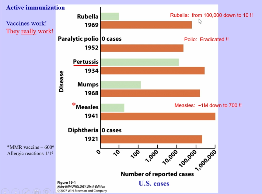
  • proof vaccines work → measles was thought to be eliminated, so people stopped vaccinating their children, however, measles is now starting to resurface

  • passive immunization (not vaccines) → administered before or immediately after exposure

    • given antibodies from someone else

    • anti-venom following spider bites

    • rhogam (anti-D) prior to birth of second baby

    • doesn’t generate an adaptive immune response, meaning after passive immunization passes, you are still susceptible

    • IVIG → immune globulin (given to people deficient in B cells)

    • during covid, people who recovered/initially exposed to covid, were asked to donate plasma to those on ventilators

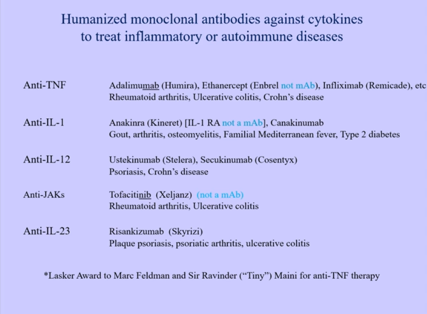
    • humanized monoclonal antibodies

  • polio → two different vaccines

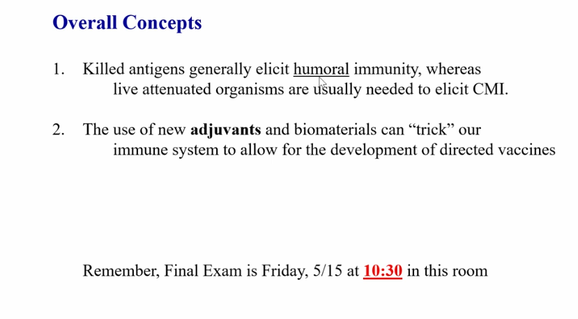
    • Live (sabin) → live attenuated vaccine, which spread from children who got the vaccine to others that were not vaccinated, giving herd immunity

      • risk to immunocompromised individuals, because the vaccine is still alive

      • Oral polio vaccine (OPV)

      • induces cell mediated immunity

    • Killed (salk)

      • inactivated polio vaccination (IPV)

      • induces humoral immunity


<ul><li><p>proof vaccines work → measles was thought to be eliminated, so people stopped vaccinating their children, however, measles is now starting to resurface</p></li><li><p>passive immunization (not vaccines) → administered before or immediately after exposure</p><ul><li><p>given antibodies from someone else </p></li><li><p>anti-venom following spider bites </p></li><li><p>rhogam (anti-D) prior to birth of second baby</p></li><li><p>doesn’t generate an adaptive immune response, meaning after passive immunization passes, you are still susceptible </p></li><li><p>IVIG → immune globulin (given to people deficient in B cells) </p></li><li><p>during covid, people who recovered/initially exposed to covid, were asked to donate plasma to those on ventilators </p></li><li><p>humanized monoclonal antibodies </p></li></ul></li><li><p>polio → two different vaccines</p><ul><li><p>Live (sabin) → live attenuated vaccine, which spread from children who got the vaccine to others that were not vaccinated, giving herd immunity </p><ul><li><p>risk to immunocompromised individuals, because the vaccine is still alive </p></li><li><p>Oral polio vaccine (OPV) </p></li><li><p>induces cell mediated immunity </p></li></ul></li><li><p>Killed (salk) </p><ul><li><p>inactivated polio vaccination (IPV)</p></li><li><p>induces humoral immunity </p></li></ul></li></ul></li></ul><p></p>
37
New cards

Pneumococcal Conjugate

  • vaccine given during early age

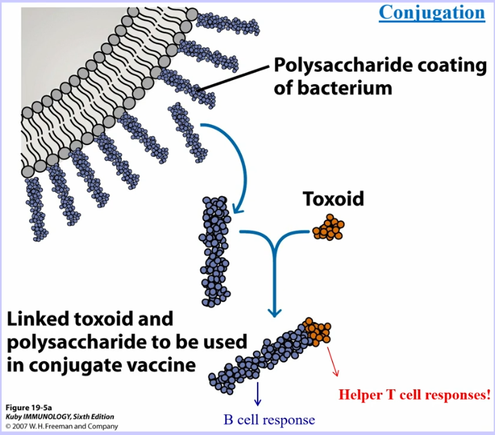
  • type of strain is determined by polysaccharide coating the bacterium (antigen used for vaccinations)

  • polysaccharides don’t fit into MHC molecules, meaning T cell help doesn’t occur

    • produces a IgM antibody response, which is less effective

  • conjugate vaccine → take tetanus toxoid (already vaccinated against) and combine it with the polysaccharide, allowing it to be presented by MHC and induce T cell help

    • produces IgG response


<ul><li><p>vaccine given during early age</p></li><li><p>type of strain is determined by polysaccharide coating the bacterium (antigen used for vaccinations) </p></li><li><p>polysaccharides don’t fit into MHC molecules, meaning T cell help doesn’t occur</p><ul><li><p>produces a IgM antibody response, which is less effective</p></li></ul></li><li><p>conjugate vaccine → take tetanus toxoid (already vaccinated against) and combine it with the polysaccharide, allowing it to be presented by MHC and induce T cell help </p><ul><li><p>produces IgG response </p></li></ul></li></ul><p></p>
38
New cards
<p>Smallpox + Tuberculosis </p>

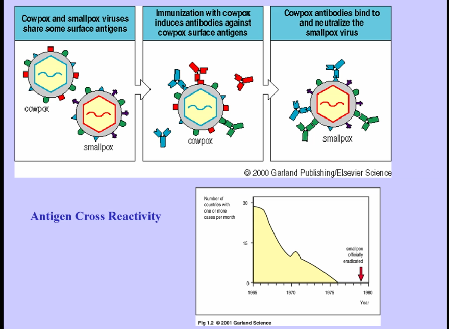
Smallpox + Tuberculosis

  • cowpox and smallpox have enough cross reactivity between them, mounting immune response to related organism

  • Tuberculosis Vaccine (BCG)

    • intracellular bacterium

    • use live attenuated vaccine to induce cell-mediated immunity

    • killed/purified protein induces humoral immune response, which is ineffective

    • effective in children, but ineffective in adult
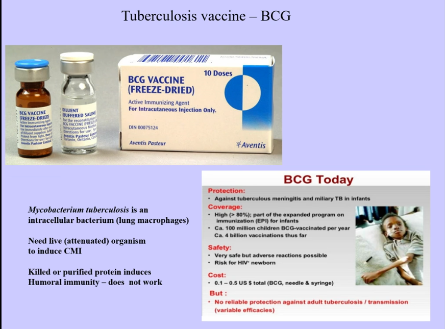

<ul><li><p>cowpox and smallpox have enough cross reactivity between them, mounting immune response to related organism</p></li><li><p>Tuberculosis Vaccine (BCG)</p><ul><li><p>intracellular bacterium</p></li><li><p>use live attenuated vaccine to induce cell-mediated immunity</p></li><li><p>killed/purified protein induces humoral immune response, which is ineffective</p></li><li><p>effective in children, but ineffective in adult</p></li></ul></li></ul><p></p>
39
New cards
<p>Attenuation </p>

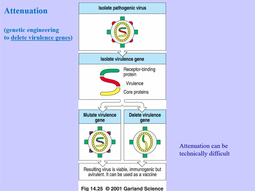
Attenuation

  • attenuation → altering organism to lower its virulence, but maintain structure and characteristics that induce immune responses

    • if altered too far, the organism is unable to cause disease

    • if altered too little, organism can mutate back, giving a very virulent pathogen
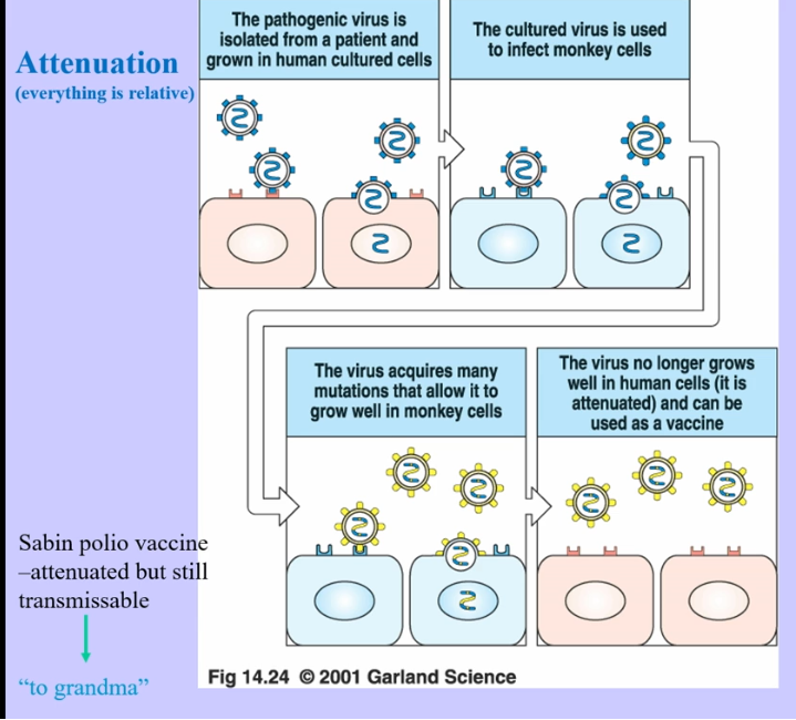

<ul><li><p>attenuation → altering organism to lower its virulence, but maintain structure and characteristics that induce immune responses</p><ul><li><p>if altered too far, the organism is unable to cause disease</p></li><li><p>if altered too little, organism can mutate back, giving a very virulent pathogen</p></li></ul></li></ul><p></p>
40
New cards
<p>DNA Vaccinations </p>

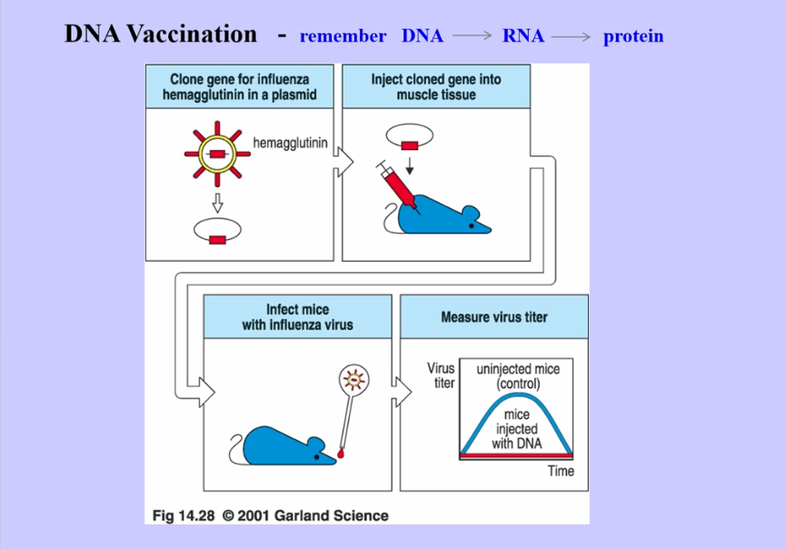
DNA Vaccinations

  • should be effective, because DNA will go through the central dogma

  • should induce CD4 and CD8 immunity

  • haven’t proven effective → potentially DNA enters into our genome and interrupts function of important genes, making it too dangerous to use
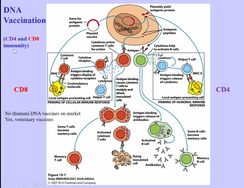
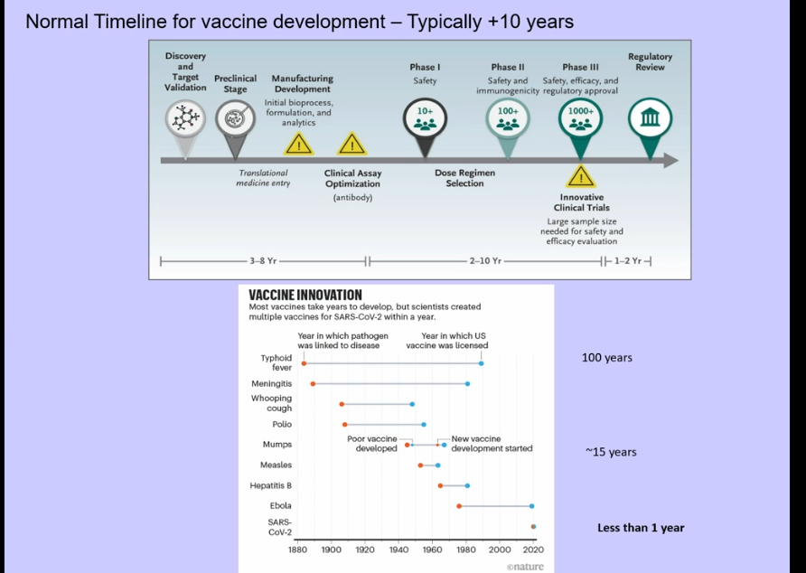
<ul><li><p>should be effective, because DNA will go through the central dogma</p></li><li><p>should induce CD4 and CD8 immunity </p></li><li><p>haven’t proven effective → potentially DNA enters into our genome and interrupts function of important genes, making it too dangerous to use </p></li></ul><p></p>
41
New cards
<p>RNA Vaccinations </p>

RNA Vaccinations

  • RNA less stable than DNA

  • RNAses → degrade RNA and present all over the body

  • first approved against SARS-CoV-2

  • Vaccine development → normally prolonged process, but took 8 months to develop SARS-CoV-2 vaccination

    • two mutations in prefusion conformation (prevented spike from springing away to postfusion form)
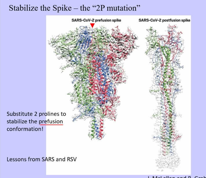

<ul><li><p>RNA less stable than DNA </p></li><li><p>RNAses → degrade RNA and present all over the body</p></li><li><p>first approved against SARS-CoV-2 </p></li><li><p>Vaccine development → normally prolonged process, but took 8 months to develop SARS-CoV-2 vaccination </p><ul><li><p>two mutations in prefusion conformation (prevented spike from springing away to postfusion form) </p></li></ul></li></ul><p></p>
42
New cards
<p>COVID Vaccines </p>

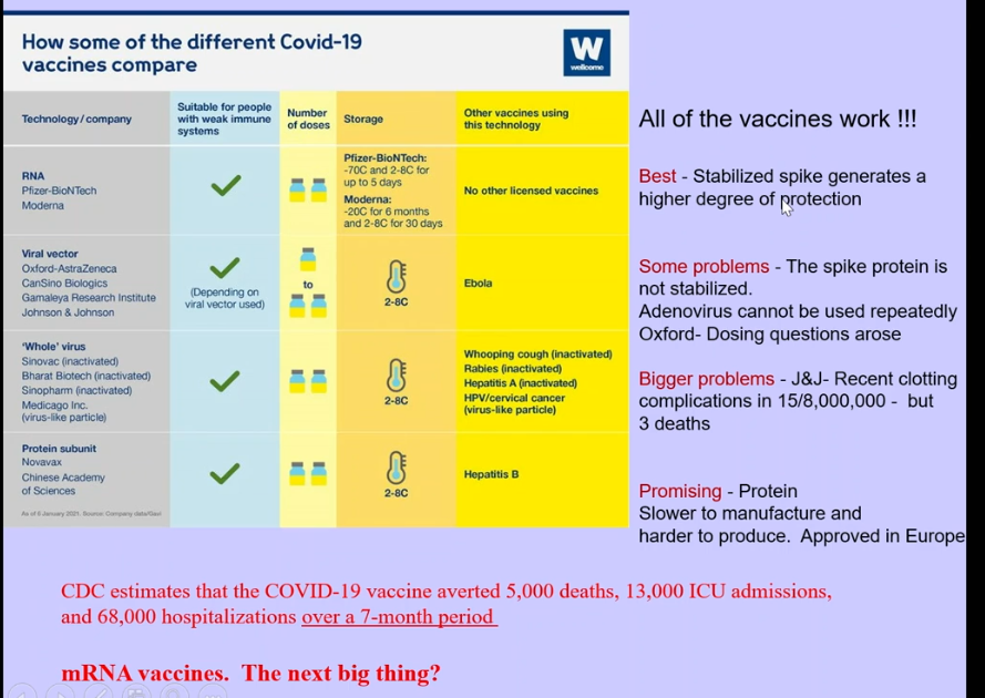
COVID Vaccines

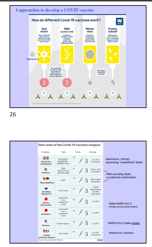
  • viral vector → nonmutated, so virus had tendency to go into post fusion conformation when isolated, when binding to ACE2, etc

  • adenovirus → majority of people have been infected with this type of virus, so antibodies already present/made towards the virus, getting rid of it, leading to less persistence

    • unstable conformation → post fusion spike

  • RNA encoding spike

    • pre-fusion conformation


<ul><li><p>viral vector → nonmutated, so virus had tendency to go into post fusion conformation when isolated, when binding to ACE2, etc</p></li><li><p>adenovirus → majority of people have been infected with this type of virus, so antibodies already present/made towards the virus, getting rid of it, leading to less persistence </p><ul><li><p>unstable conformation → post fusion spike</p></li></ul></li><li><p>RNA encoding spike</p><ul><li><p>pre-fusion conformation </p></li></ul></li><li><p></p></li></ul><p></p>
43
New cards
<p>mRNA vaccines </p>

mRNA vaccines

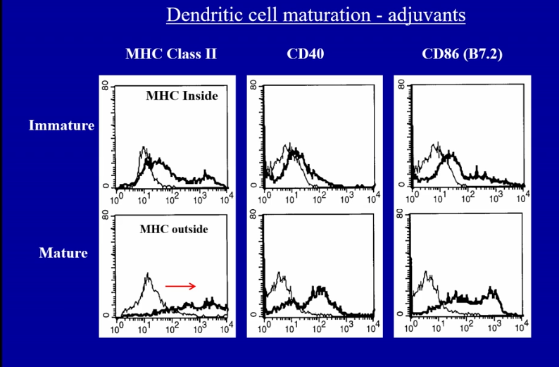
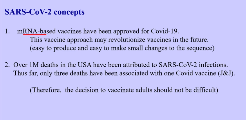
  • mRNA vaccines tend to be their own adjuvants

    • nucleic acids can be recognized as being an adjuvant

    • adjuvants upregulate co-stimulation on APC, leading to T cell help, which improves immune response

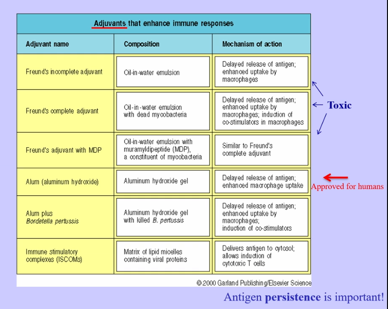
  • most purified proteins will not be immunogenic, even if the body hasn’t seen it before, unless it is perceived to be dangerous

    • perceived to be dangerous when enveloped in an adjuvant or if it binds to a pattern recognition receptor

    • freud’s adjuvant → too toxic for humans

    • alum → less toxic in humans, but still induces immune response

    • works in type 1 hypersensitivity (allergen shots), attempting to switch to a TH1 response

<ul><li><p>mRNA vaccines tend to be their own adjuvants</p><ul><li><p>nucleic acids can be recognized as being an adjuvant</p></li><li><p>adjuvants upregulate co-stimulation on APC, leading to T cell help, which improves immune response </p></li></ul></li><li><p>most purified proteins will not be immunogenic, even if the body hasn’t seen it before, unless it is perceived to be dangerous</p><ul><li><p>perceived to be dangerous when enveloped in an adjuvant or if it binds to a pattern recognition receptor </p></li><li><p>freud’s adjuvant → too toxic for humans</p></li><li><p>alum → less toxic in humans, but still induces immune response </p></li><li><p>works in type 1 hypersensitivity (allergen shots), attempting to switch to a TH1 response </p></li></ul></li></ul><p></p>
44
New cards
<p>Needed Vaccines </p>

Needed Vaccines

  • HIV (less so, because of therapies and treatments that are effective in treating HIV)

  • Malaria

  • Tuberculosis


<ul><li><p>HIV (less so, because of therapies and treatments that are effective in treating HIV) </p></li><li><p>Malaria </p></li><li><p>Tuberculosis </p></li></ul><p></p>
45
New cards

Anamnestic Response

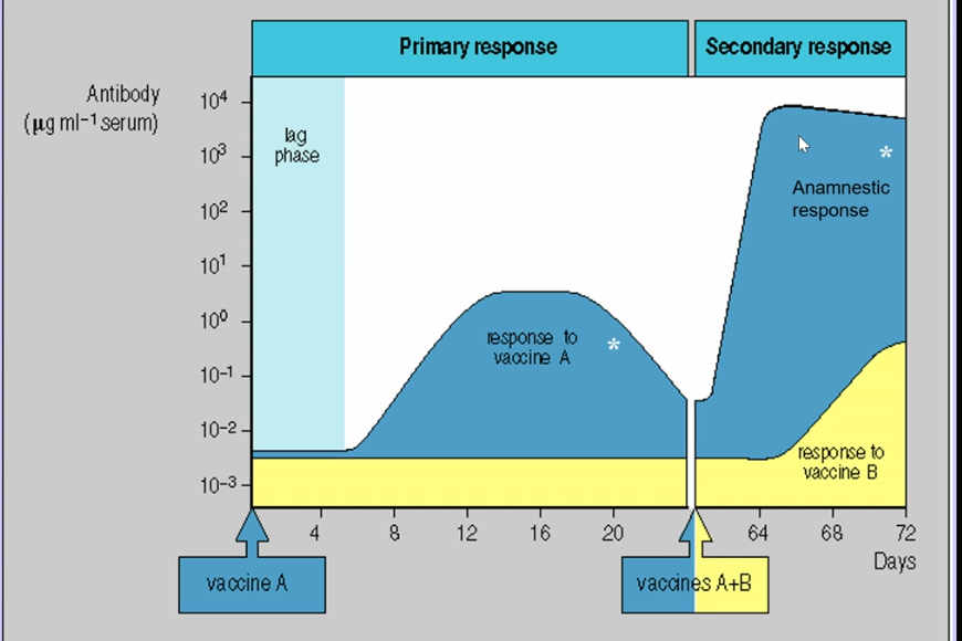
  • antigen-specific secondary response

    • shorter lag time

    • higher amount of antibodies

    • faster response produced

    • peak is extended for a longer period of time

<ul><li><p>antigen-specific secondary response </p><ul><li><p>shorter lag time</p></li><li><p>higher amount of antibodies </p></li><li><p>faster response produced </p></li><li><p>peak is extended for a longer period of time </p></li></ul></li></ul><p></p>
46
New cards
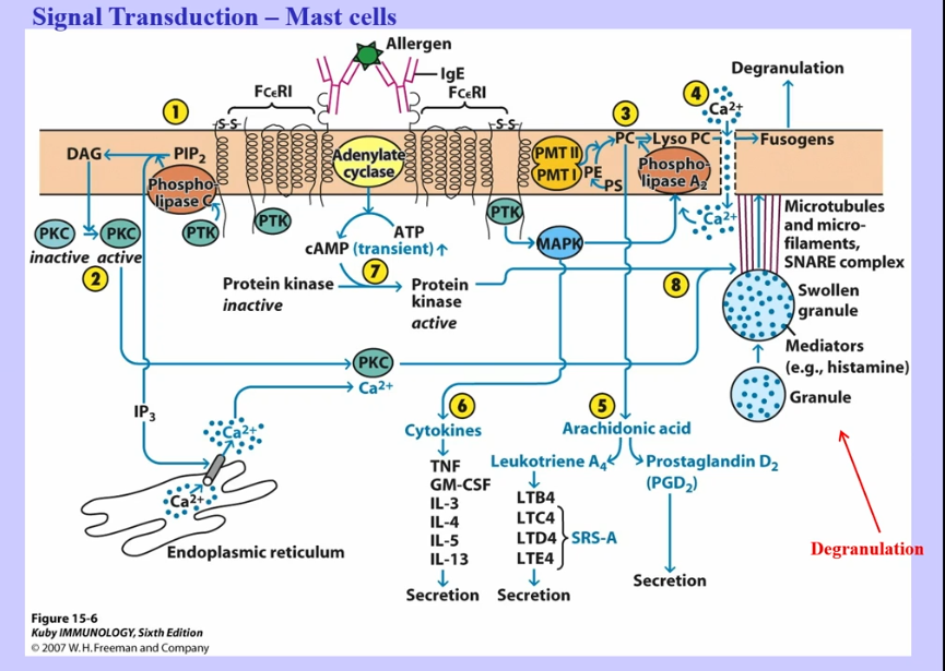
<p>Signal Transduction (Mast Cells and T cells) </p>
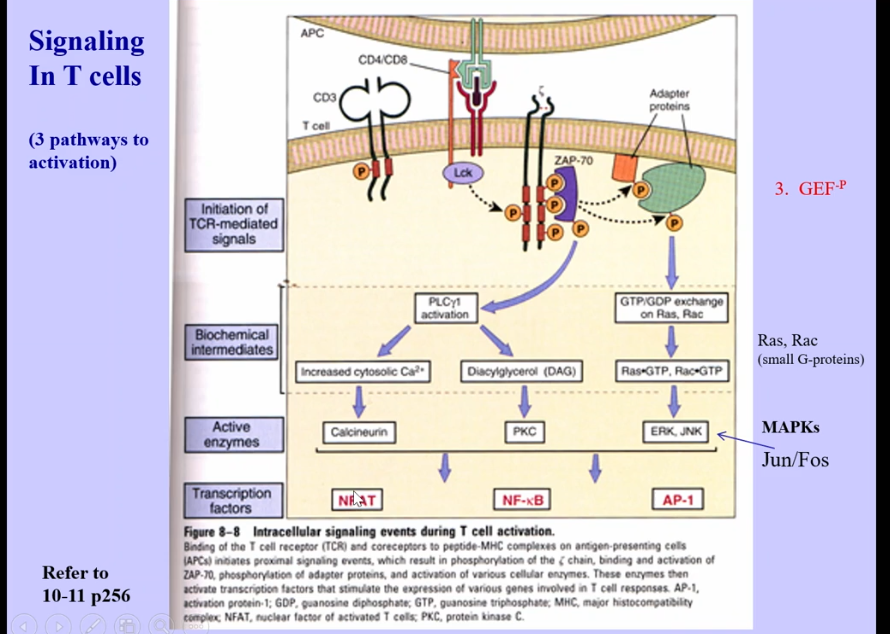
Signal Transduction (Mast Cells and T cells)

knowt flashcard image
47
New cards

NK Cells

knowt flashcard image
48
New cards
<p></p><p>TH Cells</p>


TH Cells

knowt flashcard image