7/24: Brain Anatomy
 Call Kai
 Learn
Learn Practice Test
 Spaced Repetition
 Match
Match Flashcards
 Knowt Play
1/59

There's no tags or description
Looks like no tags are added yet.
Name | Mastery | Learn | Test | Matching | Spaced | Call with Kai | Chat |
|---|
No analytics yet
Send a link to your students to track their progress
60 Terms
central nervous system
CNS → a major division the nervous system
Brain
Spinal cord
Contains sensory and motor neurons
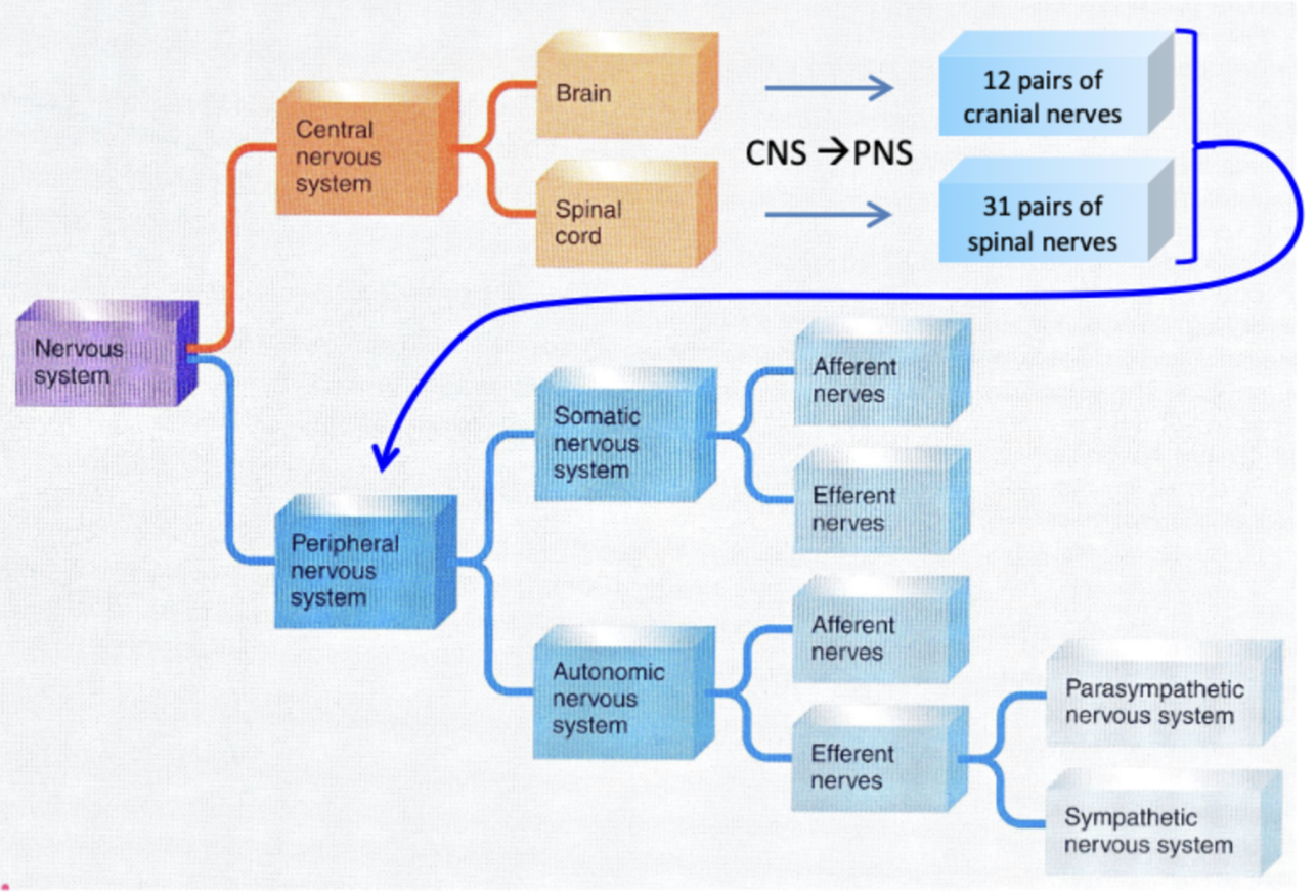
peripheral nervous system
PNS → a major division the nervous system
Nerves
Spinal
Cranial
Contains sensory and motor neurons
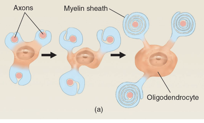
oligodendrocytes
Manufactures myelin in CNS
1 cell produces many myelin sheaths
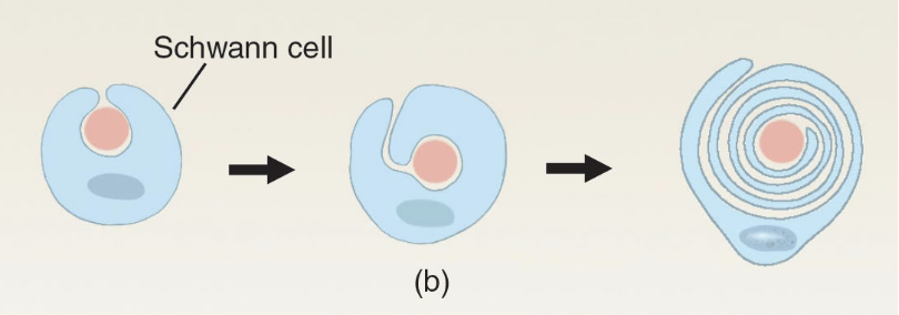
schwann cells
Manufactures myelin in PNS
1 cell produces a single myelin sheath
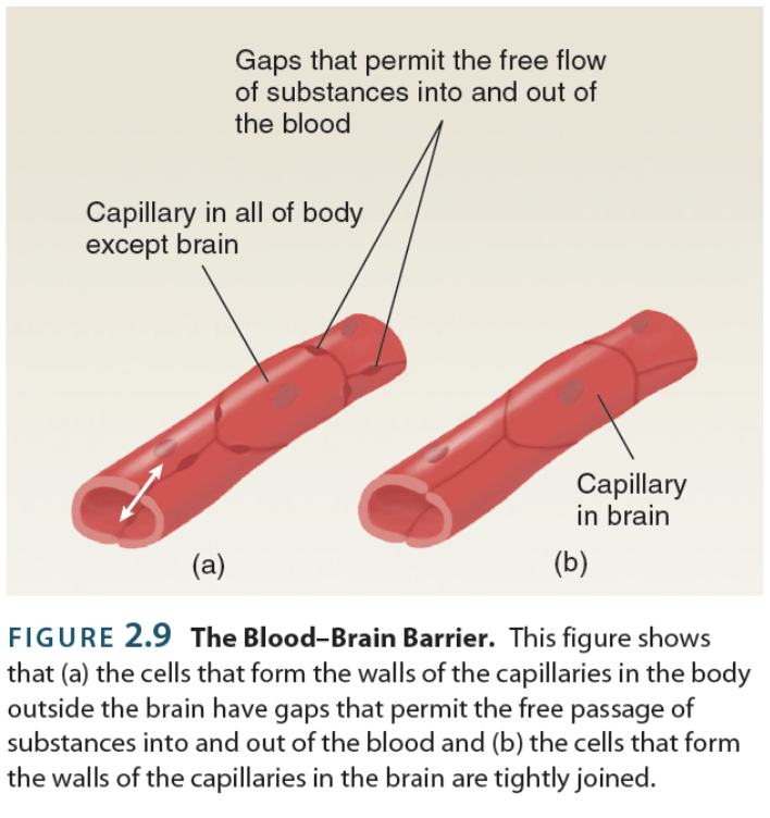
blood brain barrier
→ refers to the CNS’ lack of participation in the lymphatic system
Dye injected into bloodstream will dye all tissue except for spinal cord + brain
This is because the CNS doesn’t have open capillaries (vessels don’t have gaps or holes like other tissues, which permit free flow of substances in and out of blood)
CNS produce their own solution (CSF) by deliberately selecting components from the blood (there’s no leakage)
extracellular fluid
→ fluid that flows around cells to provide nutrients and collect waste
a.k.a interstitial fluid
Liquid part of the blood (plasma) that leaks out of all vessels except nervous ones
Collected into lymphatic system (part of immune system) = lymph vessels → lymph nodes → lymph organs
cerebrospinal fluid
CSF → extracellular solution (that isn’t plasma) deliberately made by the brain to surround / suspend organs of the CNS
Made from blood by tissue called chloroid plexus
Carries nutrients in and wastes out from the CNS’ organs
Made continuously and is fully exchanged 4x daily
Recycled from CNS into blood supply via holes in the dura mater
projection neurons
→ have axons that innervate distal areas of the brain (synapses far out from its origin)
Part of CNS
Releases glutamate (excitatory)
interneuron
→ have axons that innervate proximal areas of the brain (synapses locally about its origin)
Part of CNS
Releases GABA (inhibitatory)
motor neurons
→ nervous cells controlling muscle contraction and gland secretion
Part of PNS & CNS
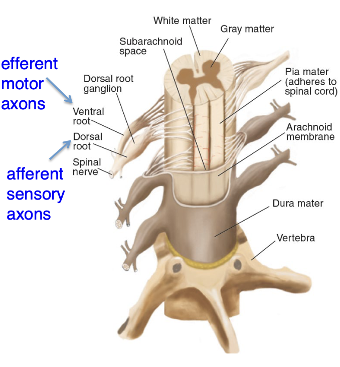
Axons called efferent fibers
efferent fibers
→ axons of motor neurons directing information away from CNS; output
sensory neurons
→ nervous cells detecting changes in external and internal environments
Part of PNS & CNS
Axons called afferent fibers
afferent fibers
→ axons of sensory neurons directing information towards CNS; inputs
nerves
→ enclosed, cable-like bundles of axons in the PNS
Spinal
Cranial
Most contain a mix of motor and sensory neurons
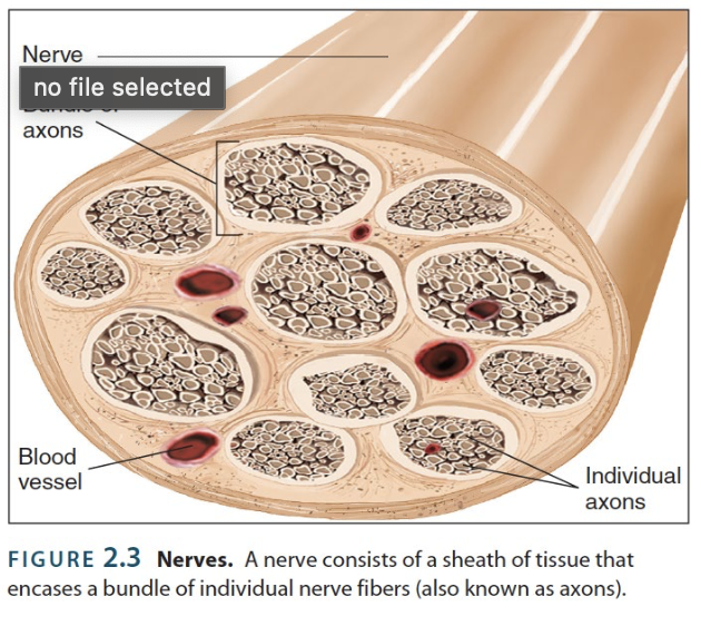
spinal nerves
→ nerves whose axons enter/leave spinal cord
Significant part of PNS
31 pairs
Nearly 1 pair for each vertabrae in spine
cranial nerves
→ nerves whose axons enter/leave brain
Significant part of PNS
12 pairs
Nearly all pairs (except 10th) process movements and sensory information around head and neck;
Vagus (X) nerve is the exception that branches extensively in the thoracic and abdominal cavities (upper half of body)
spinal cord
→ brings sensory information to the brain + connects the axons of motor neurons (efferent fibers) to effector organs throughout the body
Significant part of CNS
Maintains a certain degree of autonomy from its CNS counterpart (the brain) since it contains various reflexive control circuits
Has a long, conical structure that is approximately as thick as an adult pinkie
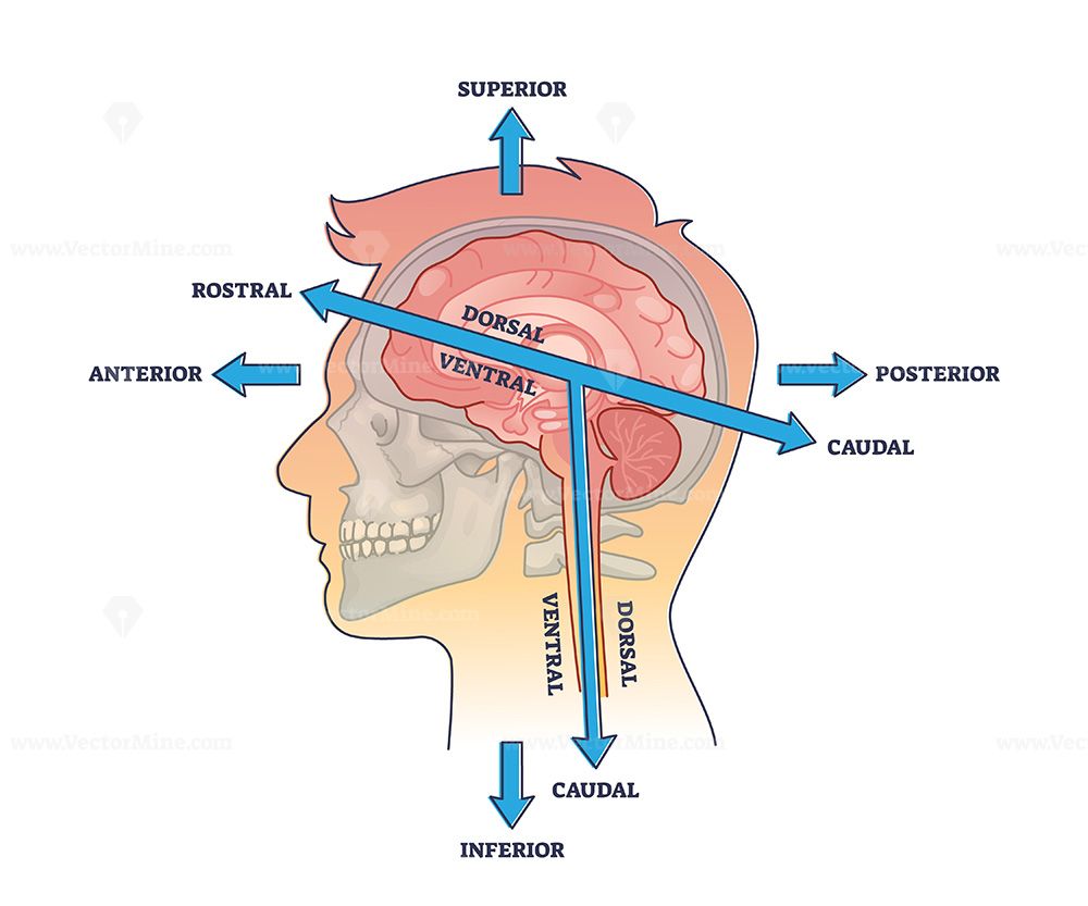
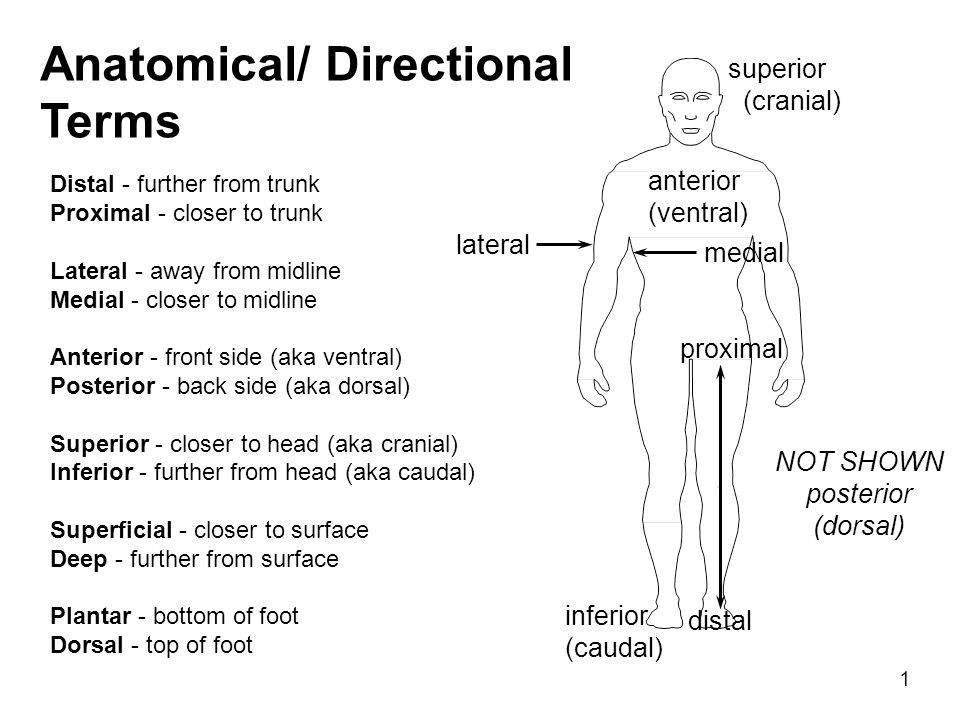
anatomical directions
Transverse plane (Y -axis, up & down)
Superior → above
Rostral → upper
Inferior → below
Caudal → lower
Sagittal plane (Z - axis, front & back)
Anterior → in front
Ventral → front-facing
Posterior → behind
Dorsal → back-facing
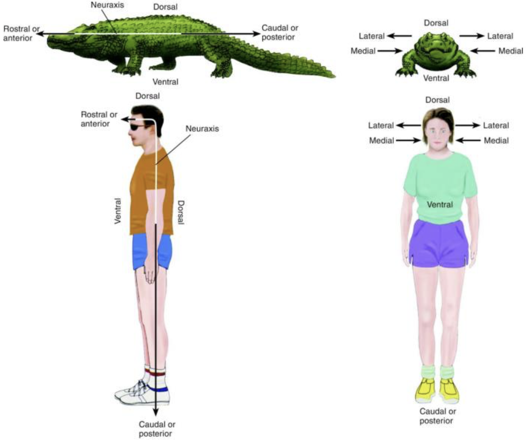
Frontal plane (X - axis, moving along left and right)
Medial → away midline
Lateral → toward midline
neuraxis
→ imaginary line that runs along the length of the CNS
Relevant for reference by anatomical directions
somatic nervous system
→ branch of PNS that interacts with external environment (processes sensory information)
Afferent nerves’ sensory signals: Body surface → CNS
Efferent nerves’ motor signals: CNS → skeletal muscles
autonomic nervous system
→ branch of PNS that regulates internal environment (senses and regulates smooth muscle, cardiac muscle, & glands)
Afferent nerves’ sensory signals: Internal organs → CNS
Efferent nerves’ motor signals: CNS → internal organs
2 divisions
Sympathetic
Parasympathetic
sympathetic division
→ division of ANS that primes the body for threatening situations
“Fight-flight-freeze” response
Always active to some extent (regulates many organs’ functions)
Strong stimulation increases blood flow to organs involved in intense physical activity / immediate survival + shunts blood flow from organs that are not ostensibly necessary
parasympathetic division
→ division of ANS supporting activities in a relaxed, neutral state
“Rest and digest” response
Also always active to some extent (also regulates many organs’ functions)
Responsible for increasing the body’s energy stores
brain nuclei
→ refers to a collection of neurons that are clustered together that regulate a shared function
ipsilateral
→ refers to structures on the same side of the body
Taste and smell are the only sensory systems that are ipsilaterally organized
contralateral
→ refers to structures on the opposite side of the body
anatomical directions for limbs
Superficial → closer to surface
Deep → further from surface
Proximal → close to trunk of body
Distal → away from trunk of body
hindbrain
→ a.k.a. brain stem
Medulla oblongata
Pons
Cerebellum
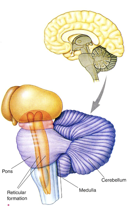
medulla oblongata
→ part of the hindbrain; the most caudal part whose brain nuclei regulate autonomic functions
Notable nuclei:
Area postrema → brain nuclei that initiates vomiting when poisons are detected
Reticular formation → regulates sleep and arousal
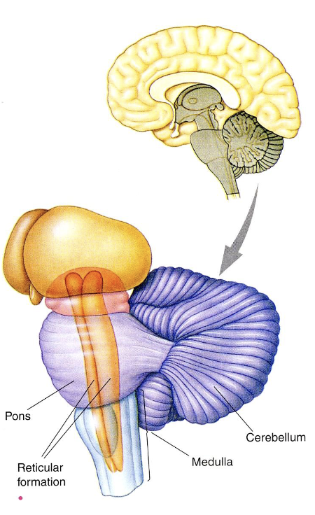
pons
→ part of the hindbrain; bulge that relays information between the cerebrum and cerebellum
Location of several cranial nerve nuclei central to perception
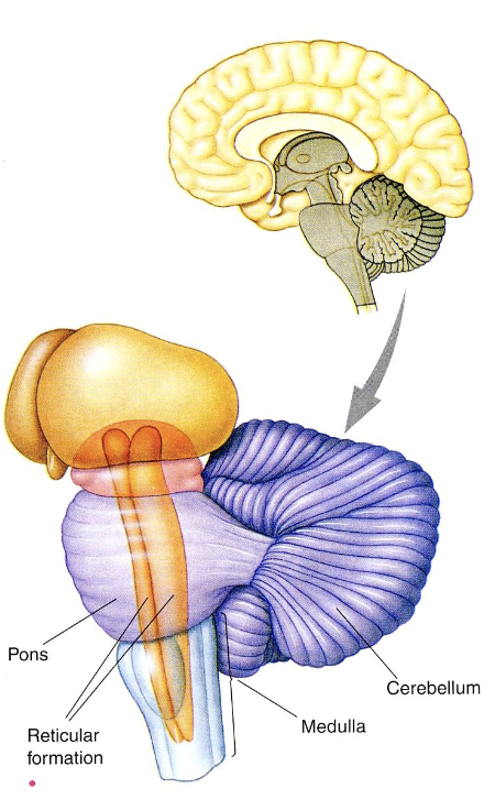
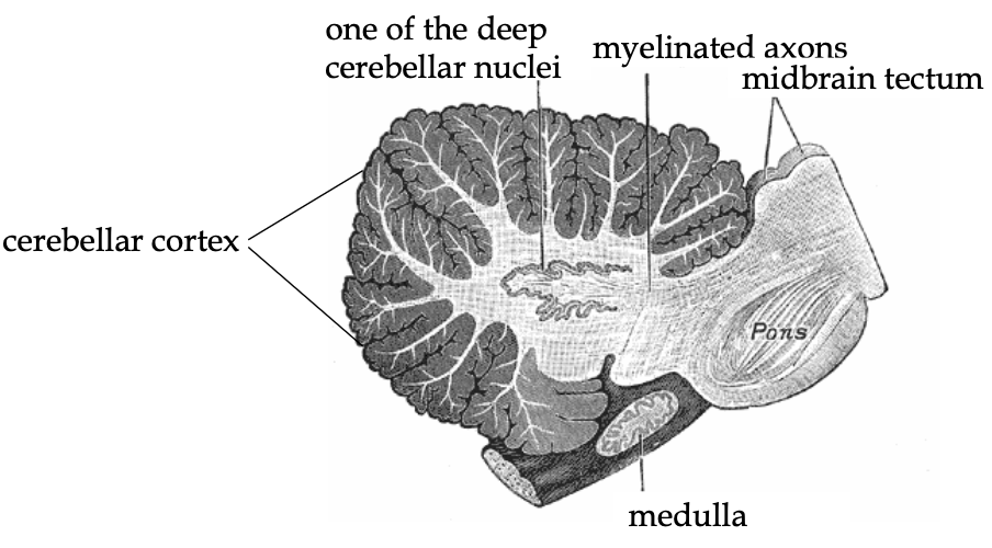
cerebellum
→ part of the hindbrain; facilitates sensory-motor integration (overall motor learning as the body grows and changes over time)
a.k.a “Little brain”
Has ↑ [ ] of neurons of any other part of the brain, but these neurons are small
Damage leads to awkward movement and poor coordination (precision and accuracy); severe cases lead to paralyzation
![<p>→ part of the <strong>hindbrain</strong>; facilitates sensory-motor integration (overall motor learning as the body grows and changes over time)</p><ul><li><p>a.k.a “Little brain”</p></li><li><p>Has ↑ [ ] of neurons of any other part of the brain, but these neurons are small</p></li><li><p>Damage leads to awkward movement and poor coordination (precision and accuracy); severe cases lead to paralyzation</p></li></ul><p></p>](https://knowt-user-attachments.s3.amazonaws.com/c36ec616-b652-44e8-acdb-9ce05a36636c.png)
superficial cerebellar cortex
→ a continuous thin layer of cells in the cerebellum tightly folded in the style of an accordion
Locale where afferent fibers of the cerebellum synapse
Neurons send axons inwards to deep cerebella nuclei, which eventually project to CNS
midbrain
→ collection of nuclei that orchestrate complex reflexive behaviours
Tectum
Tegmentum
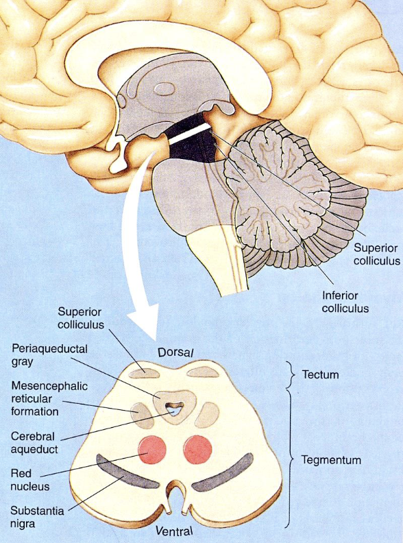
tectum
→ part of the midbrain; appears as 2 pairs of bumps on the dorsal surface
Superior colliculi → top 2 bumps involved in orienting to things in peripheral vision
Inferior colliculi → bottom 2 bumps involved in orienting to unexpected sounds
tegmentum
→ part of the midbrain; that includes several structures that coordinate and motivate complex species-typical movements
Some areas process pain + responses to threats
forebrain
Cerebrum & cerebral cortex
Basal ganglia
Limbic system
Thalamus
Hypothalamus
hypothalamus
→ part of the forebrain; connects the nervous system to the endocrine system via the pituitary gland
Considered to be the “master hormonal regulator” since the hormones it releases regulates the functions of other endocrine glands
Bilateral structure made up of several nuclei that regulate ANS activity:
Notably involved with behaviours directly related to survival (feeding, fighting, fleeing, & mating)
Body temp., circadian rhythm, hunger, sex, etc.
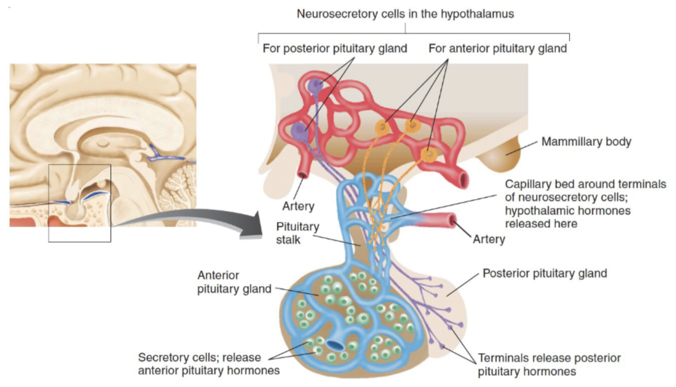
hormone
→ chemical substance that is released into the blood by an endocrine gland
endocrine gland
→ secretes hormones into the blood
Master hormonal regulator = hypothalamus
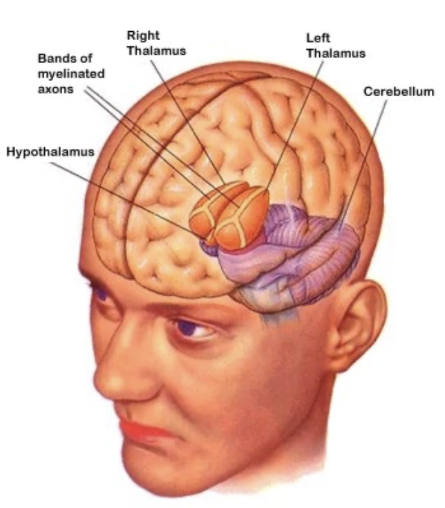
thalamus
→ part of the forebrain; structure sandwiching the hypothalamus
Bilateral structure made up of several nuclei that relay ascending sensory information to different regions of the cerebrum
Lateral geniculate nuclei (LGN) → transmits visual info
Media geniculate nuclei (MGN) → transmits auditory info
cerebrum
→ part of the forebrain interchanged with cerebral cortex; where sensory information enters conscious awareness
Primary area → input/output area to cerebral cortex
Association cortex →
Where our understanding of the world (perception) is formed
Where we decide how to move purposefully
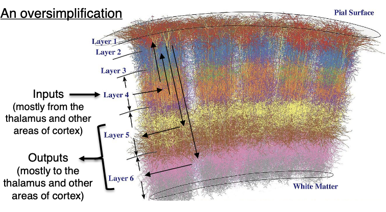
Multi-layered structure
6 layers in mammals
3 layers in reptiles
Embedded with cortical columns (neurons interconnected between layers) that are partially distinct functional units
NOT made up of distinct nuclei (except for in birds)
lobes of cerebrum
Frontal → controls movement
Parietal → process tactile info (touch)
Occipital → process visual info (vision)
Temporal → process auditory info (hearing)
Lateral fissure → junction between frontal, parietal, and temporal lobe that processes taste and smell
Insular cortex → processes gustatory info (taste)
Piriform cortex → processes olfactory info (smell)
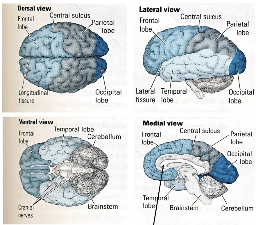
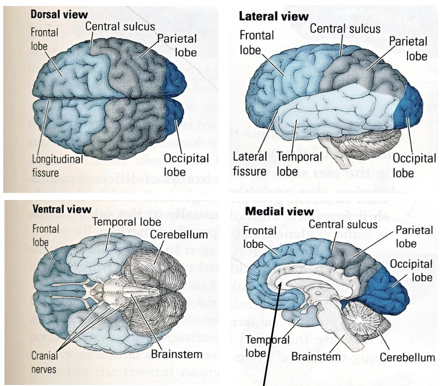
divisions between cerebrum lobes
Longitudinal fissure → separates left and right hemispheres
Visible from dorsal view
Lateral fissure → separates frontal and temporal lobe
Visible from lateral view
Central sulcus → separates frontal lobe and parietal lobe
Visible from dorsal and lateral view
Corpus callosum → large bundle of axons that connects corresponding parts of the left and right hemispheres + unifies perceptions and memories
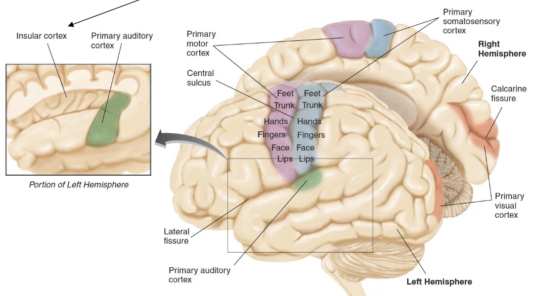
somatosensory cortex
Primary cortical area in parietal lobe
Area where tactile info enters cerebrum
Receives input from different parts of the body
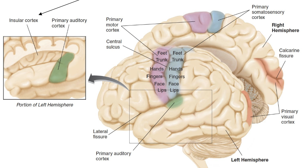
primary motor cortex
Primary cortical area in frontal lobe
Motor neurons synapse in the spinal cord
Controls different parts of the body
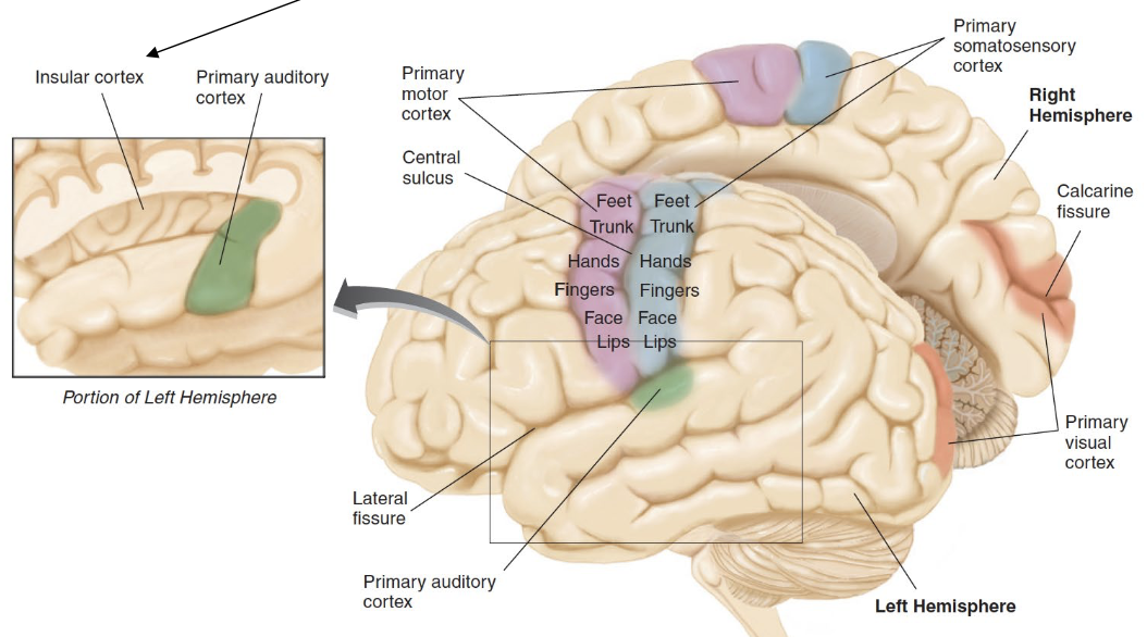
primary auditory cortex
Primary cortical area in temporal lobe
Area where auditory info enters cerebrum
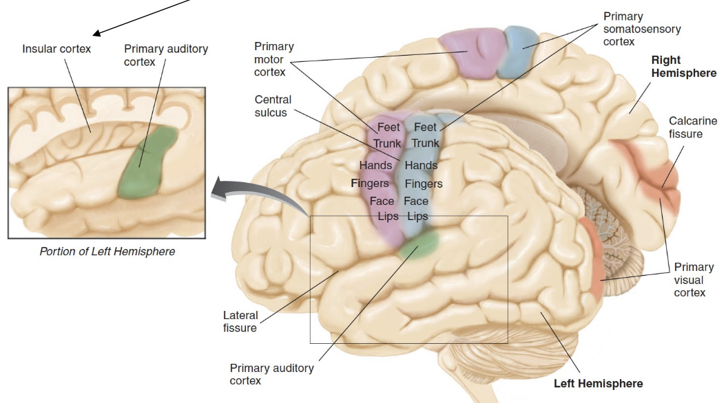
primary visual cortex
Primary cortical area in occipital lobe
Area where visual info enters cerebrum
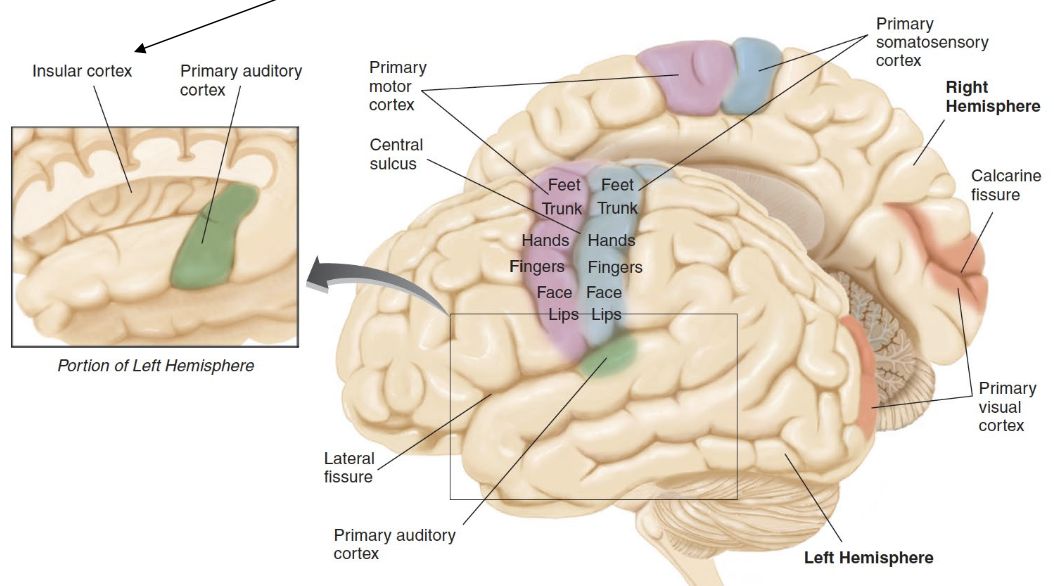
insular cortex
Primary cortical area in lateral fissure
Area where gustatory info enters cerebrum
cerebral cortex
→ surface covering the cerebrum
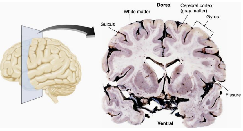
Gray matter → outermost portion of with high concentration of cell bodies
White matter → region beneath gray matter primarily composes of myelinated axons
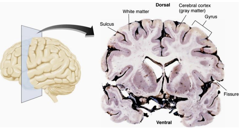
convolutions
→ increase surface area of the cerebral cortex
Sulci → small grooves
Fissures → large or major grooves
Gyri → ridges between sulci and fissures
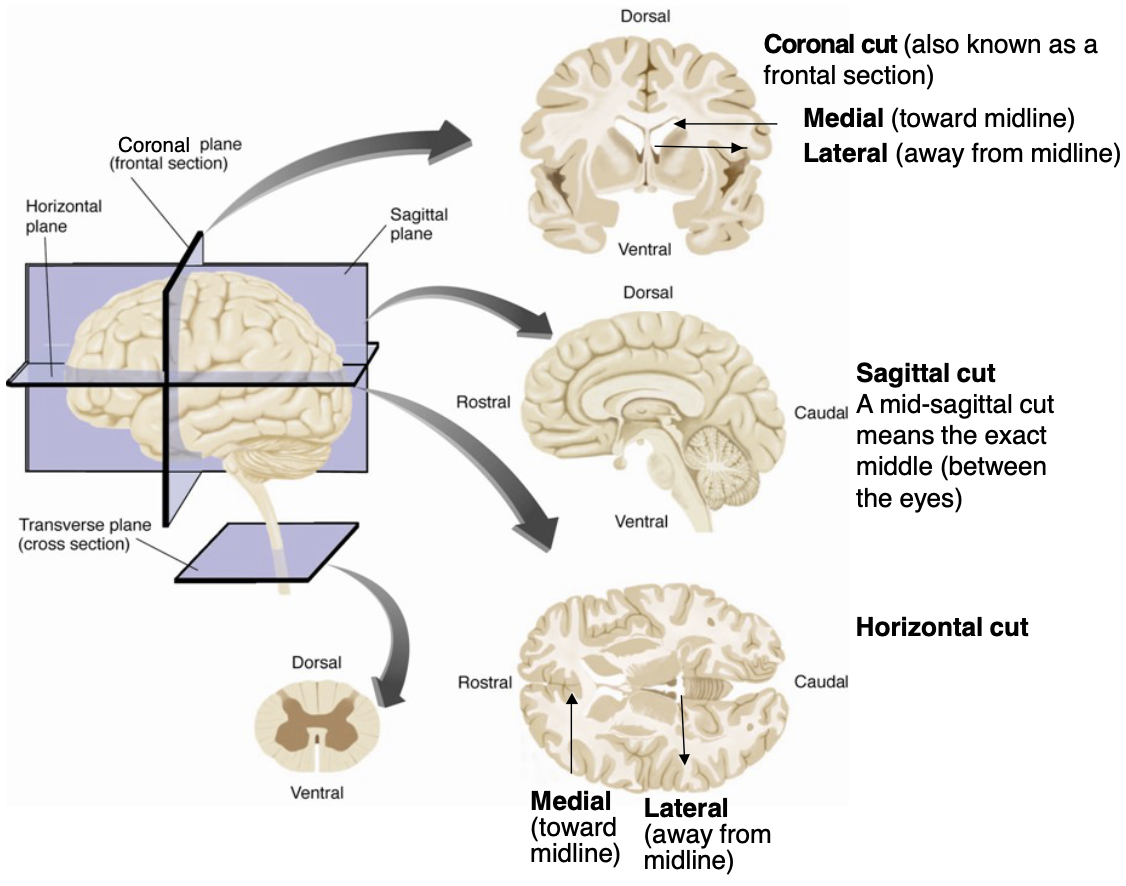
cross sections of brain
Horizontal cut → cut across y level (transverse plane)
Sagittal cut → cut down axis of symmetry (sagittal plane)
Coronal cut → cut across x level (frontal plane)
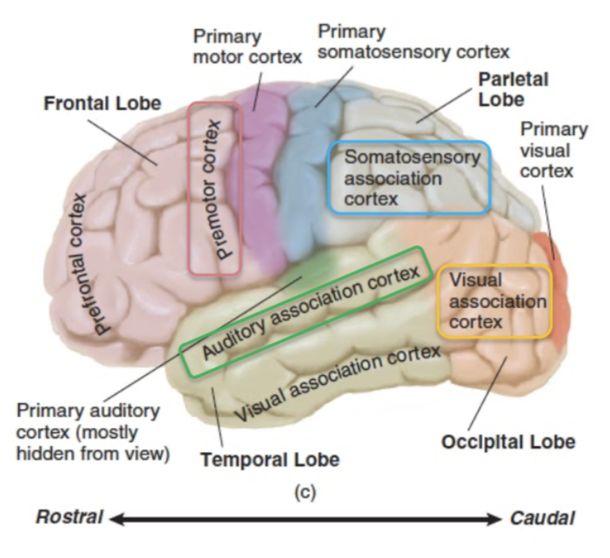
sensory association cortex
→ specific area where a sense’s perception takes and memories are stored, adjacent to every primary cortical (sensory) area
(ex. Premotor cortex is adjacent to primary motor cortex)
1 per sensory system
basal ganglia
→ subcortical structure of the forebrain; circuit of nuclei regulating voluntary movement, reinforcement learning, and habits
Input: Forebrain (especially the frontal lobe) → for movement
Output:
Ascension: Cerebrum (via thalamus) → for sensory processing and decision-making
Descension: Midbrain, hindbrain → for direct movement
Used to be called the primitive “reptilian” brain
Neurological “movement” disorders like Parkinson’s are related to the loss of signaling in this region
limbic system
→ subcortical structure of the forebrain; regulates emotions and the formation of episodic memories (personal experiences)
Hippocampus → area in the temporal critical for explicit memory formation
Amgydala → area in the temporal lobe critical for processing emotion (especially fear)
Cingulate cortex → large area connecting limbic areas that overlies the corpus callosum
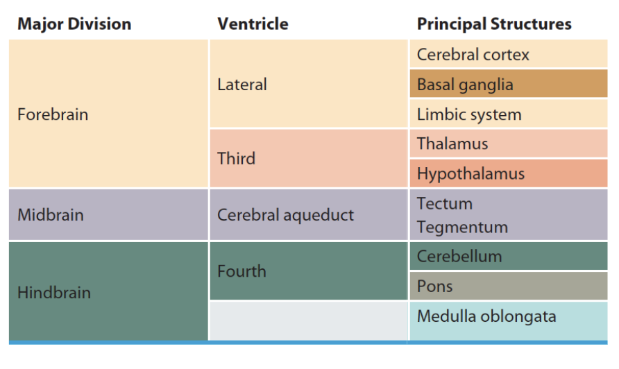
brain anatomy
Forebrain
Midbrain
Hindbrain
meninges
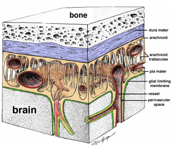
→ 3 protective layers of tissue wrapping organs of the CNS
Dura mater → outermost layer; thick, tough, unstretchable tissue
Arachnoid membrane → middle layer; web-like extensions create a soft, spongey layer filled with CSF
Subarachnoid space → area where large blood vessels and capillaries delivering nutrients and oxygen course though and form arachnoid trabeculae
Pia mater → innermost layer; saran-wrap like
ventricles of the brain
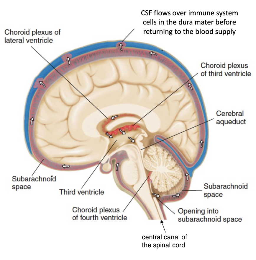
→ 4 interconnected chambers in the center of the brain filled with choroid plexus
(2) Lateral ventricles → largest chambers right underneath cerebrum
Third ventricle → lies between 2 thalamic nuclei at center of the brain
Cerebral aqueduct → long, tube-like structure connecting the third and fourth ventricles
Fourth ventricle → located in hindbrain (between pons and cerebellum)
Connects to central canal of spinal cord
brain development
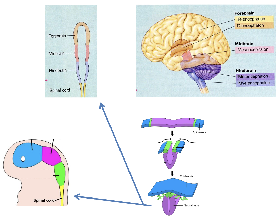
IN THE WOMB
0-4 weeks: Embryonic membrane pinches off and forms neural progenitor cells, which begin to form a hollow neural tube (frame for the CNS & CSF) via symmetrical cell division (where 2 daughter cells are also neural progenitor cells). This is forming the brain’s ventricle space.
4-8 weeks: Symmetrical cell division halts, commencing asymmetrical cell division (where neural progenitor daughter cells migrate away from the neural tube and divide into either 2 neurons or 2 glial cells instead). This is gradually forming the rest of the cerebrum.
5 months: Fetus has produced roughly 85 billion neurons in the human brain (the most we could ever have). However, many neurons die (undergo apoptosis) before birth (ostensibly due to a lack of accommodation in the network at term).
neurogenesis
→ production of new neurons (via asymmetrical cell division of neural progenitor cells)
Researchers are uncertain as to whether neurogensis occurs in adult mammals or even humans
apoptosis
→ process of programmed cell death that occurs in multicellular organisms
This highly regulated and controlled form of death suicide ensures that a dying cell doesn’t extend its illness to neighbouring cells; apoptosis prevents cancer