BIO 142 Lab practical I Study Guide
1/63
There's no tags or description
Looks like no tags are added yet.
Name | Mastery | Learn | Test | Matching | Spaced | Call with Kai |
|---|
No analytics yet
Send a link to your students to track their progress
64 Terms
Identify Pituitary, Hypothalamus, Adrenal, Thyroid and Pancreas on the models.
Identify Adrenal glands under the microscope based on structures and arrangement (or on images). Also identify the layers and there secretions:
Layer | Hormones Secreted | Function |
|---|---|---|
Zona Glomerulosa | Mineralocorticoids (mainly Aldosterone) | Regulates sodium, potassium, and blood pressure |
Zona Fasciculata | Glucocorticoids (mainly Cortisol) | Increases blood glucose and helps the body respond to stress |
Zona Reticularis | Androgens (e.g., DHEA) | Contributes to development of secondary sex characteristics |
Identify thyroid glands under the microscope based on structures and arrangement (or on images)
Identify pituitary glands under the microscope based on structures and arrangement (or on images)
Identify parathytoid under the microscope based on structures and arrangement (or on images) and define the functions of parathyroid hormone. differentiate between parathyroid and thyroid under microscope
PTH raises blood calcium by:
Taking calcium from bone
Keeping calcium in the kidneys
Absorbing more calcium from the intestine
Identify pancreas under the microscope based on structures and arrangement (or on images)
Identify exocrine and endocrine parts of pancreas under the microscope.
Exocrine Pancreas (about 98–99% of the organ)
Appears as darkly stained clusters of cells called acini.
Cells are arranged around small ducts.
Produces digestive enzymes (amylase, lipase, proteases) that are secreted into the small intestine through ducts.
Makes up most of what you see on a slide
Endocrine Pancreas
Appears as lighter-staining, round clusters of cells called the Islets of Langerhans.
Scattered throughout the exocrine tissue.
Richly supplied with blood vessels.
Secretes hormones directly into the bloodstream:
Beta cells → Insulin
Alpha cells → Glucagon
Delta cells → Somatostatin
Define the functions of thyroid hormones, parathyroid hormone, Insulin, Glucagon and
Cortisol.
Hormone | Main Function |
|---|---|
Thyroid Hormones (T3/T4) | Increase metabolism and support growth |
PTH | Raise blood calcium |
Insulin | Lower blood glucose |
Glucagon | Raise blood glucose |
Cortisol | Stress response; raises blood glucose |
Define the hormones that control the blood calcium level (three hormones: Parathyroid,
Calcitonin and Calcitriol).
Hormone | Source | Effect on Blood Calcium |
|---|---|---|
Parathyroid Hormone (PTH) | Parathyroid glands | Raises blood calcium levels |
Calcitonin | Thyroid gland (C cells) | Lowers blood calcium levels |
Calcitriol (active vitamin D) | Produced by kidneys from vitamin D | Raises blood calcium levels |
Define the hemoglobin content of blood and name the method for measuring of hemoglobin.
Group | Hemoglobin Level (g/dL) |
|---|---|
Average | 15 g/dL |
Males | 14–18 g/dL |
Females | 12–16 g/dL |
Newborn Babies | 17–23 g/dL |
(Tallquist method).
Define the method performed in the lab for determining the ABO and Rh blood groups.
agglutination
Place blood samples on a slide.
Add Anti-A, Anti-B, and Anti-Rh sera to separate samples.
Look for agglutination (clumping).
Determine blood type:
Clumping with Anti-A → Type A
Clumping with Anti-B → Type B
Clumping with both → Type AB
No clumping with A or B → Type O
Clumping with Anti-Rh → Rh+
No clumping with Anti-Rh → Rh−
Identify the blood group from a test result.
Reaction | Blood Type |
|---|---|
Clumps with Anti-A only | Type A |
Clumps with Anti-B only | Type B |
Clumps with Anti-A and Anti-B | Type AB |
No clumping with Anti-A or Anti-B | Type O |
Clumps with Anti-Rh | Rh Positive (+) |
No clumping with Anti-Rh | Rh Negative (−) |
Define universal donor and universal recipient.
Universal Donor
A person with blood type O negative (O−)
Can donate blood to all blood types (A, B, AB, O)
Reason: has no A, B, or Rh antigens on red blood cells, so it will not trigger an immune reaction.
Universal Recipient
A person with blood type AB positive (AB+)
Can receive blood from all blood types (A, B, AB, O)
Reason: has no anti-A or anti-B antibodies in the plasma, and Rh positive can accept both Rh+ and Rh− blood.
Quick Summary
O− = universal donor
AB+ = universal recipient
Rh Type | Antigen on RBC | Antibodies in Plasma |
|---|---|---|
Rh+ | Rh (D) antigen present | No anti-Rh antibodies |
Rh− | No Rh antigen | May develop anti-Rh antibodies after exposure |
Blood Type | Antigens on RBC membrane | Antibodies in Plasma (Serum) |
|---|---|---|
A | A antigen | Anti-B antibodies |
B | B antigen | Anti-A antibodies |
AB | A and B antigens | No anti-A or anti-B antibodies |
O | No A or B antigens | Anti-A and Anti-B antibodies |
Define and measure the hematocrit and the average normal value in a healthy young adult.
Percentage of blood occupied by RBCs
Method:
Microhematocrit (centrifugation) method
Fill capillary tube with blood
Centrifuge it
Blood separates into:
Plasma (top)
Buffy coat (middle)
RBCs (bottom)
Calculate: RBC layer ÷ total blood × 100
Normal Values:
Males: 42–52%
Females: 37–47%
Define the normal average of WBC count in a healthy young adult.
less then 1 percent of blood
Identify Neutrophile under the microscope and their percentages in normal condition (or image)
Neutrophile
NUCLEUS: Multi-lobed nucleus with 2–5 or more lobes
GRANULES : Small, no distinct pale lilac to neutral-staining
granules
COLOR OF CYTOPLASM : Usually pink but, sometimes has a reddish tinge
the most abundant (50% - 70%)
Identify Basophil under the microscope and their percentages in normal condition (or image)
Basophile
NUCLEUS: Nucleus is large, varied in shape;
GRANULES : Large, dark blue-purple granules
COLOR OF CYTOPLASM : Purple
Least abundant less 1%
Identify Eosinophil under the microscope and their percentages in normal condition (or image)
Eosinophile
NUCLEUS: Bi-lobed nucleus (occasionally 3 lobes)
GRANULES : Many medium, red-orange
COLOR OF CYTOPLASM : Pink
Between 2% - 4% (Increase in Parasitic infection or Allergy)
Identify Monocyte under the microscope and their percentages in normal condition (or image)
NUCLEUS: Large kidney bean or horseshoe-shaped lacy nucleus
GRANULES : NO
COLOR OF CYTOPLASM : Light blue-gray
The largest of the leukocytes.(Twice the size of RBC)
3% - 8% of total leukocytes
Identify Lymphocyte under the microscope and their percentages in normal condition (or image)
NUCLEUS: Large, round, or slightly indented nucleus that stains very dark purple
GRANULES : NO
COLOR OF CYTOPLASM : Light sky-blue cytoplasm; small cells have only a rim of cytoplasm
The smallest of the leukocytes. (About the size of RBC)
25% - 30% of WBC
Identify Platelet under the microscope and their percentages in normal condition (or image)
NUCLEUS: NO
GRANULES : Dark purple granules
COLOR OF CYTOPLASM : Difficult to see because of dark purple granules The smallest of the leukocytes. (About the size of RBC)
WBC UNDER MICROSCOPE OVERVIEW
What causes an increase in certain WBC
Define the meaning of Leukocytosis and Leukopenia.
Word Breakdown
Leukocytosis
leuko- = white
cyt/o = cell
-osis = condition (usually increase)
👉 Meaning: condition of increased white blood cells
Leukopenia
leuko- = white
penia = deficiency / decrease
👉 Meaning: decrease in white blood cells
RBC under microscope
Define the meaning of thrombocytopenia and thrombocytosis.
Thrombocytopenia
thrombo- = clot / platelets
cyto- = cell
-penia = decrease / deficiency
👉 Meaning: Abnormally low platelet count in the blood
Can lead to easy bleeding, bruising, or poor clotting
Thrombocytosis
thrombo- = clot / platelets
-cytosis = increase in cells
👉 Meaning: Abnormally high platelet count in the blood
Can increase risk of excess clot formation (thrombosis)
Identify the Pharyngeal (adenoid), palatine and lingual tonsils on head and neck or Torso models
Identify Right lymphatic duct, Thoracic duct, Spleen, Cisterna chili and different lymphatic regions on the model.
Identify different structures on microscopic slide of lymph node: Capsule, Germinal center of lymph nodule and medulla of lymph node.
Identify different structures on microscopic slide of spleen: Central vein, Red pup and white pulp and list the functions of each part.
🧠 1. White Pulp
What you see (microscope):
Darker-staining lymphoid nodules
Surrounds a central arteriole
Looks like small “islands” of dense purple/blue tissue
Function:
Immune response against blood-borne pathogens
Contains lymphocytes (T cells + B cells)
Produces antibodies
Detects and reacts to antigens in the blood
🩸 2. Red Pulp
What you see (microscope):
More abundant, lighter pink/red areas
Made of splenic cords (cords of Billroth) and venous sinusoids
Full of red blood cells
Function:
Filters blood
Removes old/damaged RBCs and platelets
Recycles iron from hemoglobin
Acts as a blood reservoir (stores blood)
________________________________________________
🔴 3. Central Arteriole
What you see (microscope):
Small artery running through white pulp
Thick smooth muscle wall compared to nearby capillaries
Function:
Supplies oxygenated blood to white pulp and red pulp
Distributes blood into splenic circulation
Helps deliver antigens to immune cells for surveillance
Identify the Mucosa Associated Lymphatic tissue (MALT) in small intestine
Peyer’s patch

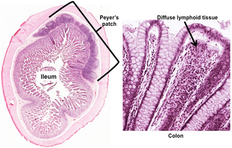
Identify lymphatic vessels under the microscope.
Identify the following heart structures on models:
a. Right & left atria and also auricle
b. Right & left ventricles
c. Anterior & Posterior interventricular sulcus
d. Coronary sinus
e. Apex & Base of the heart
g. Ascending aorta, arch of aorta & descending aorta and branches
h. Pulmonary trunk, right and left pulmonary artery
i. Pulmonary veins
j. Superior and inferior vena cava
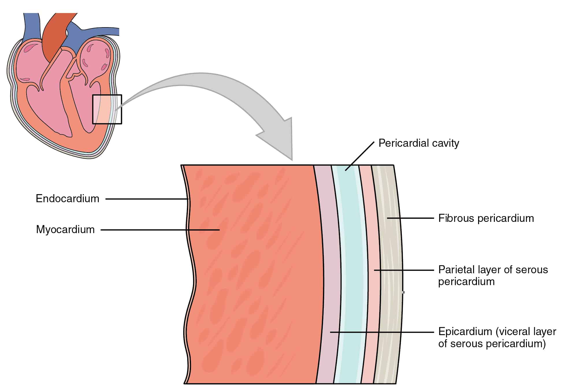
Identify Endocardium, myocardium & epicardium
Different layers of pericardium (fibrous pericardium, parietal and visceral pericardium).
Identify the following internal heart structures:
l. Bicuspid and tricuspid valves
m. Aortic and pulmonary semilunar valves
n. Interventricular septum
o. Trabeculae carneae
p. Pectinate muscles
q. Papillary muscles
r. Chordae tendinae
s. Fossa ovalis in the right atrium
t. Opening of the superior vena cava & inferior vena cava in the right atrium
u. Opening of the coronary sinus in the right atrium
v. Openings of the pulmonary veins in the left atrium

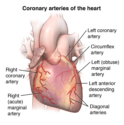
Identify the following coronary blood vessels:
a. Marginal branch & posterior interventricular branch of the right coronary artery
b. Anterior interventricular branch and circumflex branch of the left coronary artery
c. Great cardiac vein, middle cardiac vein, small cardiac vein and the coronary sinus

Identify a microscopic section of an artery and a vein, and name the layers of the wall of an
artery and a vein.
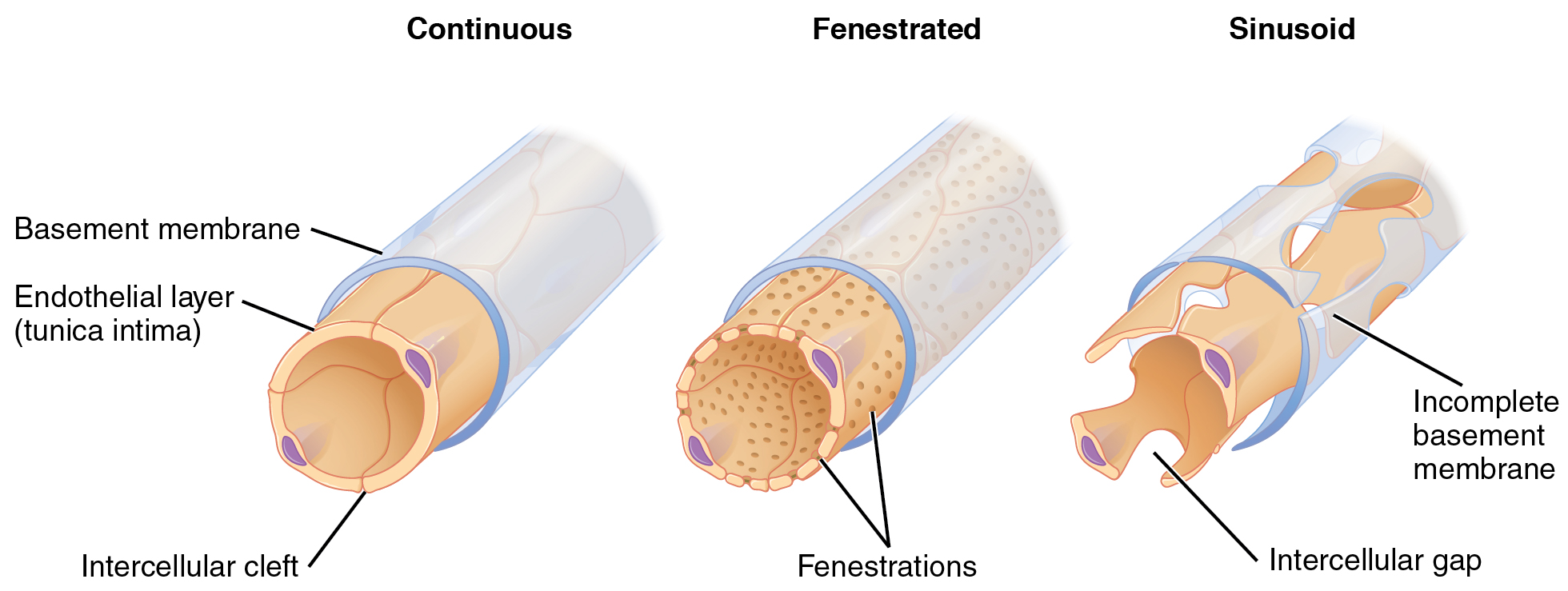
Identify three types of capillary on image.
Identify cardiac muscles on microscopic slide and identify the intercalated disk on slide.

Be able to identify the following major arteries on the blood vessel model:
a. Ascending, descending, and arch of aorta
b. Pulmonary artery
c. Coronary arteries
d. Brachiocephalic trunk
e. Left and right common carotid
f. Left and right subclavian
g. Axillary
h. Brachial
i. Celiac trunk
j. Superior mesenteric
k. Inferior mesenteric
l. Right and left common iliac
m. Right and left external iliac
n. Right and left internal iliac
o. Right and left femoral
Be able to identify the following major veins on the blood vessel model:
a. Superior and inferior vena cava
b. Right and left external jugular
c. Right and left internal jugular
d. Right and left subclavian
e. Brachiocephalic
f. Right and left common iliac
g. Right and left internal iliac
h. Right and left external iliac
i. Right and left femoral
j. Right and left renal
k. Hepatics
Contrast the pathway of blood in the pulmonary and systemic circulation
Compare the amount of oxygen (oxygen-rich or oxygen-poor blood) in the following blood
vessels: aorta, pulmonary artery, pulmonary veins, superior & inferior vena cava.
Name the device for listening of the heart sounds.
Name the process of listening of heart sound (auscultation), define the location of four areas
for auscultation.
Define the heart murmur.
Contrast the characteristics of the first and second heart sounds.
Define the process of pulse palpitation and five grades of pulse palpitation (from 0 – 4).
Describe why you should never assess both of your patient’s carotid arteries at the same time.
Name of the instrument used for measuring blood pressure.
Define systolic and diastolic blood pressures are determined using the sphygmomanometer.
Describe the cause of production and disappearance of the Korotkoff sounds.
Define the normal average blood pressure in a healthy young adult
Define the changes happened to blood pressure during sleep, in the standing position, and
after exercise.
Correlate the systolic and diastolic blood pressures with the functioning of the heart
Define the Pulse pressure and mean arterial pressure and calculate them by given systolic and diastolic pressure.
Compare the changes happened in systolic, diastolic, mean arterial pressure, pulse pressure,
pulse and Respiratory rate between before, during and after exercise.
Discuss the conduction system of the heart, different structures and define the location and
function of each of them
Identify the P wave, QRS complex and the T wave from an ECG record.
Identify the P-R interval, Q-T interval and S-T segment from an ECG record
Define what each wave signifies.
Calculate Heart rate and cardiac cycle length, the time of systole and Diastole by given an
ECG.
Define the meaning of Tachycardia and Bradicardia. Identify them by given ECG.