biochem unit 4 pt 2
 Call Kai
 Learn
Learn Practice Test
 Spaced Repetition
 Match
Match Flashcards
 Knowt Play
1/11

There's no tags or description
Looks like no tags are added yet.
Name | Mastery | Learn | Test | Matching | Spaced | Call with Kai | Chat |
|---|
No analytics yet
Send a link to your students to track their progress
12 Terms
Explain why hormonal control of metabolism across tissues and organs is necessary for efficient use of dietary energy sources in humans.
Hormonal control of metabolism is critical to prioritize fuel sources and to regulate opposing pathways.
Indicate the tissues responsible for generation of energy, storage of carbon, generation of organic molecules for other tissues and waste management. State which molecules are used to achieve this goals and indicate the relevant metabolic pathways associated with these tissue.
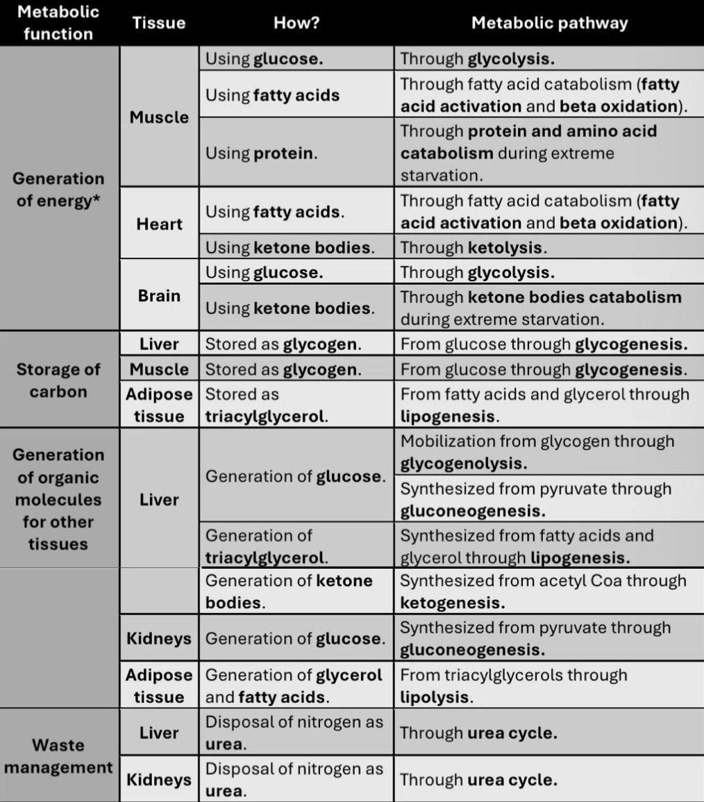
Describe the main metabolic function of the liver, muscle, kidney, adipose tissue, and brain under different metabolic conditions (normal, starvation, during exercise), as needed.
Liver: maintains the blood glucose levels. Export glucose, ketone bodies, and triacylglycerols to peripheral tissues for use as metabolic fuel; also transports high energy fatty acids between itself and adipose tissue
Skeletal muscle: “selfish”; makes its own ATP from multiple fuel sources and will use energy from other tissues, but does not provide energy to other tissues.
Kidney: uses gluconeogenesis to synthesize glucose for export to other tissues; involved in removal of nitrogenous waste via the urea cycle.
Adipose tissue: location of lipolysis and triacylglycerol synthesis; primary tissue for long term energy storage.
Brain: is a major energy drain metabolically – also does not provide energy for other tissues. Requires lots of glucose. Uses ketone bodies if there is low blood glucose levels.
Give examples and explain how some tissues are incapable of performing some metabolic functions.
Adipocytes are incapable of performing gluconeogenesis because they lack the enzymes fructose-1,6-bisphosphatase and glucose-6-phosphatase.
Muscle tissue is incapable of performing gluconeogenesis because they lack the enzyme glucose-6-phosphatase, and of performing fatty acid synthesis because they lack the enzyme fatty acid synthase.
State the main source of metabolic energy for each organ under different metabolic conditions.
Liver: glucose under normal conditions, or fatty acids when there is low blood glucose levels.
Brain: glucose under normal conditions, or ketone bodies when there is low blood glucose levels.
Skeletal muscle:
• During resting state, the muscle uses free fatty acids released from adipose tissue as a source of energy.
• When muscle contraction is required for a very short burst of activity, the cells make use of intracellular ATP pool.
• If a more sustainable level of exercise is needed, additional ATP is quickly synthesized by the enzyme creatine kinase, using phosphocreatine as the phosphoryl group donor.
• Glycogen stores in the most active muscle groups become depleted after an hour of continual use, whereas liver glycogen maintains safe blood glucose levels for 12-18 hours.
• As muscle glycogen levels decline with continual use, muscle cells use fatty acids or ketone bodies for energy.
• Muscle tissue lack fatty acid synthase and glucose-6-phosphatase, which means that fatty acid synthesis and gluconeogenesis will not happen. Instead, the reduced carbon molecule will be used to produce ATPs.
• In other words, selfishly, muscle will use energy from other tissues but normally will not provide energy to other tissues.
• However, under extreme starvation, amino acids from muscle protein can be used to make energy.
Heart: acetyl-CoA (from ketone bodies and fatty acid oxidation)
Describe the phosphocreatine shuttle system and state the role of phosphocreatine in muscle.
Phosphocreatine is a phosphate reservoir that can be used to create additional ATP in skeletal muscle. The phosphocreatine shuttle itself allows for the transport of ATP between the mitochondria and into the cytosol.
Explain the Cori cycle, Glutamine/alanine transport system and triacylglycerol cycle.
• The Cori cycle is a process by which chemical energy in the form of lactate can be transferred from tissue to the liver, where it is used to make glucose to be distributed back to the other tissues.
• The glutamine transport system transports nitrogen from the breakdown of proteins in tissue to the liver.
• The alanine transport system is used by muscles performing anaerobic glycolysis and protein catabolism and the same. Glycolysis yields pyruvate, pyruvate can be converted to Alanine and transported to the liver.
• The triacylglycerol cycle circulates fatty acids and triacylglycerols between adipose tissue and the liver. This pathway is extremely important when blood glucose levels are low
Describe how glycerol is synthesized under normal conditions and low blood glucose levels.
Normal: glycerol can be synthesized from glucose via glycolysis.
Low blood glucose levels: first, DHAP must be synthesized from amino acids or lactate via gluconeogenesis. Because adipocytes do not contain certain gluconeogenic enzymes, all DHAP generated will be used to exclusively synthesize glycerol.
Recognize that insulin and glucagon are peptide hormones that respond to high and low blood glucose concentrations, respectively.
Insulin signals for high blood glucose; glucagon signals for low blood glucose.
Describe in your own words the mechanisms of insulin and glucagon, starting from the change in blood concentration and ending in the change in metabolism.
When blood glucose rises, pancreatic beta cells detect this change and induce the release of insulin into the bloodstream. The release of insulin is triggered by a change in ATP/ADP, inducing a membrane depolarization, an influx of calcium ions and then the exocytosis of the insulin vesicles. Insulin travels in the bloodstream to different tissues and binds to the extracellular domain of insulin receptors on the membranes of different cells in the body. This binding event induces a conformational change in the intracellular domain of the same receptor, causing an autophosphorylation event. In turn, a protein cascade is triggered intracellularly, which will lead to a metabolic change via either gene expression or the activation of more proteins.
When blood glucose falls, pancreatic alpha cells detect this change and induces the release of glucagon into the bloodstream. The release of glucagon is triggered by a change in ATP/ADP, inducing a membrane depolarization, an influx of calcium ions and then the exocytosis of the glucagon vesicles. Glucagon travels through the bloodstream and binds to the glucagon receptor on the cell membrane. This binding event induces a conformational change in the intracellular domain of the same receptor. The activated receptor induces a signal transduction, which will lead to a metabolic change via either gene expression or the activation of more proteins.
Predict the effect (increase or decrease) of insulin on the rates of metabolic pathways such reviewed in class.
Gluconeogenesis: decrease
Glycolysis: increase
Glycogenesis: increase
Glycogenolysis: decrease
Lipogenesis: increase
Lipolysis: decrease
Fatty acid synthesis: increase
Fatty acid oxidation: decrease
Ketogenesis and ketolysis: decrease
Explain how metabolic fuel is used during starvation.
1. Initially blood glucose levels are maintained by mobilization of liver glycogen but is quickly depleted.
2a. Gluconeogenic pathway in the liver and the kidneys are stimulated to generate glucose for brain cells and erythrocytes.
2b. In parallel, fatty acids are used as the primary metabolic fuel in almost all other tissues, through beta oxidation.
3. By the 6 day, ketogenesis is induced, providing ketone bodies that act as a secondary fuel for the brain.
4. Proteins will be used for energy minimally until is absolutely necessary, through protein catabolism