Cells and Methods to Observe Them Ch3
 Call Kai
 Learn
Learn Practice Test
 Spaced Repetition
 Match
Match Flashcards
 Knowt Play
1/52

There's no tags or description
Looks like no tags are added yet.
Name | Mastery | Learn | Test | Matching | Spaced | Call with Kai | Chat |
|---|
No analytics yet
Send a link to your students to track their progress
53 Terms
Microscopes
most important tool for studying microorganisms
different types
light microscope
electron microscope
atomic force microscope
Compound light microscopes
use visible light to observe objects
compound - 2 sets of magnifying lenses
ocular lens (5x, 10x)
objective lenses - 4x, 10x, 40x, 100x
microscope parts and their function (8)
ocular lens: eye piece
can see the image, magnifies the image, 10x
specimen stage
stage clip holds slide in place
condenser lens
focuses the light up to the specimen, collects all the light, shines it up to the stage to the specimen
iris diaphragm lever
controls the amount of light that enters the objective lens, works like a shutter on the camera, helps control and adjust the amount of light focused on the specimen
objective lens
has a opening on the bottom, with 10x its pretty big and then inc gets smaller
a selection of lens options provides different magnifications. the total magnification is the product of the magnifying power of the ocular lens and the objective lens
light source
rheostat
controls the brightness of the light
side circle
course and fine adjustment focus nob, used for low power → doesn’t let you run into the slide and instead slowly go into it

principles of light microscopy
visible light passes through specimen and then series of magnifying lenses, illuminator provides light
How good an image is, depends on: magnification, resolution, contrast
Magnification
apparent increase in size
two lens: objective and ocular
total magnification TM = power of ocular lens x power of objective lens (10x * 100x) = 1,000x
Bacteria - only seen with 100x (oil immersion) objective lens
always start at lowest objective lens
Resolution
resolving power
ability to distinguish 2 objects that are very close together (how sharp the image is)
depends on quality and type of lens, wavelength of light (visible light, uv rays, beams), and specimen preparation, the shorter the wavelength the higher the resolution
maximum resolving power of light microscope is 0.2 micrometer (using this resolving power, if u have 2 things that are 0.2 microns apart, you’ll see them as separate, if they’re closer, they’ll blend together and the image isn’t as sharp)
minimum distance between 2 points at which those points can be observed as separate
100x lens - need immersion oil to enhance resolution, wont be able to get in focus without it ever
immersion oil: reduces light refraction, has nearly same refractive index as glass
as magnification inc, the objective lens opening becomes smaller, so fewer light rays can enter the lens (but we want a lot), air has a lower refractive index than glass, so light bends as it passes from the glass slide → air → objective lens, causing some light rays to be lost, immersion oil has a refractive index similar to glass so there is less bending of light between the slide and OL, this allows more light rays to enter the objective lens → improves resolution → produces a clearer image
refractive index: a measure of how much a material slows down and bends light as light passes thru it
low refractive index: light bends MORE when entering/leaving another material
similar refractive index: light bends less
glass + immersion oil = similar RI
Contrast
determines how easily cells can be seen against background
live bacteria lack contrast bcuz they have no color, difficult to see against colorless background
increase contrast by:
staining: increases contrast but kills microbes
using phase-contrast and darkfield microscopy, creates areas of different color between the organism and background
Light Microscope
uses visible light, magnifies images approx 1,000x
3 types of light microscopy for different uses: brightfield, darkfield, phase-contrast
Brightfield: used to view colored or stained specimen, most common type of microscopy, the background is bright/white, red perimysium
Darkfield: increases contrast of live specimen, cells appear as bright objects against dark background, used to view transparent live organisms
Phase-contrast Microscope: Uses special optics in the condenser and objective lens to enhance differences in refractive index within a specimen. It amplifies differences between the cell's structures and the surrounding medium, creating contrast. Denser areas of the cell generally appear darker, while less dense areas appear lighter/brighter. Allows you to see internal structures of living, unstained cells.
Unlike dark-field microscopy, which mainly shows a bright specimen against a dark background, phase-contrast microscopy can show both lighter and darker areas within the cell.
Electron Microscopes
Uses electron beams, a lot shorter in wavelength and bcuz of this we can achieve greater resolving power
Resolving power of ~0.3nm (know mm, nm etc)
magnify images up to 100,000x
Two types:
SEM - used to observe surface details, scanning electron microscope, produces 3d like image for texture and surface details
TEM - used to view internal details, transmission electron microscope, we take our bacteria and embed it in a plastic block and then slice the block into tiny pieces, pic- new cell wall and membrane revealing that its about to divide

Scanning Probe Microscopes
uses metal probe
detailed images of surfaces
ex. atomic force microscope
resolving power much greater than that of EM
sharp probe moves across sample’s surface
feels bumps, valleys of atoms
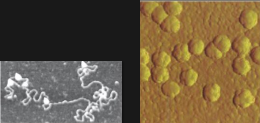
pic: a protein, enzyme that’s a dna or rna structure
yellow: virus, surface of the cell, the bumps are where the virus that has infected the cell is gonna come from
Preparing Specimens for Light Microscopy
Wet mount uses a drop of liquid specimen
live bacteria - hard to see due to lack of contrast
higher contrast is achieved by
using different types of microscopies
specimen staining
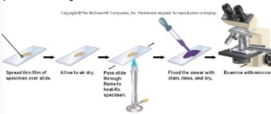
Prepare a smear
smear: a thin layer of cells dried and fixed onto a slide before staining
fixing: in the lab using heat from a bunsen burner, thru the flame a couple times → first it will kill the bacteria and preserve it…, then it denatures the protein on the surface of bacteria, then attaches bacteria on the slide
Dyes and stains (coloring agents)
made of organic salts ( + or - charge) → bacteria has a slight neg charge
basic dyes - carry pos charge, colored ion has a +
bond to cell
commonly stain the cell, bcuz its attracted to the neg charged bacteria
more commonly used than acidic dyes
include:
methylene blue, crystal violet, safranin (red), malachite green
acidic dyes - carry neg charge
repelled by cell, bcuz its repelled by the neg charged bacteria
commonly stain the background
nigrosin - black dye
Both used in different staining procedures: simple, differential, special
Simple staining procedure
Uses one stain to stain cell, background or bacterial cell
increases contrast → size (can observe the different types if there are different ones), shape, arrangement of cells
all cells are stained same color
no differentiation between cell types
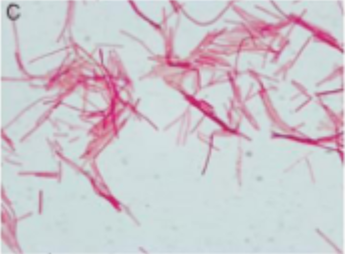
but we can see that its safranin rod-shaped bacteria and they are connected together in a string (strepto)
Differential Staining procedure
used to distinguish different types of bacteria
uses a series of reagents
2 most common differential stains: gram stain, acid-fast stain
Gram Stain** tested on it in the lab
most widely used staining procedure for bacteria - differential
developed by Dr. Hans Christian Gram,
a series of reagents and stains and separates bacteria into 2 major groups based on cell wall structure and chemistry
does not work for all bacteria
one of the reasons why acid fast stain was developed
purple - positive & red/pink - negative
can tell difference cuz when mixed its separate

Gram Stain Procedure and Result
Gram - Positive (has nothing to do with charge but whether they retain the primary dye or they dont)
stained purple (retains primary dye)
Gram - Negative
stained red or pink (loses primary dye)
Between each step have to rinse with ample amount of water for 1 min until there’s no more dye
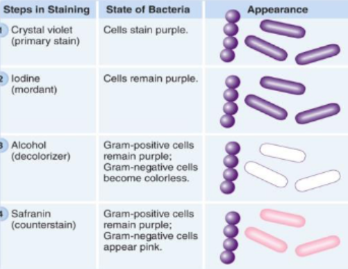
1st step: make a smear and heat fix it
1st stain used is CV; binds to both types of bacteria
2nd is iodine for 1 min, helps to enhance the purple color by creating the CV Iodine complex, binds to CV in bacteria to form CVI complex which is more purple than the crystal alone (intensifies it) → both cells will be purple
3rd: most important step, the differentiating step, add enough but not too much, allows us to differntiate between g+ and g- cells, alcohol wash, decolorizing step. when alcohol is added it removes the dye from gram - cells and become colorless, gram + should still be purple if done carefully bcuz of their cell wall structure
4th: stains the gram - cells that were colorless, g+ already have purple so won’t pick up red from safranin
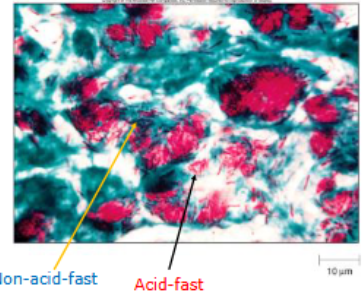
Acid-fast Stain
Used to detect members of genus Mycobacterium (doesnt stain well with gram stain)
includes causative agents of TB and leprosy - these are caused by mycobacterium
cell wall contains high concentrations of mycolic acid
waxy fatty acid that prevents uptake of dyes, dyes don’t penetrate and stain well
harsher methods needed - once stained difficult to decolorize
used for presumptive identification of agents (those pathogens) in clinical specimens for diagnosis
somebody thought to have TB, will take the sputum and do an acid fast stain, if it comes back with a acid fast bacteria, they will be given an antibiotic right away
Acid-fast stain procedure
requires multiple steps
Primary dye (carbol fuchsin)
colors all bacteria red
decolorizer (acid alcohol)
removes red stains from non acid-fast bacteria
counter stain (methylene blue)
colors non acid-fast bacteria blue, so acid fast is red
Capsule Stain
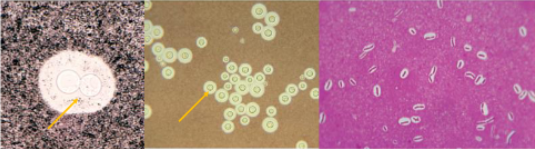
capsule: gel like polysaccharide layer surrounding cell
stains poorly, negative stain often used, use acidic dyes
allows capsule to stand out around organism
Endospore stain
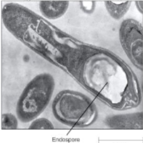
endospore: resistant, dormant structure formed by species of bacillus Clostridium, resists gram stain, often appears as clear object
endospore stain: uses heat to facilitate uptake of the primary dye malachite green by endospore, counterstain (usually safranin) used to visualize vegetative cells

Flagella stain
flagella: used for prokaryotic motility, too thin to be seen with light microscope, presence and distribution can help in identification
flagella stain: dyes and coats flagella to thicken and make visible
bacterial shapes
…
binary fission
most prokaryotes divide by bf, cells may stick together following division, form characteristic groupings or arrangements depending on plane of division
planes
plane
structures external to cell wall
gel like layer outside cell wall
capsule: organized, attached to cell wall
slime layer: unorganized, loose
composed of sugar (glycocalyx) and/or polypeptides
excreted by organism, not always present
function
protection from host defenses (phagocytosis)
attachment to surfaces
both increase virulence of pathogens**
protection against drying (desiccation)
reserve of nutrients
capsules
dental plaques: oral streptococci use capsular slime to adhere to surfaces of teeth and gums —> biofilm
Pili
protein appendage, not essential, but give advantage
shorter, thinner than flagella; found only on gram negative bacteria
fimbriae:
hair-like, facilitates attachment
contributes to pathogenicity
Neisseria gonorrhoeae
sx pilus
attachment to another cell
transfer of dna between cells during conjugation
Flagella
protein appendage, not essential, but give advantage
long protein structure for motility
typically, on rod-shaped bacteria
some important in bacterial pathogenesis
helicobacter pylori penetration through mucous coat
has three basic parts:
filament: extends to exterior, made of proteins called flagellin
hook: connects filament to cell
basal body: anchors flagellum into cell wall

Flagellar arrangement
numbers and arrangements vary and help with characterization
motility
movement is a series of runs and tumbles
chemotaxis: bacteria sense chemicals and move accordingly
nutrient - acts as attractant
toxic compound - acts as repellent
cell wall
strong, rigid structure
functions: maintain shape of bacteria, protects cell from osmotic lysis (bursting)
major site of actions for most antibiotics
unique chemical structure: distinguishes gram positive from gram negative bacteria
differences in cell wall account for differences in staining characteristics
PTG - peptidoglycan
cell wall made of peptidoglycan (PTG), found only in bacteria
basic structure of PTG: polymer of NAG and NAM, cross-linked by tetrapeptide chains
a good target of antimicrobials since unique to bacteria
interferes with synthesis of or break down of PTG → cell lysis
examples include
penicillin: prevents cross-linking of adjacent glycan chains??
lysozyme: breaks bonds linking glycan chain
gram positive cell wall
has thick layer of PTG (as many as 30)
teichoic acid
no outer membrane
small periplasm
gram + bacterium:
retains CV-I complex after decolorization
decolorizer dehydrates thick layer of PTG
gram negative cell wall
more complex than gram +
has thin layer of PTG
no teichoic acids
has large periplasmic space with degradative enzymes
has outer membrane: lipopolysaccharide layer (LPS)
portions medically significant
o-specific polysaccharide side chain → antigenic
used to identify species or stains
E. coli O157:H7
lipid A → endotoxin
gram negative infection of bloodstream
gram - bacterium:
loses CV-I complex
decolorizer damages outer membrane; thin layer of PTG
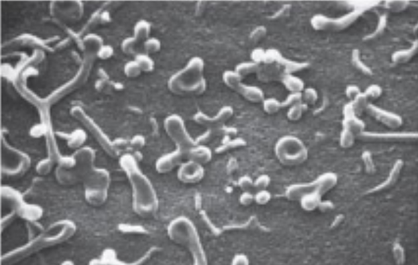
bacteria that lack a cell wall
mycoplasma species:
have extremely variable shape
antimicrobial directed towards cell wall ineffective: penicillin, lysozyme
sterols in membrane give strength to membrane
other types of prokaryotic cell wall
primary difference between members of domain bacteria and domain archaea
domain archaea do not contain PTG but rather pseudopeptidoglycan (lack NAM)
structures internal to cell wall
the cytoplasmic membrane
defines boundary of cell
phospholipid bilayer embedded with proteins
functions:
ATP production: ETC
serves as a selective barrier between cell and external environment
simple diffusion
molecules move freely across cytoplasmic membrane
moves from area of high concentration to area of low concentration until equilibrium is reached
no energy required
water (osmosis), certain gases, small hydrophobic molecules
facilitated diffusion
moves molecules across membranes with help of transporter proteins
movement down concentration gradient - no energy required
active transport
moves molecules against a concentration gradient using a transporter protein
requires energy
group transport
transport mechanism that chemically alters molecule during passage
requires energy
phosphorylation of glucose
cytoplasm
substance inside plasma membrane
about 80% water
internal components - chromosome
single, circular, double stranded DNA
contains all genetic information
packed tightly - forms the nucleoid
internal components - plasmids
extrachromosomal, circular, dsDNA
independently replicating
encode characteristic potentially enhancing survival
antimicrobial resistance
internal components - ribosome
involved in protein synthesis
composed of 2 subunits:
prokaryotic
large = 50s and small = 30s, total = 70s??
eukaryotic - bigger
large = 60s and small = 40s, total = 80s
target of many antibiotics due to this difference
internal components - cytoskeleton
internal protein framework
bacterial protein analogous to those in eukaryotic cytoskeleton
actin (MreB)
tubulin (FtsZ)
intermediate filaments (crescentin)
function:
controls cell shape
involved in cell division
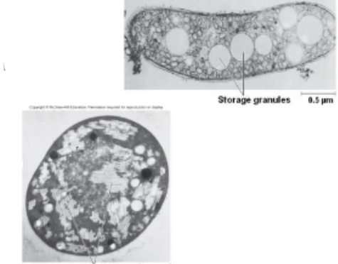
internal components - storage granules vs gas vesicles
SG: store excess nutrient (carbon, energy sources)
ex. glycogen granules (glucose), poly-B-hydroxybutyrate
GV: small protein compartments containing gas, provides buoyancy to cell
internal components - endospores
unique dormant* structures, produced by members of bacillus clostridium, thick coat resisting damage conditions
heat, desiccation, chemicals, uv light
found at different positions in cell
central, terminal, subterminal
sporulation: endospore formation
germination: return to vegetative state
differences between prokaryotes and eukaryotes
P
one circular chromosome, not in a nucleus
no organelles
PTG cell walls
reproduce by binary fission
E
multiple paired chromosomes, in nuclear membrane
membrane bound organelles
no cell wall except plants (polysaccharide)
reproduce by meiosis and mitotic spindle
origins of mitochondria and chloroplasts
endosymbiotic theory: mitochondria and chloroplasts were derived from bacteria
over billions of years each partner became indispensable (abs necessary) to the other
key supportive evidence:
similar in size and shape to bacteria
a circular chromosome
70s ribosomes
divide by binary fission
surrounded by a double membrane