Lecture 3 - peripheral and central afferent pathways
1/42
There's no tags or description
Looks like no tags are added yet.
Name | Mastery | Learn | Test | Matching | Spaced |
|---|
No study sessions yet.
43 Terms
Somatosensory afferent neurons
mecahnoreceptors are specialized cells that project to an afferent neuron or are actually part of the afferent neuron
Factors that determine how fast the afferent information reaches the CNS:
Amount of insulation around the axon - insulation prevents ions from leaking out of the axons and forces ions to move along the axon
The diameter of the axon of the afferent neuron - A larger diameter allows ions to flow unobstructed along the axon (faster conduction)
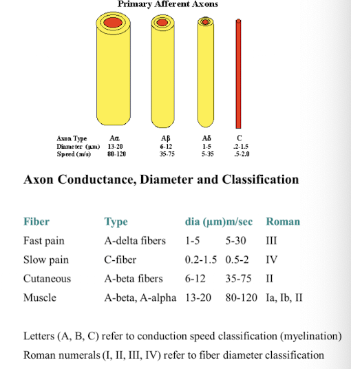
Receptor type: Cutaneous mechanoreceptor
Afferent fiber classification: Group II, A-beta
receptor type: Muscle spindles
Afferent fiber type: Group Ia/A-alpha
Receptor type: Dynamic Static
Afferent fiber: Group II/A-beta
receptor type: Golgi tendon organs
Afferent fiber type: Group Ib/A-alpha
receptor type: joint receptors
afferent fiber type: group II/A-beta
receptor type: free nerve endings
Afferent fiber type: Group III and Group IV/A-delta and C fibers, which respond to pain, temperature, and crude touch.
Afferent projections to the CNS
travel to gray matter regions in the CNS specific to that sensory system
sensory info from mult systems converge in association areas to generate a unified percept or action
Divergence
the same input separates/disperses to multiple locations
1 input —> many outputs
Convergence
multiple inputs project to a common location
many inputs —> 1 output
Topographic mapping
ordered projection of a sensory surface, like the retina or the skin, within the nuclei (neurons) in the CNS
topography is found throughout all levels of the CNS
Ipsilateral spatial mapping
belonging to or occurring from the same side of the body
Contralateral spatial mapping
belonging to or occuring for the opposite of the body.
Geniculostriate visual system
90% of fibers
primary visual pathway
retina —> lateral geniculate (LGN) thalamic nucleus —> Primary visual cortex
Tectopulvinar visual system
10% of fibers
the secondary visual pathway
retina —> superior colliculus —> pulvinar nucleus of the thalamus —> visual areas of the cortex
Retinotopic organization
is the orderly mapping of the visual field onto the visual cortex such that adjacent areas in the retina correspond to adjacent areas in the cortex.
how would your perception change if: you had a lesion of the right optic nerve
loss of peripheral aspect of the right visual field
how would your perception change if: you had a lesion of the optic chiasm
loss of peripheral vision in both sides —> tunnel vision, loose all lateral portions of vision
how would your perception change if: you suffered a lesion to the right optic tract
loss of the left visual field, affecting both peripheral and central vision in that area. 3
visual field
the part of the visual environment that can be detected by both eyes
Hemifield
the left or right half of the visual field
optic chiasm
the point in the brain where the optic nerves cross, allowing visual information from both eyes to be integrated.
Left hemifield
right thalamus/V1
right hemifield
left thalamus/V1
Dorsal stream
A pathway in the brain that relates the visual environment (spatial location, motion) to the body and guides actions related to spatial awareness.
The “where” or “how” pathway
Ventral stream
A pathway in the brain responsible for processing visual information related to object recognition and form.
It is often referred to as the "what" pathway.
prosopagnosia
A neurological condition characterized by the inability to recognize faces, often due to damage in the fusiform gyrus.
what cranial nerve carries vestibular (balance) information to the brain?
Vestibulocochlear nerve (CNVIII)
Do cranial nerves run through the spinal cord?
No, they go directly to the brain stem
Where do vestibular axons project directly to in the brain?
the ipsilateral vestibular nuclei (pons and medulla)
the ipsilateral cerebellum
how many nuclei make up the vestibular nuclear complex?
four nuclei
where do somatosensory nerves enter the spinal cord?
dorsal horn
what do somatosensory nerve do once they enter the spinal cord?
Ascending pathways are formed by layered neurons with divergent projections for reflexes and local processing.
posterior column pathway
A major somatosensory pathway that transmits fine touch, proprioception, and vibratory sensations from the lower body and trunk to the brain.
third order neuron
Neuron that relays information from the thalamus to the sensory cortex.
second order neuron
neuron that crosses the body midline in the medulla and projects to the thalamic nucleus
first order neuron
axon that projects from the receptor to the medulla
Spinothalamic somatosensory pathways
anterior spinothalamic tract
more anterior-medial part of the spinothalamic tract
crude, poorly localized information about touch
lateral spinothalamic tract
more lateral part of the spinothalamic tract
info abt painful stimuli and temperature
Dorsal spinocerebellar tract (DSCT)
projects to the inferior peduncle of the cerebellum
proprioceptive information from the lower limbs (spindle fibers via Ia afferent neurons)
Rostral spinocerebellar tract (RSCT)
projects to the inferior peduncle of the cerebellum
proprioceptive information from the upper limbs
Ventral spinocerebellar tract (VSCT)
Projects to the superior peduncle of the cerebellum
carries proprioceptive information from the lower limbs, integrating sensory feedback for motor coordination (golgi tendon organs via Ib afferent neurons).