Medical Instrumentation 4-7
 Call Kai
 Learn
Learn Practice Test
 Spaced Repetition
 Match
Match Flashcards
 Knowt Play
1/93

Earn XP
Description and Tags
final test
Name | Mastery | Learn | Test | Matching | Spaced | Call with Kai | Chat |
|---|
No analytics yet
Send a link to your students to track their progress
94 Terms
EEG (Electroencephalogram) use
to detect brain electrical activity
typical EEG frequencies
Gamma (stimulated) 22-30Hz
Beta (awake/alert) 13-22Hz
Alpha (relaxed) 8-13Hz
Theta (asleep) 4-8Hz
Delta (deep sleep) 0.5-4Hz
Phonocardiogram use
records heart sounds
sounds of contraction, relaxation, valves and blood flow
Diseases tested for with phonocardiogram
Split sound
Murmurs
Split sound (phonocardiogram)
S1 is split, mitral and tricuspid valve closes asynchronously
Murmurs (phonocardiogram)
caused by stenoses and regurgitation at aortic, pulmonary and mitral valve
S1 of phonocardiogram (time and frequency)
t: 20-100ms
f: 30-100Hz
S2 of phonocardiogram (time and frequency)
t: 25-50ms
f: >100Hz
electromyography use
detect electrical signals used to contract muscles
evaluates the health of muscle and nerve cells
defibrillator use
applies external charge to effectively stop that heart, for SA node to trigger normal conduction cycle
defibrillator paddles size
8-10cm in diameter
formula for energy stored after charging defibrillator
w = ½CV²
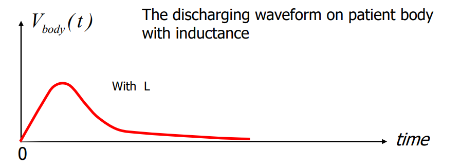
what is the point of inductor in discharge circuit
to obtain desired discharge waveform
AED (automatic external defibrillator) use
visually and vocally instructs operators, measures ECG and can provide defibrillation if needed
ICD (implantable cardio-verter defibrillator) use
implanted into the chest to give time continuous ECG readings, and can deliver defibrillation if needed
Pacemaker use
regulates heart rhythm and rate
Different kinds of pacemakers
asynchronous
synchronous
asynchronous pacemaker
functions regardless of patient’s natural rhythm
synchronous pacemaker
detects ECG and applies stimulus pulse in response to feedback
battery for pacemaker
normally lithium iodide cell
energy = rating (A-H) x voltage
can last as long as 12 years
haemodialysis machine use
to remove metabolic waste products from blood of kidney failure or uremic patients
basic working principles of haemodialysis
osmosis: movement of water through a semi-permeable membrane to the more concentrated side
diffusion: movement of solute through a semi-permeable membrane to the more diluted side
ultra-filtration: bulk flow of fluid by hydrostatic pressure through a semi-permeable membrane
how does haemodialysis work
uses dialysate (same concentration as healthy blood) on one side and patients blood on the other side of the membrane
after prolonged exchange across membrane, patient’s blood plasma achieves same concentration as dialysate, hence becoming healthy
various substances in dialysate can be chosen to achieve required speed of exchange
how does peritoneal dialysis occur
dialysate is pumped into peritoneal cavity to slosh about
visceral peritoneum and parietal peritoneum allows for exchange of molecules between dialysate and capillaries behind membranes
after some time dialysate is drained
ventilator use
provides artificial ventilation for patients with reduced of failed breathing, and achieves and maintains desired arterial partial pressure of CO2 (PaCO2) and arterial partial pressure of O2 (PaO2)
can be used in anesthesia as well
kinds of ventilators
positive pressure ventilator
negative pressure ventilator
what is negative pressure ventilator
simulates negative pressure from diaphragm contraction during inspiration
what is positive pressure ventilator
applies positive pressure into airways
inspiration phase:
inspiratory flow delivery system delivers positive pressure, exhalation control system is closed
exhalation phase:
inspiratory flow delivery system is closed, exhalation control system opens to allow airway pressure to balance with atmospheric pressure
modes of positive pressure ventilator
mandatory
spontaneous
mandatory mode of positive pressure ventilator
for patients with no independent control over breathing
controls all breathing parameters
spontaneous mode of positive pressure ventilator
for recovering patients with limited independent ability to breathe
responds to patient’s effort to breathe independently
what does an x ray machine consist of
high voltage generator
generates high voltages to emit electrons by thermionic emission
x ray tube
produces x rays when electrons hit metal on anode
collimator
restricts x rays with its aperture
intensifying screen and film
primary radiation strike screen phosphor, resulting in light being exposed on the film
grid
traps secondary radiation
CT (computer tomography) scan use
generates anatomical images that are digitally reconstructed
main components of CT
x ray tube
detector
computer system
gantry and patient table
how does a CT work
source and detector pair get rotated at a small angle, and an x ray is taken
repeats until full 180°
image is reconstructed using x ray from detectors and computers
MRI (magnetic resonance imaging) use
visualizes detailed internal structures
main components of MRI
main magnet
gradient coils
radiofrequency coils
computer system
PROS of MRI
non ionizing rays (unlike radiation)
can build image in any plane (unlike CT)
very low incidents of side effects
provides good contrast between different soft tissues (especially useful for brain muscle, heart and cancer)
how does an MRI work
uses 3 magnetic fields (very strong magnetic field, gradient magnetic field & radiofrequency field)
strong magnets generate magnetic fields to align atoms in the body
radio waves disrupt that alignment
radio waves stop and atoms return to their original position, emitting signals
signals are used to create MRI images
Ultrasound frequency band
uses ultrasounds from 1MHz to 15MHz
main components of Ultrasound
transducer/prob
processing unit
display
how an ultrasound works
piezoelectric effect is applied to crystals with dipoles
changing polarity of electrode plates beside the piezoelectric crystal causes its molecules to constantly twist to align with the produced magnetic field, causing expansion and contraction, making mechanical vibrations (sound)
ultrasonic beams enter the body and gets echoed eventually
piezoelectric crystal receives returning echo and deforms, producing a voltage
processing unit processes signal for display
impedance formula for ultrasound
Z=ρc
Z is impedance
ρ is density
c is acoustic velocity
reflection coefficient and impedance formula for ultrasound
Rcoeff = (Z2-Z1)/(Z2+Z1)
pressure and reflection coefficient formula for ultrasound
Pr = Rcoeff x Pi
P is pressure
4 major sections of medical labs
chemistry
haemotology
microbiology
blood bank
what does chemistry sections of medical labs do
perform analysis on blood, urine, cerebrospinal fluid and other fluids
what does haemotology sections of medical labs do
measures (electrical and optical) blood and its elements
what does microbiology sections of medical labs do
studies various body tissue and fluids
uses devices to automatically monitor status of blood cultures
uses devices to semi-automatically monitor sensitivity of microorganisms to antibiotics
what does blood banks sections of medical labs do
not much electronic instruments used, except for automatic classification of blood group (ABO)
shelve lives of blood and temperature storage in blood banks
whole blood ~35 days (1-6°C)
RBC ~10 years (-80°C)
platelets 5 days (20-24°C)
blood components and their measurements
plasma - 55%
WBC and platelets (leukocytes and thrombocytes) - <1%
RBC (erythrocytes) - 45%
functions of blood
transport dissolved substances
regulate pH, ions and temperature
restricts blood loss
defends against toxins and pathogens
Erythrocytes (RBC)
uses haemoglobin to transport O2 and CO2
normal range
males: 4.6-6.2 ×10⁶ /µL
females: 4.2-5.4 ×10⁶ /µL
size: 8×3 microns
haemoglobin
binds to oxygen and some carbon dioxide
normal range
male: 13.5-18g/dL
female: 12-16g/dL
leukocytes (WBC)
helps to defend against infections
normal range
male & female: 4500-11000 /µL
size: 10 µm diameter
disease
numbers will increase
immature and malignant WBCs may also appear
platelets (thrombocytes)
clots bleeding
normal range
male & female: 140,000-400,000 /µL
size: 2-3 µm diameter
blood plasma consists of:
proteins: organic repair substances
nutrients: energy storing substances
regulatory and protective substances
electrolytes: acid, base for nerve impulse transportation
metabolic waste substances: urea, uric acid and CO2
spectrophotometer use
determines the level of absorption/emission of photons at different wavelengths to find presence and quantity of specific substances
components of spectrophotometer
light source
wavelength selector
cuvette
detector and readout
light source of spectrophotometer
UV —————— Visible —————— Infrared ———>
λ<380nm 380-750nm 750-2000nm
wavelength selector of spectrophotometer
isolates desired λ
2 classes
filters (non-dispersive)
monochromators (dispersive)
filters for wavelength selector
selectively absorbs/transmits wave lengths
e.g. blue absorbs red (high λ) and transmits blue to green (lower λ)
consists of one of more layers to achieve low/high/band pass
monochromator for wavelength selector
prism
glass (λ>350nm)/quartz (λ<350nm UV)
shorter λ gets more bent than longer λ
diffraction grating
disperses light as a function of wavelength through interference
cuvette of spectrophotometer
contains sample solution
Beer’s law for light intensity of spectrophotometer
It = Ii ×10^(⁻µCL)
µ is extinction coefficient
C is concentration of material
L is length of material
Beer’s law of absorption of light of spectrophotometer
A(λ) = lg(Ii/It) = µCL
A(λ) is absorption of light
flame atomic spectrophotometer
detects traces of pure metals
sample feeds into nebulizer with flame, which replaces cuvette and is now before wavelength selector
uses hollow cathode lamp
how does flame atomic spectrophotometer work
flame provides thermal energy to dissociate atoms from chemical bonds, atoms then emit light when returning back to ground state
hollow cathode tube: produces light of characteristic wavelength of atoms of interest
amount of absorption is proportional to the amount of atoms present
automated cell counters principles
electrical impedance principle
light scattering principle
electrical impedance principle of automated cell counter (coulter principle)
change in resistance/impedance every time a cell passes through an aperture of similar size
change in impedance is directly proportional to cell volume
light scattering principle of automated cell counter
light produced by light source gets deflected when cell passes through
deflection is detected by the photodetector
gives number of cells present in sample’s volume
pH formula
pH is determined by concentration of H⁺ ions
pH = -lg[H⁺]
normal range of blood pH
pH 7.38 - 7.44
how does the pH electrodes work
glass electrodes generate electric potential when solutions of different pH are placed on 2 sides of membrane
H⁺ ions from the sample solution diffuse past the ion selective membrane into filling solution, until charge is balanced
voltage across membrane formula
v = k(C₂ - C₁)
glucose
main circulating carbohydrate in the body
insulin promotes absorption of glucose into skeletal muscles and adipose
O₂ electrode of blood glucose sensor
current flows as a function of O₂ concentration
platinum cathode: reduction
O₂ + 2H₂O + 4e⁻ → 2H₂O₂ + 4e⁻ → 4OH⁻
Ag/AgCl anode: oxidation
4Ag + 4Cl⁻ → 4AgCl + 4e⁻
enzymatic reaction of glucose oxidase
glucose + O₂ ←→ gluconic acid + H₂O₂
how does an enzymatic glucose sensor work
one O₂ electrode is placed into gel without oxidase (I1) and another into gel with oxidase (I2), and their difference ΔI = I1 - I2 is directly proportional to amount of glucose present
major problems with enzymatic glucose sensor
instability in immobilized enzyme
fouling of membrane surface in physiological conditions
most operate effectively for a short period of time only
more highly selective membranes must be developed
pulse oximetry use
accesses tissue oxygenation, SaO₂
measured during anesthesia, pulmonary functions test, intensive care and oral surgery
one Hb can bind to 4 oxygen molecules, a full 4 is considered saturated
SaO₂ formula for pulse oximetry
SaO₂ = (O₂Hb)/(O₂Hb + Hb)
pulse oximetry transducer
940nm infrared light
660nm red light
(the 2 are alternately switched on and interleaved)
photodetector
absorption of Hb formula
A(λ) = Aₒ₂ₕ₆(λ) + Aₕ₆(λ)
kinds of absorption observed in pulse oximetry
dc - constant absorption (bone, tendon, tissue, venous blood)
ac - variable absorption due to arterial pressure changes (arterial blood)
modulation ratio of red and infrared light
R = (acλred / dcλred) / (acλir / dcλir)
R is negative proportional to SaO₂
Instrumental amplifier with gain formula
Vo = (1+2R₂/R₁)(Rբ/R₃)(V₊ᵥₑ - V₋ᵥₑ)
What is the high impedance of a buffer amplifier used for
Since the buffer has a very high impedance, it takes up negligible amounts of current from the input, hence not affecting the signal.
What is common mode rejection ratio
Differential amplifier removes common mode signals (noise).
Since both inputs have the common noise, the noise will be removed after passing through the differential amplifier, as they are common between them.
Cut off frequency formula
1/2πRC
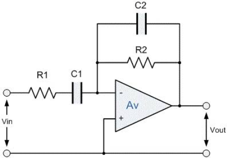
Which part of this active band pass filter is high and low cut off
high pass frequency: 1/2πR₂C₂
low pass frequency: 1/2πR₁C₁
How to know if its high or low pass filter
If parallel to capacitor: low pass filter
If parallel to resistor: high pass filter
time constant for charging defibrillator formula
T = (Rin + Rex) (C)