Flashcards Cardiology: L8 Traumatic infectious and inflammatory | Quizlet
 Call Kai
 Learn
Learn Practice Test
 Spaced Repetition
 Match
Match Flashcards
 Knowt Play
1/109

There's no tags or description
Looks like no tags are added yet.
Name | Mastery | Learn | Test | Matching | Spaced | Call with Kai | Chat |
|---|
No analytics yet
Send a link to your students to track their progress
110 Terms
What is the leading cause of death and disability among young people in the US?
Trauma
Penetrating Traumas
- missile or knife wound to thorax or upper abdomen
- low, medium, or high velocity
- cardiac penetration injuries are highly lethal
Low velocity cause
Knife wounds
Medium velocity cause
Shotgun injuries
High velocity cause
Rifle or shrapnel injuries
What part of the heart is most frequently injured with Penetrating Trauma
Right ventricle due to anterior position
Penetrating Traumas: Primary presentation
Hemorrhage and tamponade
How does tamponade occur
When clot and surrounding pericardial fat partially seal the pericardial defect
Penetrating Traumas: dx gold standard
Subxiphoid window
Penetrating Traumas tx
- most can be repaired through a left thoracotomy
- emergent thoracotomy if unstable or pulseless from low velocity trauma
- relieve tamponade and stop life threatening hemorrhage
Blunt Cardiac Trauma causes
- motor vehicle collision
- rapid deceleration mechanism or a direct blow to the precordium
- falls from heights
- sports related injuries
Blunt Cardiac Trauma dx
Difficult due to asx on initial presentation
Blunt Cardiac Trauma: EKG
- nonspecific ST and T wave changes
- arrhythmias self-limiting
Blunt Cardiac Trauma: dx for unstable pts
Echocardiogram
Blunt Cardiac Trauma Management
Manage by trauma team and consults with cardio and cardiothoracic surgery teams
Great Vessel Injury
- 90% from penetrating trauma; high mortality
- blunt trauma can cause cardiac rupture
- ~20% w/ descending aortic transections survive to reach medical care
Pericarditis
- inflammation of the pericardium
- can be from infection, systemic dz, neoplasm, radiation, drug toxicity, etc
Types of Pericarditis (4)
- acute
- Incessant
- Recurrent
- Chronic
Acute Pericarditis
new episodes lasting
Incessant Pericarditis
>4-6wks but
Recurrent Pericarditis
recurrence after sx free interval ≥4-6 wks
Chronic Pericarditis
> 3mo
Infectious Pericarditis Causes (3)
- viral (mcc): enterovirus
- purulent: S. aureus, S. pneumoniae
- TB
Non-infectious Pericarditis Causes (5)
- early post MI
- Dressler Syndrome
- uremic
- neoplastic
- radiation/drug induced
Early Post MI Pericarditis
occurs within several days of a transmural MI due to inflammation adjacent to myocardial necrosis
Dressler Syndrome Pericarditis
delayed immune=-mediated pericarditis occurring wks-mo after MI
Pericarditis sx (5)
- substernal CP: acute, sharp and pleuritic
- improves with sitting upright
- worsens when taking a deep breath
- can radiate to neck, shoulder, back, or epigastrium
- friction rub early; disappears with increased pericardial fluid
Pericarditis: if pericardial effusion develops sx (2)
- dyspnea
- rapid respiratory rate
Pericarditis: if pericardial tamponade develops sx (2)
- pulsus paradoxus
- JVD
Pericarditis Work up (4)
- ECG
- CXR
- Echo
- Labs: CBC, BMP, cardiac enzymes, ESR, CRP
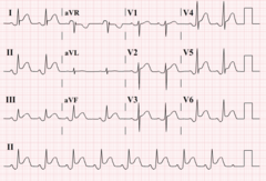
Pericarditis ECG
diffuse ST elevation and PR depression
Pericarditis dx (at least 2/4 required)
- sharp, pleuritic CP
- pericardial friction rub
- consistent changes on ECG
- new or worsening pericardial effusion
Pericarditis Viral tx (2)
- NSAIDs or ASA x 1-2wks + Colchicine x 3mo
- PPI for gastroprotection
Pericarditis NSAIDs you can use (3)
- Ibuprofen 600-800mg TID 1-2wks
- Indomethacin 25-50mg TID 1-2wks
- ASA 750-1000mg TID 1-2wks
Pericarditis Purulent tx
- emergency surgical drainage
- IV Abx (vancomycin, ceftriaxone, and cipro)
Pericarditis TB tx
RIPE regimen
Pericardial Effusion
accumulation of fluid in the pericardial cavity
Pericardial Effusion: assessment of size
Transthoracic Echocardiography (TTE)
Pericardial Effusion: sizes (3)
- small:
What is a large effusion w/ acute pericarditis associated with a higher risk of?
Development of a cardiac tamponade
Pericardial Effusion with what has a poorer prognosis?
Pulmonary HTN
Pericardial Effusion: what does the rate of fluid accumulation determine?
Hemodynamic consequences
Does a larger Pericardial Effusion have a greater chance of causing tamponade?
No it depends on the rate of fluid accumulation, not the size
Pericardial Effusion sx (6)
- dyspnea (mc)
- tachypnea (mc)
- pleuritic chest discomfort
- cough/hoarseness
- fatigue
- hiccups
What sx is concerning for tamponade?
Syncope
Pericardial Effusion PE (7)
- normal PE unless tamponade present
- tachycardia
- hypotension
- muffled heart sounds
- elevated JVP
- pulsus paradoxus
- electrical alternans
What triad are Pericardial Effusion and Tamponade associated with?
Beck Triad
Beck Triad
hypotension
JVD
muffled heart sounds
Pericardial Effusion and Tamponade dx Labs (5)
- cardiac enzymes
- ESR
- CRP
- thyroid panel
- rheumatoid factor
Pericardial Effusion and Tamponade Imaging
- CXR
- Cardiac CT/MRI
- TTE
- EKG
- pericardiocentesis (unstable; last resort)
- pericardial fluid anaysis
When should pericardiocentesis be performed? (3)
- cardiac tamponade
- large pericardial effusions
- unstable pts
Pericardial Effusion and Tamponade CXR (2)
- enlarged cardiac silhouette (water bottle heart)
- pericardial fat stripe

Pericardial Effusion and Tamponade EKG (3)
- low voltage QRS
- ectopy (extra or skipped beats)
- tachycardia
Constrictive Pericarditis
- a thickened fibrotic pericardium impedes normal diastolic filling and produces chronically elevated venous pressures
- similar to restrictive cardiomyopathy
Constrictive Pericarditis mc cause
Tuberculosis
Constrictive Pericarditis other causes (5)
- viral or idiopathic
- immune mediated or neoplastic
- purulent bacterial (20-30%)
- post surgery/radiation
- drug induced
Constrictive Pericarditis sx (8)
- slowly progressive dyspnea
- fatigue/weakness
- chronic edema
- hepatic congestion ascites
- Elevated JVP
- Kussmaul sign
- pericardial "knock" in early diastole
- A.Fib
Kussmaul sign
Increase JVP with inspiration
Constrictive Pericarditis dx
- CXR
- Echo
- Cardiac CT/MRI
- Uncertain: Invasive hemodynamics (swan cath)
Constrictive Pericarditis CXR
normal heart size of cardiomegaly
Constrictive Pericarditis Echo
Septal "bounce" reflecting rapid early filling
Constrictive Pericarditis tx (3)
- tx underlying cause
- diuretics for venous congestion/edema
- surgical pericardiectomy (if diuretics don't work)
What sign can you see in Pericarditis on catheterization?
Square root sign: abrupt stop in diastolic filling
Rheumatic Fever
- GAS pharyngitis/tonsillitis without abx treatment
- 5-15yo
Rheumatic Fever sx (6)
- high fever, malaise, fever
- erythema marginatum "target lesions"
- subcutaneous nodules
- migratory polyarthritis
- valvulitis/pancarditis
- chorea (1-8mo after)
Rheumatic Fever dx
Jones Criteria
Jones Criteria: initial ARF (2)
2 major manifestations
or
1 major + 2 minor
Jones Criteria: recurrent ARF (3)
2 major
or
1 + 2 minor
or
3 minor
Jones Criteria: Major criteria in low risk (5)
- carditis
- polyarthritis
- chorea
- erythema marginatum
- subcutaneous nodules
Jones Criteria: Major criteria in mod-high risk (5)
- carditis
- monoarthritis or polyarthritis
- chorea
- erythema marginatum
- subcutaneous nodules
Jones Criteria: Minor criteria in low risk (4)
- polyarthralgia
- fever
- ESR ≥60mm in 1st hr and/or CRP ≥ 3.0 mg/dL
- prolonged PR interval
Jones Criteria: Minor criteria in mod-high risk (4)
- monoarthralgia
- fever
- ESR ≥ 30mm/h and/or CRP ≥ 3.0 mg/dL
- prolonged PR interval
Infective Endocarditis
- infection of the heart valves or mural endocardium that leads to the formation of vegetations which destroys the cardiac tissue
- >age, M>F
- cardiac devices, prosthetic valves
- IV opioid epidemic
Infective Endocarditis: mc valves
Mitral valve and aortic valve
Infective Endocarditis: what valve is most common with IV drug use?
Tricuspid valve
2 Forms of Infective Endocarditis
- acute infective endocarditis
- sub acute infective endocarditis
Acute Infective Endocarditis (3)
- infection of previously normal heart valve
- days to weeks
- S. aureaus + aggressive + attack normal valve
Sub Acute Infective Endocarditis
- infection by organism of lower virulence of an already damaged/deformed valve
- weeks to months
- S. Viridans + indolent + usually abnormal valve
Infective Endocarditis Pathogenesis
Valve injury --> platelet/fibrin --> bacteremia --> vegetation
Native Valve Endocarditis Pathogens (2)
- S. aureaus
- Streptococci Viridans
IV Drug (lemon juice) use Endocarditis Pathogen
S. aureus
Prosthetic Valve Endocarditis pathogen (3)
- S. aureus
- S. epidermidis
- gram (-) and fungi
Mouth pathogen
Viridans
Skin/Blood pathogen
S. aureus
GI/GI pathogen
Enterococcus
Colon pathogen
S. gallolyticus
Infective Endocarditis sx mneumonic
FROM JANE
Infective Endocarditis sx: FROM JANE
- Fever (mc)
- Roth spots
- Osler nodes
- Murmur
- Janeway lesions
- Anemia
- Nail-bed hemorrhages
- Emboli
Osler Nodes + Janeway lesion

Roth Spots

What is Infective Endocarditis until proven otherwise?
New/acute aortic regurgitation
When should you think Infective Endocarditis?
Stroke + fever
Infective Endocarditis dx (2)
- blood cultures
- Echo TTE
Infective Endocarditis: How to obtain blood culture
- 3 sets of blood cultures from separate venipuncture sites BEFORE Abx
- 10mL
Infective Endocarditis: dx criteria
Duke - ISCVID Criteria
Duke - ISCVID Criteria: Definite IE
2 major
or
1 major + 3 minor
or
5 minor
Duke - ISCVID Criteria: possible IE
1 major + 1 minor
or
3 minor
Duke - ISCVID Criteria: Major categories (3)
- microbiologic
- imaging
- surgical
Duke - ISCVID Criteria: Microbiologic (2)
- typical IE organisms from ≥2 culture sets
- less typical organisms from ≥ 3 culture sets
Duke - ISCVID Criteria: Imaging (3)
- echo or cardiac CT showing vegetations, abscess, perforation, aneurysm, or fistula
- significant new valvular regurgitation
- abnormal FDE-PET/CT involving valve/prosthetic material