MSK Week 1 - Lec 1 - Anatomy and Skeletal System
1/34
There's no tags or description
Looks like no tags are added yet.
Name | Mastery | Learn | Test | Matching | Spaced | Call with Kai |
|---|
No study sessions yet.
35 Terms
what is anatomy? how is it best studied in humans?
anatomy is the study of a structure and the relationships among structures
in humans, these relationships are best revealed by dissection
what are the different subdivisions of anatomy?
surface anatomy
gross anatomy
systemic anatomy
regional anatomy - easier to understand, main focus of the course
radiographic anatomy
developmental anatomy (embryology)
what is systemic anatomy?
study of the body by organ systems, examining the structures and functions of each system as a whole rather than region by region
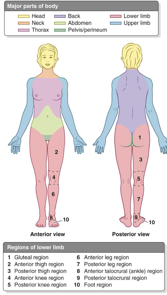
what major regions of the body are studied in regional anatomy?
head (cephalic)
neck (cervical)
trunk (thoracic)
thoracic - chest
abdominal - abdominal organs
pelvis - genitourinary organs
upper limbs
lower limbs
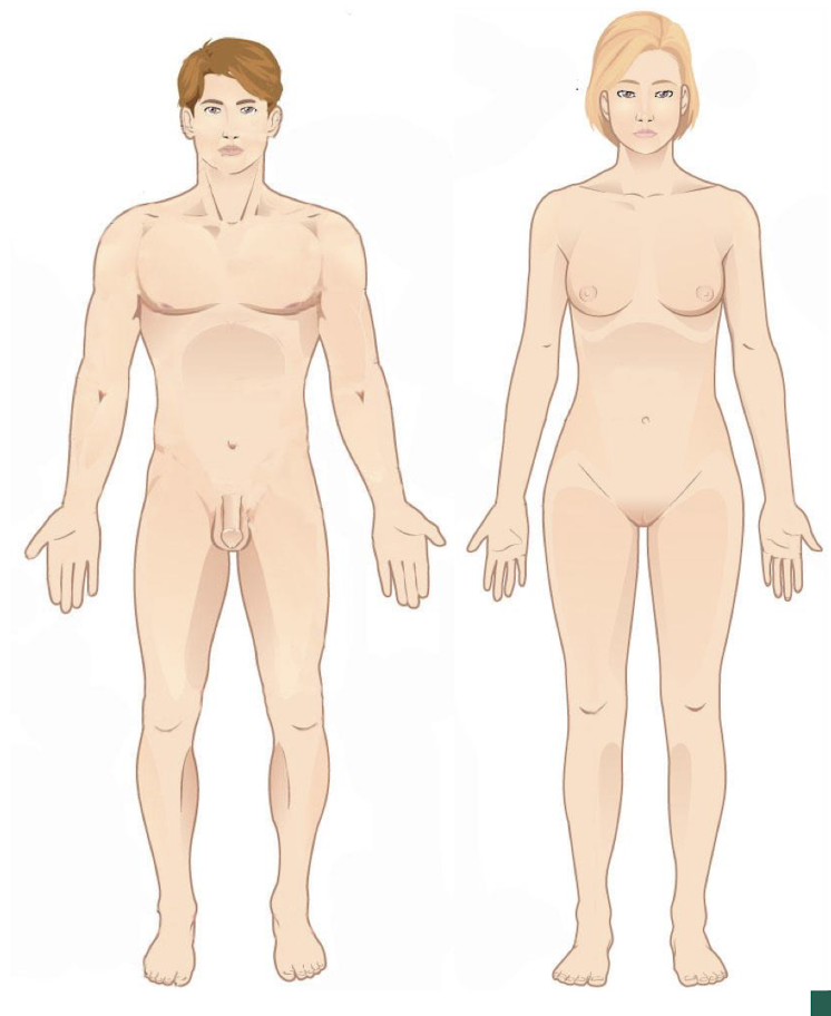
define anatomical position
anatomical position is a standardized method of observing or imaging the body that allows precise and consistent anatomical references
body must be upright
standing erect facing the observer
head and eyes facing forward
feel flat on the floor and forward
upper limbs to the sides, palms turned forward
descriptions of movements are always explained as if the body is in this position, no matter the actual body position
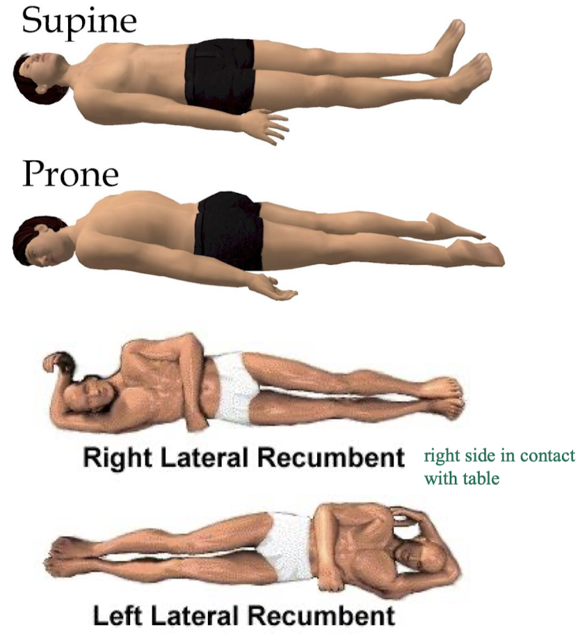
positional terminology - supine, prone, and lateral decubitus
supine - patient is lying face up (think “lying on spine” - supine)
prone - patient is lying face down
lateral decubitus (recumbent) - pt is lying on their side
on right side - right lateral decubitus (or recumbent)
on left side - left lateral decubitus (or recumbent)
what do we call the anatomical midline of the body?
the midline or the median plane - important reference point
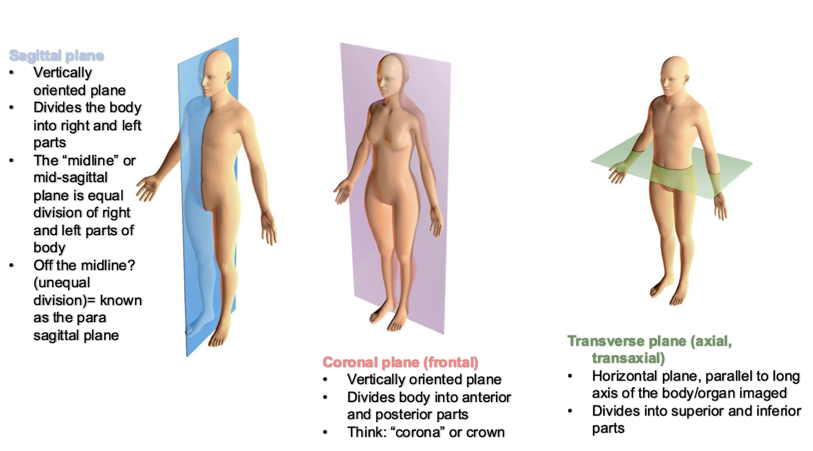
planes and sections of the body - sagittal, coronal, and transverse
sagittal plane
vertically oriented, divides body into left and right
mid-sagittal - equal divisions of left and right
off midline - para sagittal plane
coronal plane (frontal)
vertically oriented, divides body into anterior and posterior parts
transverse plane (axial, transaxial)
horizontal plane parallel to long axis of the body/organ imaged
divides into superior and inferior parts
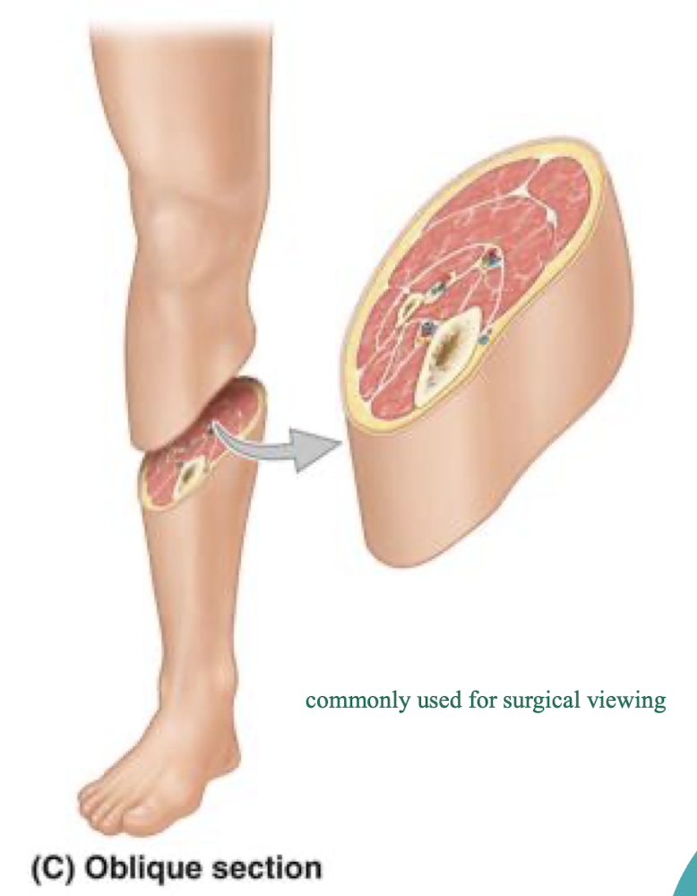
planes and sections of the body - oblique
oblique plane - passes through the body or an organ at an angle, commonly used for surgical viewing
combining features of transverse + sagittal or transverse + frontal planes because it is a diagonal slice at an angle
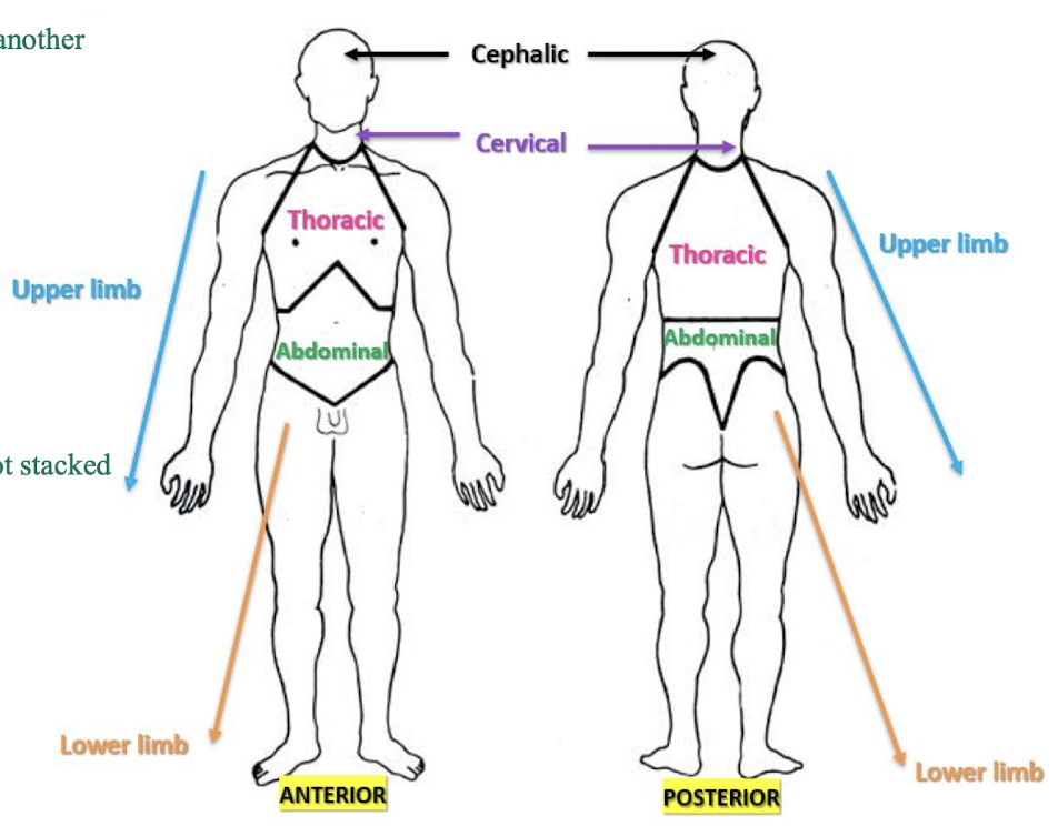
what are the primary axes of the body and what do they include?
vertical axis - they stack on top of one another
cephalic (head)
cervical (neck)
thoracic (chest)
abdominal (stomach)
oblique axis - away from body, not stacked on top of one another
upper limb
lower limb
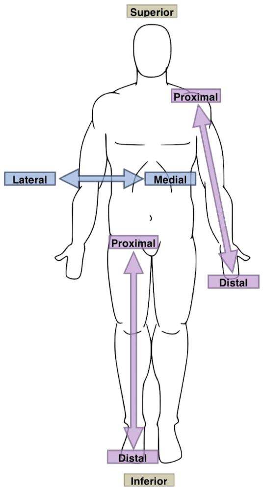
directional terminology relating to the vertical axis of the body
superior, cranial, cephalic - towards the head
inferior, caudal - towards the feet
anterior, ventral - towards the front of the body
posterior, dorsal - towards the back of the body
for example, the shoulder is inferior and posterior to the nose

more directional terminology relating to the vertical axis of the body
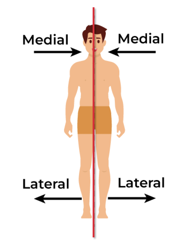
medial - towards the midline of the body (i.e. nose is medial to eyes)
lateral - away from midline of the body (i.e. eyes are lateral to nose)
directional terminology relating to the oblique axis of the body
proximal - closer to the the point of attachment of the limb to the trunk (i.e. elbow is proximal to the wrist because it is closer to the shoulder than the wrist is)
distal - farther from the point of attachment of the limb to the trunk (i.e. ankle is distal to the knee because it is farther from the hip joint than the knee is)
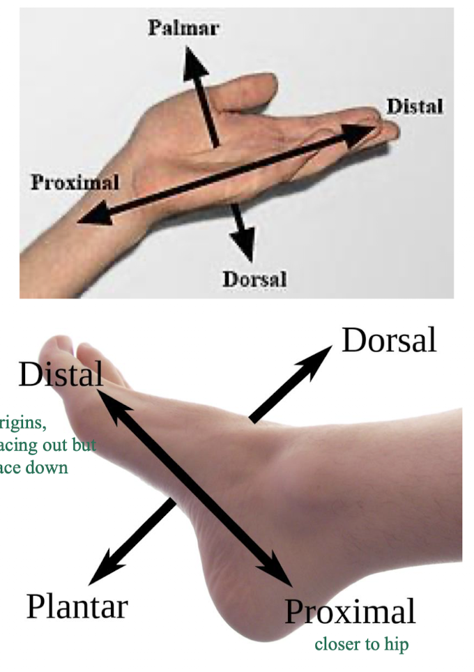
hand and foot specific directional terms
hand - the anterior surface of the hand is palmar, the posterior surface is dorsal
foot - bottom of the foot is the plantar surface of the foot (anterior surface), and top of the foot is the dorsal surface of the foot (posterior surface)
THESE ARE RELATIVE TO ANATOMICAL POSITION
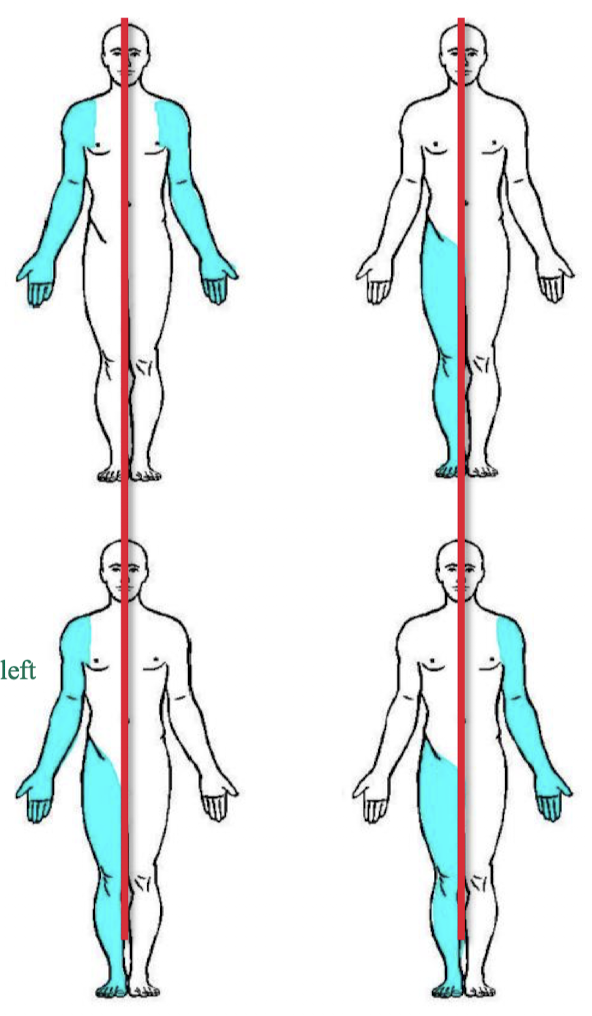
directional terms of laterality
right and left - always must describe from the position of the patient, must cross sides
bilateral - describing paired structures (e.g structures that you have both left and right like arms, kidneys, lungs, etc.)
unilateral - describing one side of the body (i.e. heart is unilateral left)
ipsilateral - describing structures on the same side of the body (i.e. spleen is ipsilateral to the stomach, both are on left side of the midline)
contralateral - describing structures on opposite sides of the body
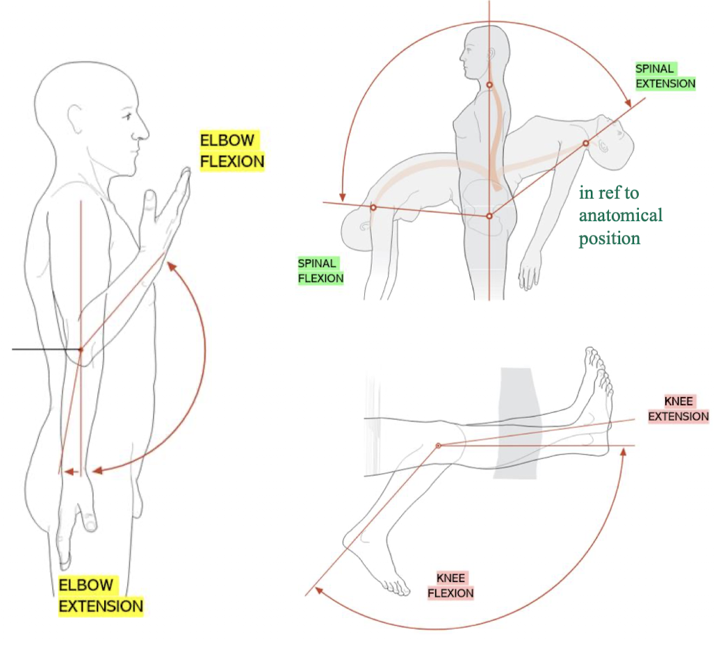
terms of movement - flexion and extension
flexion
for extremities - movement that decreases the angle between two body parts
for the spine - forward bending
extension
for extremities - movement that increases the angle between two body parts
for the spine - bending backwards
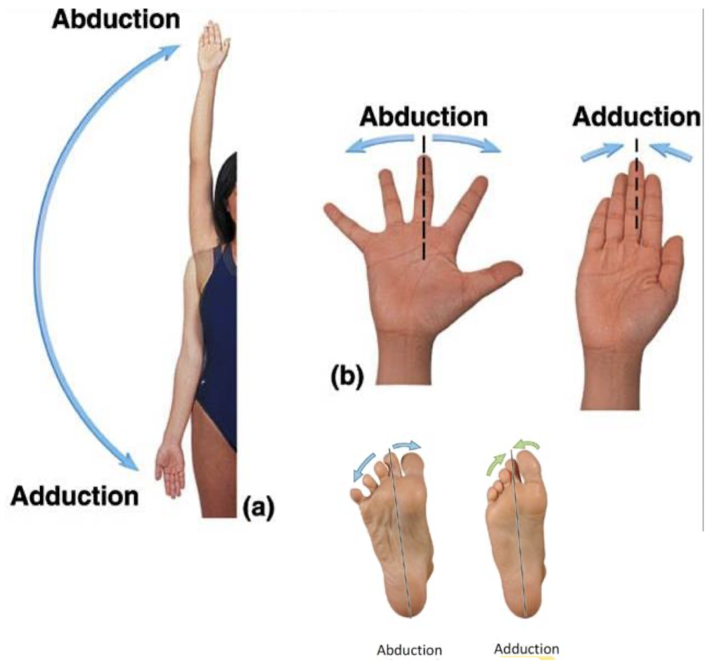
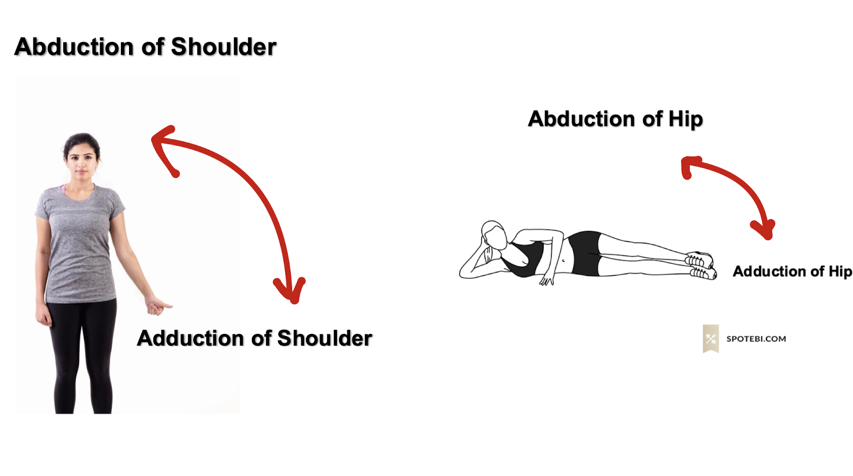
terms of movement - abduction and adduction
abduction - movement away from midline
in hand - movement away from midline of hand (3rd digit)
in foot - movement away from midline of foot (2nd digit)
adduction - movement toward from midline
in hand - movement toward midline of hand (3rd digit)
in foot - movement toward midline of foot (2nd digit)
what does the abduction of the shoulder and hip look like?
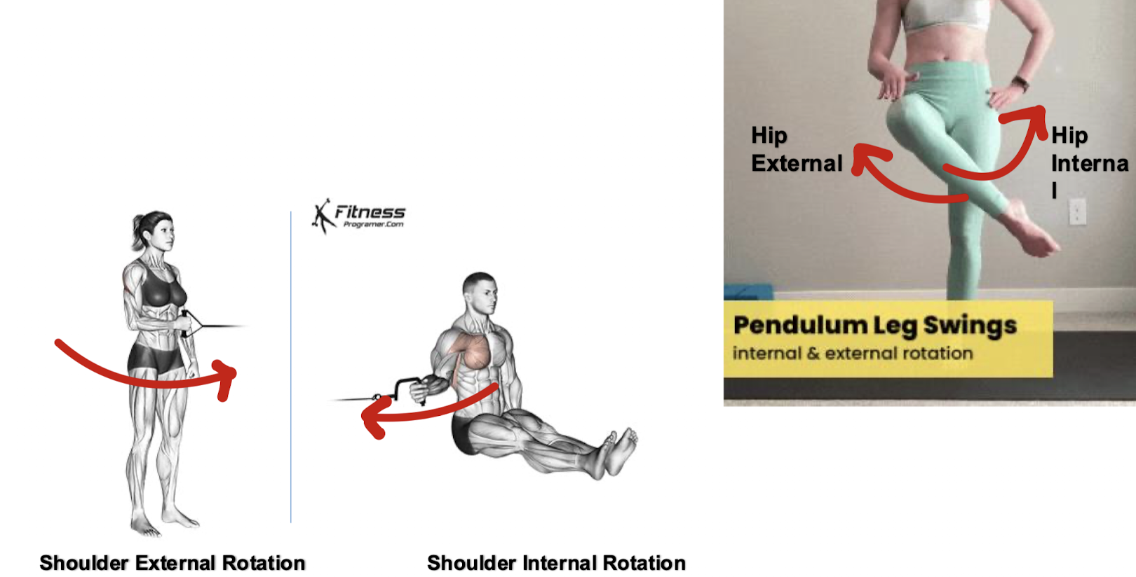
medial and lateral rotation
medial rotation - rotation toward the midline of the body
lateral rotation - rotation away from midline of the body
*NOTE: rotating your whole arm at the shoulder is called circumduction - it is a biaxial movement because the arm moves in more than one plane; can’t be called rotation; rotation happens only in one plane
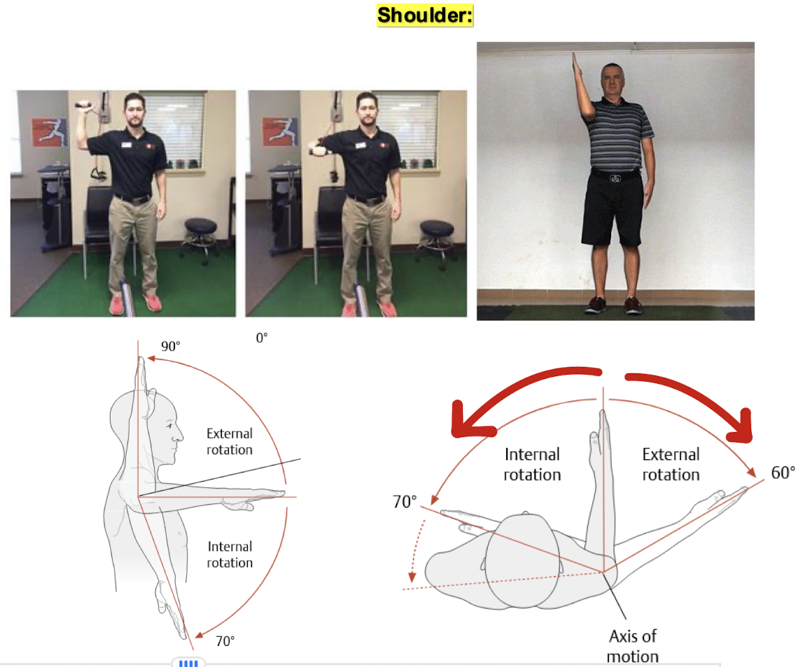
why does internal and external rotation look like for the hip and the shoulder?
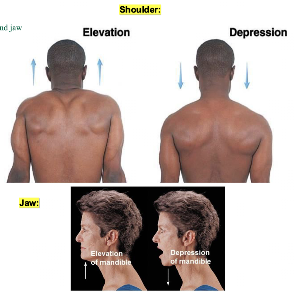
elevation and depression
we only see these movements at the shoulder blades and jaw
elevation - movement in a superior direction
depression - movement in an inferior direction
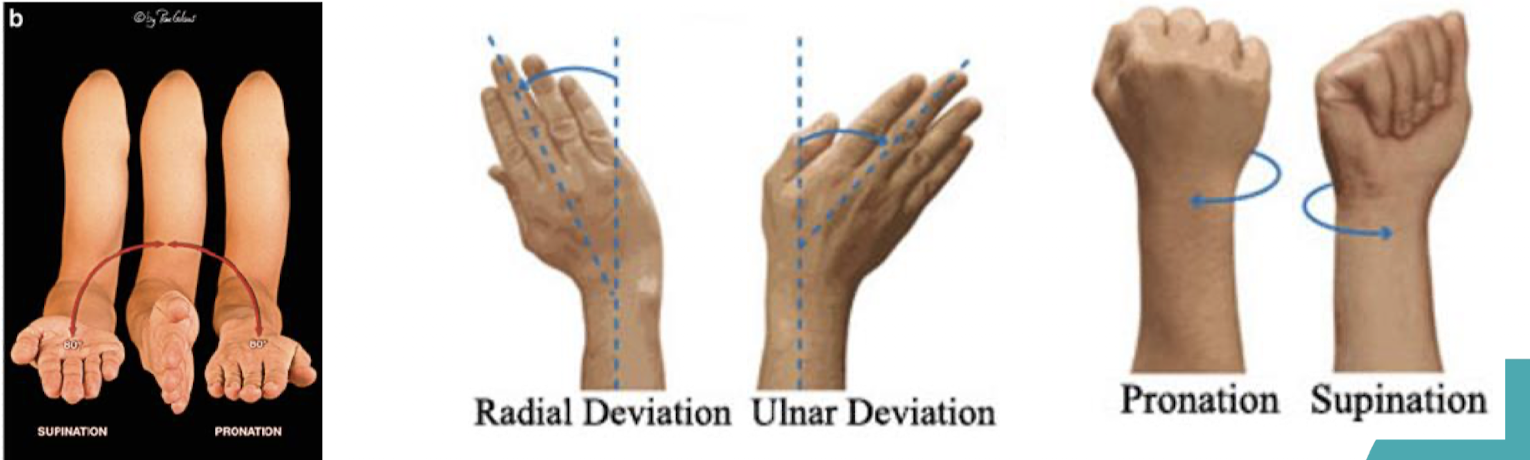
movement terms of the hand
supination - the forearm is rotated laterally (palm up)
pronation - the forearm is rotated medially (palm down)
ulnar deviation - adduction of wrist (pointing wrist towards ulna)
radial deviation - abduction of wrist (pointing wrist towards radius)
opposition - thumb moves medially to meet the other fingers
reposition - put the thumb back to neutral position
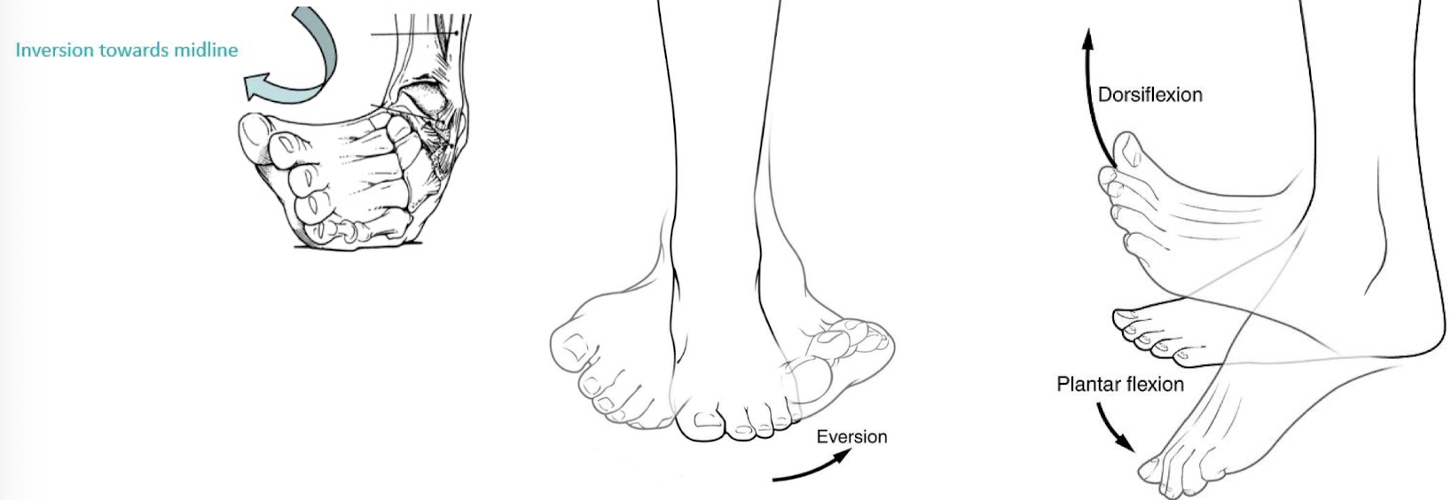
movement terms of the foot
eversion - plantar surface of the foot moves laterally
inversion - plantar surface of the foot moves medially
dorsiflexion - bending ankle towards the shin (toes point up)
plantar flexion - bending ankle towards posterior/back of body (toes point down)
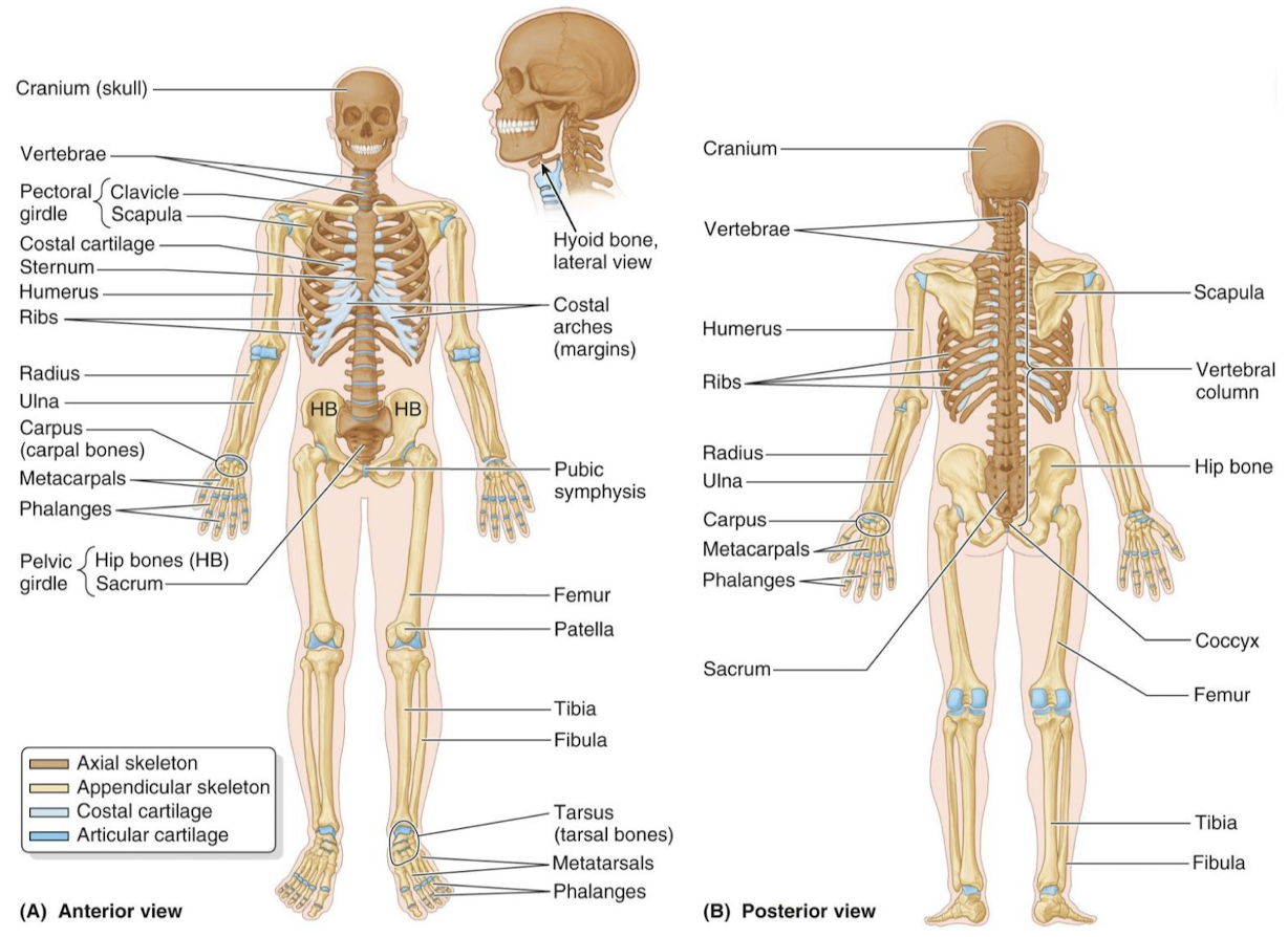
what is the skeletal system comprised of? what are the 2 basic subdivisions?
includes bone and cartilage
2 basic subdivisions
axial - central core of skeletal system
appendicular - includes bones of upper and lower limbs and bones that attach the limbs to axial skeleton
what forms the musculoskeletal system?
the bones, muscles, and joints
bones that make up the axial and appendicular skeleton
axial
80 bones
lie along longitudinal axis
appendicular
126 bones
upper + lower limbs and pelvic + pectoral girdles
functions of the skeletal system
protection for vital structures
support the body and vital cavities
movement
mineral deposition (like calcium, phosphorus, etc., deficiencies in these lead to bone break down)
blood element production (hematopoiesis)
energy storage (yellow marrow has adipose cells (fat - used for energy)
cartilage and its role
skeletal system is composed of cartilage and bones
cartilage is semi rigid and is found where flexibility is needed (i.e. costal cartilage and ribs)
types of cartilage
hyaline (joints) - most common, precursor to bone, referred to as articular because allows for friction and free movement
elastic (ear) - springy, moldable, holds shape
fibrocartilage (discs of spinal column) - very specific roles and areas of body
describe bone
bone is living tissue that is hard makes up most of the skeleton
bone is subject to constant rebuilding the same as any other living tissue
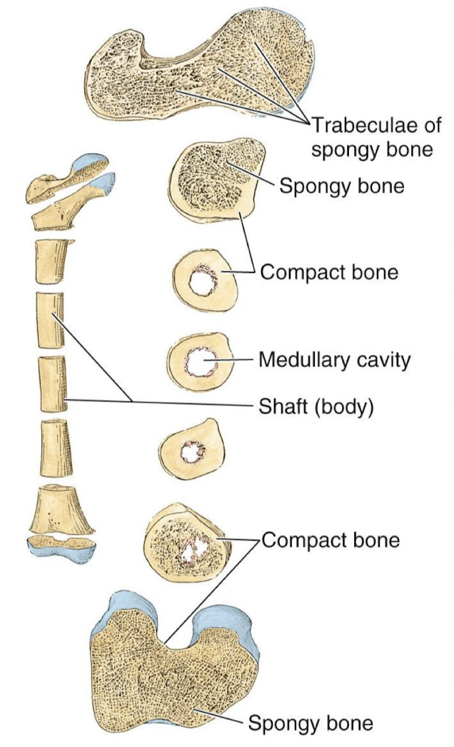
bone composition
compact bone - superficial thin layer provides the strength to bones for weight bearing, larger proportion of compact bone is found at ends of bones
spongy/cancellous/trabecular bone - found deep to compact bone and superficial to marrow cavity if present, contains many small spaces
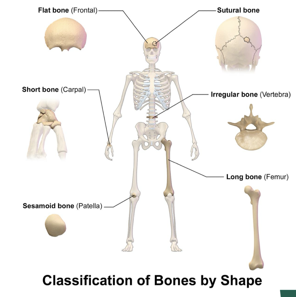
5 basic bone types
long - greater length than width (most common)
does not necessarily mean physically long, but has to fit the structural description of top, middle, end
short - almost cube-shaped and are nearly equal in length and width (i.e. some carpal bones)
needed in areas where we need a lot of mobility but not enough room
flat - thin bones
in areas where we need to spread out forces for protection, multiple bones share the burden
irregular - variable shape i.e. vertebrae
sesamoid - develop in tendons (patella)
reduce strain and muscles that travel in a straight line at the knee
sutural - in joint between skull bones
tightly adhere skull bones together
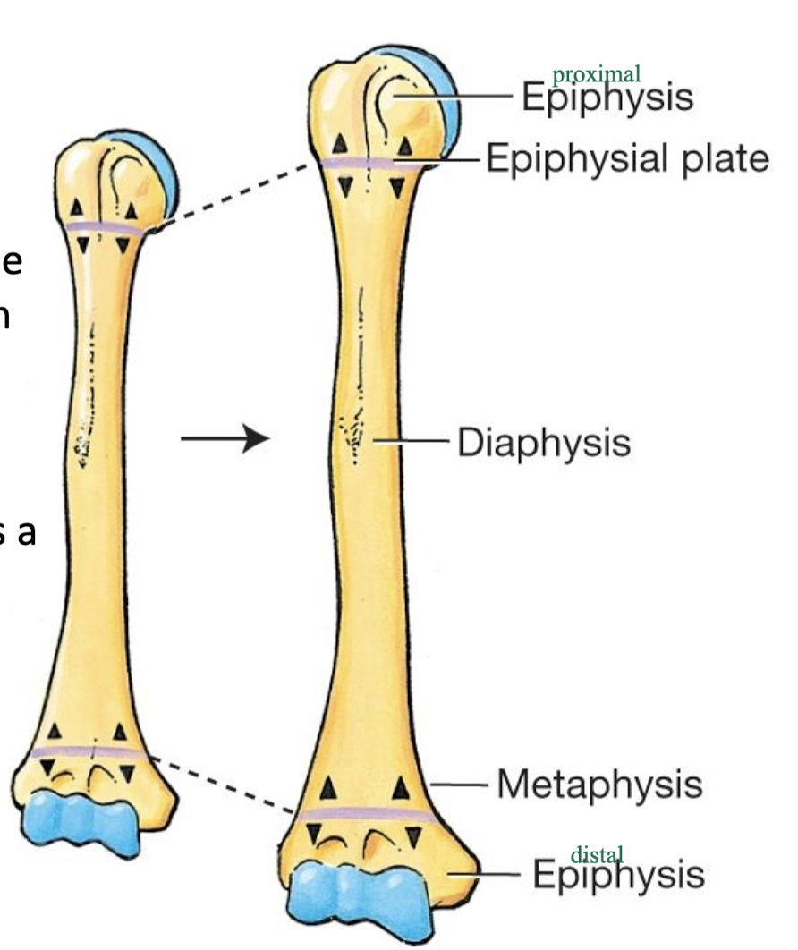
parts of a typical long bone
epiphysis - end of long bone - form articulation with other bones
metaphysis - the region between the epiphysis and the diaphysis
diaphysis - the shaft of the long bone - where bone marrow is found
epiphyseal plate - bone located within metaphysis, in adults this is a remnant line where the epiphyseal (growth) plate was in the child or developing
bone articulations
ends of long bones will usually form an articulation with another bone
these surfaces are covered with articular cartilage to offer smooth movement at joint surface
head - rounded projection at end of bone, articulates with base of adjacent bone
neck - supports head in articulation
condyle - large rounded protuberance at end of bone
trochlea - groove at end of bone, houses moveable tendon
facet - smooth flat articular surface
two major types of bone surface markings
depressions and openings - participate in joints or allow passage of soft tissue - called fossa, sulcus, foramen, fissure, canal
processes - projections or outgrowths that either help form joints or serve as attachment points for connective tissue - called trochanter, tubercle, tuberosity, spinous, crest