radiographic procedures chest p1
 Call Kai
 Learn
Learn Practice Test
 Spaced Repetition
 Match
Match Flashcards
 Knowt Play
1/48

There's no tags or description
Looks like no tags are added yet.
Name | Mastery | Learn | Test | Matching | Spaced | Call with Kai | Chat |
|---|
No analytics yet
Send a link to your students to track their progress
49 Terms
bony thorax
protective framework
respiratory system
lungs and airway
mediastinum
space between lungs
bony thorax structures
Sternum
Manubrium
Body
Xiphoid process
Clavicles
Scapulae
12 pairs of ribs
12 thoracic
vertebrae
Respiratory System structures
Four divisions:
Pharynx
Trachea
Bronchi
Lungs
Mediastinum structures
Four
structures:
Trachea
Esophagus
Thymus gland
Heart and great vessels
Pharynx
common passage for both air and food, used by both digestive and respiratory system
Thyroid Gland
located anteriorly in neck just below the larynx-very radiosensitive
right bronchi measurements
2.5cm long, 25 deg angle of divergence. wider
left bronchi measurements
5cm long, 37 deg angle of divergence
what level is Carina at
t5
right lungs
3 secondary bronchi, 3 lobes
left lungs
2 secondary bronchi, 2 lobes
parenchyma
highly elastic tissue allows for expansion and contraction of lungs
pleura
membranes containing lungs
parietal pleura
outer layer of pleural sac
Pulmonary or Visceral pleura
inner layer covers surfaces of lungs
pleural cavity
space between-contains lubricating fluids
pleural effusion
accumulation of fluid in pleural cavity
hemothorax
accumulation of blood in pleural cavity
pneumothorax
air or gas present in pleural cavity
diaphragm
primary muscle of inspiration
hemidiaphragm
each half of diaphragm
As diaphragm moves down (inspiration)
-volume of thoracic cavity
increases
– Intrathoracic pressure
decreases

A?
Trachea

B?
Apex

C?
carina

D?
Base of lungs

e?
diaphragm

F?
costophrenic angle

g?
hilum(root region) blood vessels

A?
apex

B?
upper lobe
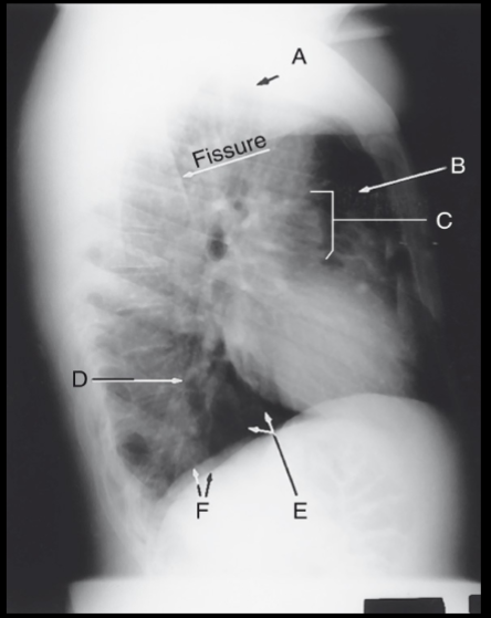
C?
hilum

D?
lower lobe
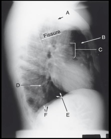
E?
base of lung
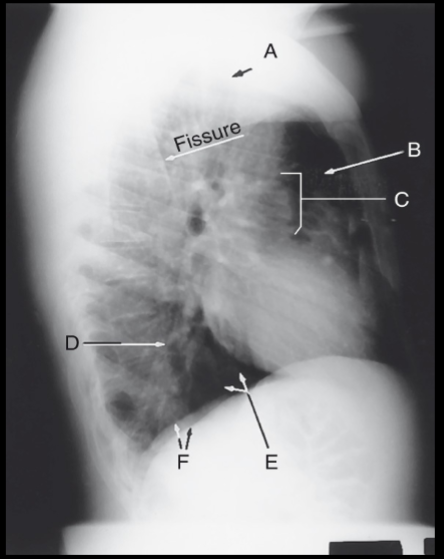
F?
diaphragm
the ? affects what lung and how
liver causes right lung to be 1 inches shorter
Evaluate body habitus of patient to determine whether the IR should be placed…
lengthwise or crosswise
The width of the “average” PA or AP chest is ?
than the vertical dimension
greater
Recumbent chests typically require the IR to be placed
?
crosswise
what should be shown after a inspiration scan
10 posterior ribs above diaphragm (ideal)
For certain conditions both inspiration & expiration images are taken bc..?
pneumothorax, lack of movement of diaphragm, foreign body, and the need to distinguish between opacity in the rib and one in the lung
Radiation Protection collimation-
reduces dose and improves quality
radiation protection back scatter-
Additional shield between pt and IR
the most repeated xray scan?
chest
kVp for chest xray
High kV (110 to 125)
why does chest have high kVp
it allows for low contrast for lung markings (more shades of gray)
Evaluating mAs
visualize faint outlines of at least the
mid and upper vertebrae
posterior ribs through the heart
Mediastinum