Ortho - Upper Extremity Part 2
 Call Kai
 Learn
Learn Practice Test
 Spaced Repetition
 Match
Match Flashcards
 Knowt Play
1/121

There's no tags or description
Looks like no tags are added yet.
Name | Mastery | Learn | Test | Matching | Spaced | Call with Kai | Chat |
|---|
No analytics yet
Send a link to your students to track their progress
122 Terms
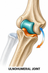
Which articulation of the elbow provides primary flexion/extension and high intrinsic bony stability?
Ulnohumeral joint (humerus trochlea + ulna olecranon/coronoid).
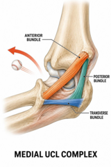
What is the primary stabilizer of the elbow against valgus stress?
Anterior bundle of the Medial Collateral Ligament (MCL).
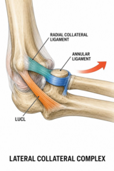
What structure is the primary stabilizer preventing posterolateral rotatory instability (PLRI) of the elbow?
Lateral Ulnar Collateral Ligament (LUCL).
Where is the ulnar nerve most superficial and vulnerable at the elbow?
In the cubital tunnel posterior to the medial epicondyle.

What motor and sensory deficits characterize Posterior Interosseous Nerve (PIN) palsy?
Finger and thumb extension weakness with NO sensory loss.

Where does the median nerve become compressed in pronator syndrome?
Between the two heads of the pronator teres muscle.
Which vascular structure is at highest risk during supracondylar fractures and elbow dislocations?
Brachial artery.
What is the normal carrying angle of the elbow in adults?
5° to 15° valgus.
What does a focal posterior 'goose egg' swelling at the elbow suggest?
Olecranon bursitis.
What visual finding on inspection indicates a diffuse elbow joint effusion?
Loss of the normal posterolateral concavity/hollow lateral to the olecranon.
What range of motion loss is the most sensitive indicator of elbow pathology?
Loss of elbow extension.
What is the functional range of motion arc for elbow flexion/extension and forearm rotation?
Flexion/extension: 30-130°; Pronation/Supination: 50° to 50°.
Which nerve roots and muscle carry out primary elbow extension?
Triceps muscle via the radial nerve (C7).
What muscle is the strongest supinator of the forearm?
Biceps brachii.
How is ulnar nerve motor function tested on physical exam?
Finger abduction/adduction and key pinch strength.
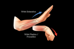
How is Cozen's test performed and what indicates a positive result?
Resisted wrist extension; positive if pain occurs at the lateral epicondyle.
What special test produces lateral epicondyle pain during resisted middle finger extension?
Maudsley's test.
How is Mill's test performed for lateral epicondylitis?
Passive wrist flexion with the elbow fully extended.
How is the Valgus Stress Test performed on the elbow?
Elbow flexed 20-30° with a valgus force applied to test the MCL.

What finding indicates a positive Pivot Shift test for Posterolateral Rotatory Instability?
Apprehension or a clunk as the radial head subluxes and reduces.
Where is sensory testing performed for the superficial radial nerve?
Dorsal aspect of the first web space.
What specific muscle tendon origin is primarily affected in lateral epicondylitis?
Extensor carpi radialis brevis (ECRB).

What is the underlying histological pathology of lateral epicondylitis?
Angiofibroblastic degeneration (tendinosis), not acute primary inflammation.

What activities typically aggravate lateral epicondylitis pain?
Gripping, wrist extension, palm-down lifting, and turning doorknobs.
Where is the exact point of maximum tenderness in lateral epicondylitis?
1 to 2 cm distal and anterior to the lateral epicondyle.
What is the evidence regarding corticosteroid injections for lateral epicondylitis?
Provides short-term relief (6-12 weeks) but may worsen long-term outcomes.
Where should a counterforce brace be worn for tennis elbow?
2 to 3 cm distal to the lateral epicondyle.
What nerve entrapment syndrome can mimic lateral epicondylitis?
Radial tunnel syndrome (compression of the posterior interosseous nerve).
Which tendons are primarily affected in medial epicondylitis (golfer's elbow)?
Pronator teres and flexor carpi radialis (common flexor-pronator origin).
What nerve pathology coexists in 20% to 50% of medial epicondylitis cases?
Cubital tunnel syndrome (ulnar nerve compression).
What is the most common organism isolated in septic olecranon bursitis?
Staphylococcus aureus (~80% of cases).
What physical exam features distinguish septic from non-septic olecranon bursitis?
Septic presents with moderate-severe pain, warmth, erythema, fever, and restricted ROM.
What aspirate WBC cutoff strongly suggests septic olecranon bursitis?
WBC >2,000/μL (frequently >50,000/μL).
What anatomical structure forms the roof of the cubital tunnel?
Osborne's ligament (arcuate ligament).
What positions or activities exacerbate symptoms of cubital tunnel syndrome?
Prolonged elbow flexion (e.g., sleeping, holding a phone, driving).
How is the Elbow Flexion Test performed for cubital tunnel syndrome?
Maximally flex the elbow for 60 seconds to reproduce ulnar paresthesias.
What motor physical findings signify late or severe cubital tunnel syndrome?
Weakness of finger abduction/adduction and atrophy of the first dorsal interosseous.
What conservative night positioning is recommended for cubital tunnel syndrome?
Night splinting with the elbow maintained at 45° extension.
What is the most common direction for an elbow dislocation?
Posterior or posterolateral (~90% of cases).
Which ligamentous structures are universally disrupted in a simple elbow dislocation?
Both Medial Collateral Ligament (MCL) and Lateral Collateral Ligament (LCL).
What physical exam step is mandatory before and after reducing an elbow dislocation?
Complete neurovascular examination (median/ulnar/radial nerves and distal pulses).
What plain radiographic views must be obtained post-reduction of an elbow dislocation?
AP and lateral radiographs to confirm concentric joint reduction.
What three injuries comprise the 'Terrible Triad' of the elbow?
Elbow dislocation, radial head fracture, and coronoid process fracture.
What is the typical injury mechanism for the Terrible Triad of the elbow?
Fall on outstretched hand (FOOSH) with axial load, valgus stress, and supination.
What is the management requirement for a Terrible Triad elbow injury?
Surgical treatment (radial head fixation/replacement, coronoid repair, LCL repair).
What is the most common elbow fracture in adult patients?
Radial head fracture (~30% of all elbow fractures).
Describe Mason Type I, II, and III radial head fractures.
Type I: Non-displaced (
What radius-ulna-wrist complex injury is associated with radial head fractures?
Essex-Lopresti lesion (interosseous membrane disruption and DRUJ injury).
What plain radiographic sign indicates an occult elbow fracture when no fracture line is seen?
Posterior fat pad sign.
What physical exam test confirms disruption of the extensor mechanism in an olecranon fracture?
Inability to extend the elbow against gravity.
What is the standard surgical management for displaced supracondylar humerus fractures in adults?
Open Reduction and Internal Fixation (ORIF) with dual column plating.
What anatomical concept dictates that a shortened or angulated single-bone forearm fracture implies additional joint injury?
The forearm functions as a closed mechanical ring.
What is the standard of care for adult both-bone forearm fractures?
ORIF with separate plate fixation of both the radius and ulna.
What critical anatomical feature of the radius must be restored during forearm ORIF to preserve rotation?
Radial bow.
What is a Monteggia fracture-dislocation?
Fracture of the proximal ulna shaft with dislocation of the radial head.
What is Bado Type I Monteggia fracture-dislocation?
Anterior radial head dislocation with apex anterior ulna shaft fracture.
What radiographic rule prevents missing a radial head dislocation on forearm X-rays?
The radiocapitellar line must bisect the capitellum on every view.
What is a Galeazzi fracture-dislocation?
Fracture of the distal radius shaft with disruption of the distal radioulnar joint (DRUJ).
Why is a Galeazzi fracture termed a 'fracture of necessity'?
It requires surgical ORIF of the radius in adults due to high failure rates with casting.
What X-ray findings suggest DRUJ disruption in a distal radius fracture?
DRUJ widening, ulnar styloid fracture, or >5mm radial shortening relative to ulna.
How is an unstable DRUJ managed after successful ORIF of a Galeazzi radius fracture?
Reduce DRUJ and pin across joint (K-wire) in supination for 4-6 weeks.
What is a Nightstick fracture and how is it caused?
Isolated ulna shaft fracture caused by a direct defensive blow to the forearm.
When can a Nightstick fracture be managed non-operatively?
When displacement is
What is the ischemic timeframe for irreversible muscle necrosis in acute compartment syndrome?
Begins at 3 hours, irreversible by 6 to 8 hours.
What is the earliest and most reliable clinical sign of compartment syndrome?
Pain out of proportion to injury and pain with passive muscle stretch.
What are late clinical findings in acute compartment syndrome?
Paresthesias, motor weakness/paralysis, pallor, and pulselessness.
How is Delta Pressure (ΔP) calculated for compartment syndrome, and what threshold confirms diagnosis?
ΔP = Diastolic BP - Compartment Pressure; ΔP
What immediate non-operative interventions must be performed when compartment syndrome is suspected?
Remove all circumferential dressings/casts completely and maintain limb at heart level.
Why should a limb with suspected compartment syndrome NOT be elevated?
Elevation reduces arterial inflow and perfusion pressure to the ischemic compartment.
What is the definitive surgical treatment for acute compartment syndrome?
Emergent fasciotomy of all affected fascial compartments.
What classic deformity results from untreated delayed forearm compartment syndrome?
Volkmann's ischemic contracture (flexed wrist, extended MCP, flexed IP joints).
Name the eight carpal bones in order.
Proximal: Scaphoid, Lunate, Triquetrum, Pisiform. Distal: Trapezium, Trapezoid, Capitate, Hamate.
Which carpal ligament is most commonly injured in wrist sprains/trauma?
Scapholunate ligament.
Describe a classic Colles' fracture and its characteristic physical deformity.
Distal radius fracture with dorsal displacement/angulation; presents with 'Dinner fork' deformity.
Describe a Smith's fracture and its physical deformity.
Distal radius fracture with volar displacement/angulation; presents with 'Garden spade' deformity.
What is a Barton's fracture?
Intra-articular distal radius fracture-dislocation involving the dorsal or volar rim.
What is a Chauffeur's fracture?
Avulsion fracture of the radial styloid.
What nerve complication commonly occurs acutely alongside a distal radius fracture?
Acute carpal tunnel syndrome (median nerve compression).
What are the radiographic thresholds for acceptable non-operative distal radius fracture alignment?
Radial inclination >15°, radial height loss
Which carpal bone is most frequently fractured?
Scaphoid bone (~70% of carpal fractures).
What vascular anatomical feature makes scaphoid proximal pole fractures prone to avascular necrosis?
Retrograde blood supply (blood enters distally and flows proximally).
What physical exam finding requires treating a patient for a scaphoid fracture regardless of initial X-rays?
Point tenderness in the anatomic snuffbox following trauma.
What special X-ray view is ordered to evaluate a suspected scaphoid fracture?
PA wrist view with ulnar deviation (scaphoid view).
If scaphoid fracture X-rays are negative but clinical suspicion is high, what are the two management options?
Immobilize in thumb spica and re-X-ray in 10-14 days, OR order immediate MRI.
What type of cast is used for non-displaced scaphoid waist fractures and for how long?
Thumb spica cast for 8 to 12 weeks.
What severe post-traumatic arthritic condition results from untreated scaphoid nonunion?
Scaphoid Nonunion Advanced Collapse (SNAC wrist).
What structures form the roof and floor of the carpal tunnel?
Roof: Transverse carpal ligament. Floor: Carpal bones.
What anatomical structures pass through the carpal tunnel?
9 flexor tendons (4 FDS, 4 FDP, 1 FPL) and the median nerve.
Why is sensation over the thenar eminence preserved in carpal tunnel syndrome?
The palmar cutaneous branch of the median nerve passes superficial to the carpal tunnel.
What classic symptom behavior is characteristic of carpal tunnel syndrome?
Nocturnal paresthesias waking the patient, relieved by 'shaking out' hands (flick sign).
How is Phalen's test performed for carpal tunnel syndrome?
Maximal wrist flexion for 60 seconds to reproduce paresthesias in the median distribution.
What physical motor sign indicates advanced carpal tunnel syndrome requiring surgical release?
Thenar atrophy and weakness of thumb abduction (Abductor Pollicis Brevis).
What is the initial conservative management for carpal tunnel syndrome?
Neutral wrist splinting worn at night.
Where is TFCC tenderness localized on physical examination?
In the 'fovea soft spot' between the ulnar styloid, flexor carpi ulnaris, and pisiform.
What imaging modality is gold standard for diagnosing TFCC tears non-surgically?
MRI or MR arthrogram.
How are central vs peripheral TFCC tears managed surgically?
Central tears are debrided; peripheral tears are surgically repaired.
What insertion sites distinguish flexor digitorum superficialis (FDS) from flexor digitorum profundus (FDP)?
FDS inserts on the middle phalanx (PIP flexion); FDP inserts on the distal phalanx (DIP flexion).
What is the most common metacarpal fracture?
Fifth metacarpal neck fracture ('Boxer's fracture').
What physical examination finding in a metacarpal fracture absolute mandates operative reduction?
Rotational deformity (finger scissoring or overlapping during composite flexion).
How much sagittal angulation is acceptable in a 5th metacarpal neck fracture (Boxer's fracture)?
Up to 40° to 50° of angulation.