A&P NERVOUS
 Call Kai
 Learn
Learn Practice Test
 Spaced Repetition
 Match
Match Flashcards
 Knowt Play
1/78

There's no tags or description
Looks like no tags are added yet.
Name | Mastery | Learn | Test | Matching | Spaced | Call with Kai | Chat |
|---|
No analytics yet
Send a link to your students to track their progress
79 Terms
two major regions of nervous system
central and peripheral nervous systems
the nervous system region composed of the brain and spinal cord
central nervous system (CNS)
the nervous system region composed of everything except the brain and spinal cord
peripheral nervous system (PNS)
nervous tissue contains which two basic types of cells:
neurons and glial cells
one of a variety of cells that provide a framework of tissue that supports the neurons and their activities
glial cell
the more functionally important cell in terms of the communicative function of the nervous system
neuron
cell body of a neuron
soma
extension of the neuron
process
a specific extension of a neuron, which is the fiber that connects a neuron with its target
axon
a type of process that branches off from the soma of the neuron
dendrite
responsible for receiving most of the input from other neurons
dendrite
region within the nervous system largely composed of cell bodies and dendrites
gray matter
region within the nervous system largely composed of axons
white matter
lipid-rich substance that gives white matter its color
myelin
a localized collection of neuron cell bodies in the CNS
nucleus
a cluster of neuron cell bodies in the PNS
ganglion
a bundle of axons, or fibers, found in the CNS
tract
a bundle of axons, or fibers, found in the PNS
nerve
the three basic functions of the nervous system
sensation, integration, and response
receiving information about the environment around us
sensation
generating responses to sensory input
motor responses
combines sensory perceptions and higher cognitive functions such as memories, learning, and emotion to produce a response
integration
a change from homeostasis or a particular event in the environment that is registered by the sensory functions of the nervous system
stimulus
how can responses be divided
voluntary or involuntary
______ responses are governed by the somatic nervous system
voluntary
_______ responses are governed by the autonomic nervous system
involuntary
the processing of stimuli that are received by sensory structures
integration
the nervous system can be divided into two parts on the basis of a functional difference in responses:
somatic nervous system (SNS) and autonomic nervous system (ANS)
this division of the nervous system is responsible for conscious perception and voluntary motor responses
SNS
which kind of motor response would reflexes be considered
somatic
this division of the nervous system is responsible for involuntary control of the body, usually for the sake of homeostasis
ANS
this third division of the nervous system is responsible for controlling the smooth muscle and glandular tissue in your digestive system
enteric nervous system (ENS)
specialized areas of contact between two neurons, where information is passed onwards
synapses
why are neurons considered to have a polarity
they flow in one direction: dendrites, cell body, axon
where the axon emerges from the cell body; tapering of the cell body toward the axon fiber
axon hillock
within the axon hillock, the cytoplasm changes to…
axoplasm
the axon hillock narrows and transitions into the beginning of the axon, called the…
initial segment
action potentials are generated in the trigger zone, which is a combination of…
the axon hillock and initial segment
many axons are wrapped by this insulating substance
myelin
what type of cell is myelin made from
glial cells
the gaps in the myelin covering an axon; important to the way that electrical signals travel down the axon
node of Ranvier
the length of the axon between each gap (nodes of Ranvier), which is wrapped in myelin
axon segment
the end of the axon, where there are usually several branches extending toward the target cell
axon terminal
the extensions of the axon terminal each end in an enlargement called a ________; these bulbs make the connection with the target cell at the synapse
synaptic end bulb
supporting cells with functions directed at helping neurons complete their function for communication
glial cells, or neuroglia, or glia
list the 6 types of glial cells and where they are found
CNS: astrocyte, oligodendrocyte, microglia, ependymal cell
PNS: satellite cell, schwann cell
the astrocyte is to the CNS what the ________ is to the PNS
satellite
the oligodendrocyte is to the CNS what the _____ is to the PNS
schwann cell
star-shaped glial cell that provides support to the neurons of the CNS; has many processes extending from main cell body, which extend to interact with neurons, blood vessels, or the connective tissue covering the CNS (pia mater)
astrocyte
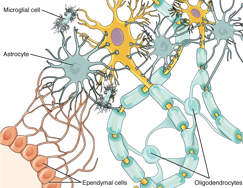
a physiological barrier that keeps many substances that circulate in the rest of the body from getting into the central nervous system, restricting what can cross from circulating blood into the CNS
blood-brain barrier (BBB)
what kind of molecules can pass through the BBB
nutrient molecules, such as glucose or amino acids
the glial cell type that insulates axons in the CNS; there are a few processes that extend from the cell body, and each one reaches out and surrounds an axon to insulate it in myelin
oligodendrocyte
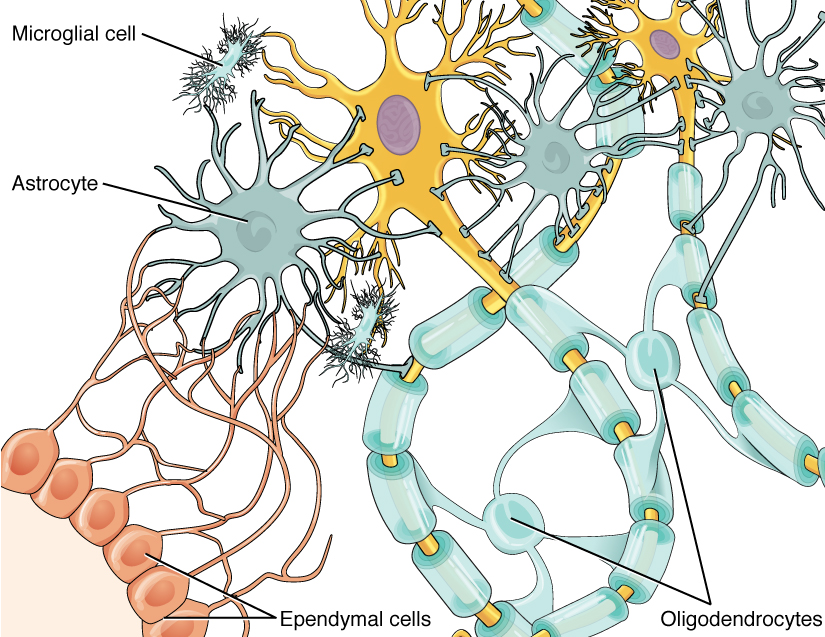
also referred to as CNS-resident macrophages; smaller than most other glial cells; act like macrophages: ingest and digest diseased or damaged cells
microglia
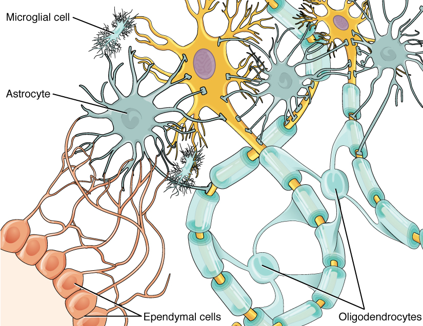
a glial cell that filters blood to make cerebrospinal fluid (CSF); ciliated; can be considered a component of the BBB
ependymal cell
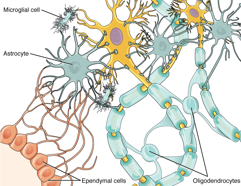
a specialized structure in the ventricles where ependymal cells come in contact with blood vessels and filter and absorb components of the blood to produce cerebrospinal fluid
choroid plexus
one of two types of glial cells found in the PNS; found in sensory and autonomic ganglia, where they surround the cell bodies of neurons; provide support, performing similar functions as astrocytes
satellite cell
one of two types of glial cells found in the PNS; insulates axons with myelin; wraps around a portion of only one axon segment and no others; nucleus and cytoplasm of this cell are on the edge of the myelin sheath
schwann cell
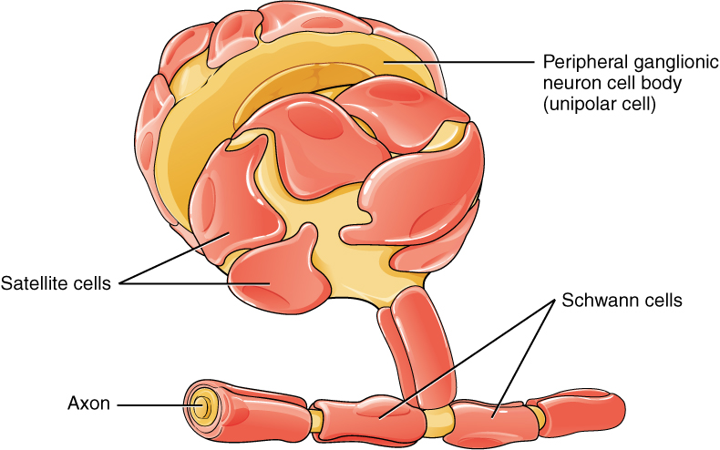
facilitates the transmission of electrical signals along the axon
myelin sheath
a type of sensory receptor that is sensitive to temperature
thermoreceptor
the change in membrane potential; dependent on the strength of the stimulus
graded potential
the voltage at which an electrical signal is generated
threshold
the electrical signal resulting from crossing the threshold
action potential
the process of the action potential traveling along the axon from the axon hillock to the axon terminals and into the synaptic bulb
propagation
the sodium/potassium pump moves sodium ions (NA+) ________ the cell
out
the sodium/potassium pump moves potassium ions (K+) ________ the cell
into
is the concentration of NA+ higher outside or inside the cell
outside
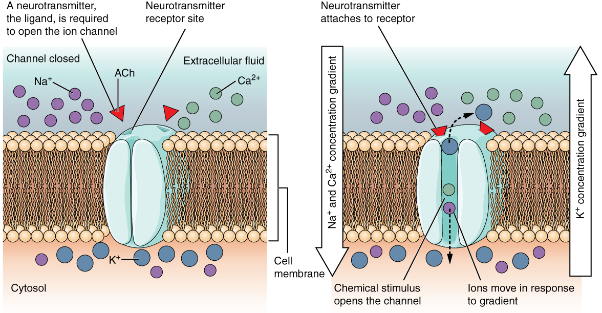
this type of gated protein channel opens because a signaling molecule, a ligand, binds to the extracellular region of the channel
ligand-gated channel
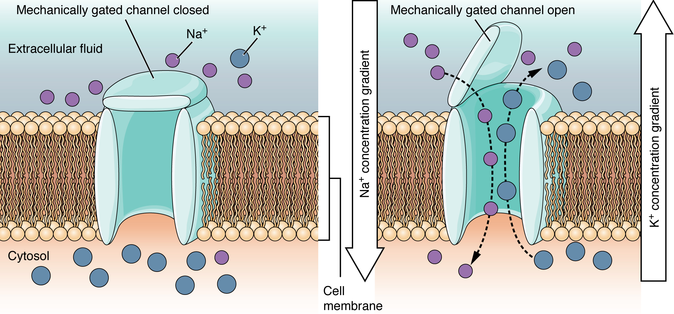
this type of gated protein channel opens because of a physical distortion of the cell membrane
mechanically gated channel
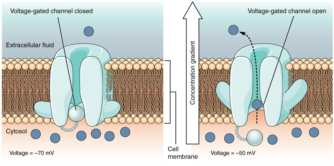
this type of gated protein channel responds to changes in the electrical properties of the membrane in which it is imbedded; when the voltage becomes less negative, the channel begins to allow ions to cross the membrane
voltage-gated channel
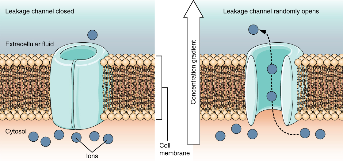
this type of gated protein channel is randomly gated, meaning it opens and closes at random
leakage channel
what is the resting membrane potential value
-70 mV
what is the membrane potential once fully depolarized
+30 mV
what is the membrane potential during hyperpolarization
< -70 mV
what is the membrane potential once fully repolarized
-70 mV
in this type of synapse, a chemical signal (neurotransmitter) is released from one cell and it affects the other cell
chemical synapse
in this type of synapse, there is a direct connection between the two cells so that ions can pass directly from one cell to the next
electrical synapse
all synapses have these common characteristics:
presynaptic element
neurotransmitter (packaged in vesicles)
synaptic cleft
receptor proteins
postsynaptic element
neurotransmitter elimination or re-uptake
briefly explain the release of a neurotransmitter
action potential reaches axon terminals, voltage-gated CA2+ channels in the membrane of the synaptic end bulb open
concentration of CA2+ increases inside the end bulb, and the CA2+ associates with proteins in the outer surface of neurotransmitter vesicles
CA2+ facilitates merging of the vesicle with the presynaptic membrane so the neurotransmitter is released through exocytosis into the synaptic cleft
travels to the postsynaptic membrane where it interacts with neurotransmitter receptors (fit together lock and key)
the small gap between two cells where the neurotransmitter is released
synaptic cleft