Support and movement
 Call Kai
 Learn
Learn Practice Test
 Spaced Repetition
 Match
Match Flashcards
 Knowt Play
1/141

There's no tags or description
Looks like no tags are added yet.
Name | Mastery | Learn | Test | Matching | Spaced | Call with Kai | Chat |
|---|
No analytics yet
Send a link to your students to track their progress
142 Terms
what are the 2 categories that lead to developmental defects
genetic mutations
teratogens
teratogenic
agents that lead to genetic defects in an embryo
how do we stop developmental defects
stop breeding the animals with the mutant gene
eliminate the teratogen
what factors affect the severity of developmental defects
dose of the teratogen
the amount of time the embryo is exposed to the teratogen
what are common factors of reproductive failure
chromosomal abnormalities
teratogenic factors
poor conditions (nutrition and stress)
idiopathic
co-dominant traits
both genes are expressed (blood types)
haploinsufficiency
when only 1 allele is able to be expressed and the other is null leading to the dominant gene only being partially expressed
single gene mutations
when a mutation occurs on a single gene (albinism, dwarfism, myotania congeita)
Malignant hypothermia in pigs
autosomal dominant mutation of RYR1
affects calcium channel in muscles
point mutated gene is dominant over wild type allele causing production of bad protein
how are we able to observe chromosomal abnormalities
using a microscope
aneuploidy
having an abnormal amount of chromosomes
monosomt
lose of a chromosome (X0)
trisomy
having an extra chromosome (XXY)
how does selective breeding lead to genetic consequences
over selecting for 1 gene can cause deleterious affects on other genes (incel chicken)
teratogen agent categories
environmental pollutants
mycotoxins
latrogens (injury from medical treatment)
ionizing radiation
infection
bacteria
protozoa
metabolic imbalance
iatrogenesis
harm caused by medical treatment
what are the six principles of teratology
severity depends on the genotype
depends on when the embryo is affected
how the teratogen gets to the embryo
teratogens act in specific ways
manifestations of development (death, malformation, growth retardation, functional defecct)
low levels of teratogen = low effect
what stage of development have low sensitivity to teratogens
fertilization and zygote
what stage of development has high sensitivity to teratogens
blastocyst, embryo, and fetus
palate, urigeneital, repro system and cerebellum remain developing into late gestation
what are the 5 components of the musculoskeletal system
Bone
Muscle
tendon
ligament
joints
what are the functions of bone
provide support and structure
protection of organs
movement
hematopoiesis
endocrine regulation (calcium)
sesamoid bone occur where
>90 degrees range of motion
significant force at the end of motion
Function of a sesamoid bone
protects the tendon and aid with muscle leverage
what are the 4 classifications of bones
long bones (femur and humerus) longer than they are wide
short bones (carpal and tarsal bone)
flat bones (skull and ilium)
irregular bones (vertebrae and mandible)
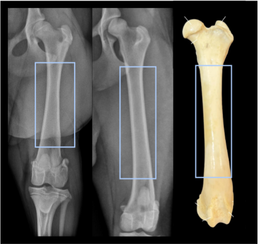
diaphysis
shaft of the long bone
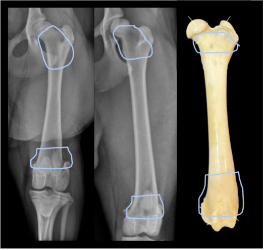
metaphysis
neck of the bone
between diaphysis and epiphysis
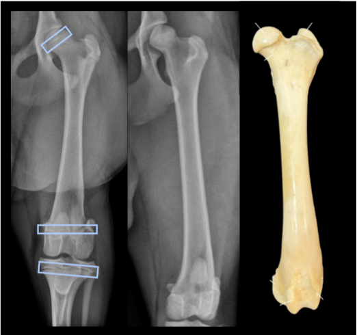
physis
growth plate
cartilagenous
only seen while bone is growing (only juveniles)
epiphysis
region between physis and joint of the bone

apophysis
site of tendon or ligament attachment
non articular
large bony marking when mature
separate center of ossification

compact bone
solid inner part of the bone
found in the diaphysis of the bone
spongy bone
found in the metaphysis and epiphysis of the bone
medullary cavity
hollow space within the bone
site of hemtopoiesis
periosteum
outer surface of the bone
innervated (lots of sensation)
fibrous outer layer
inner osteogenic cell layer
endosteum
thin layer lining medullary cavity
helps to build and maintain bone
fossa
depression in bone
foramen
hole in bone
what are the 3 types of joints
fibrous
cartilaginous
synovial
ligament
connects bone to bone
made of collagen
strong but poor blood supply
what are the components and functions of synovial joints
synovial cavity with synovial fluid
joint capsule
synovial lining
hyaline cartilage
subchondral bone
high motion
fibrous joints
connective tissue junction
limited movement
suture joints
found in the skull
dense irrgeular connective tissue

syndesmoidal joints
ligamental sheet to connect long bones

gomphosis
ligamentous connection
connect bone to jaw
cartilaginous joints
connective tissue junction
limited junction
cartilage present
synchondrosis
bones connected by cartilage (ribs to sternum)
symphysis
bones connected by cartilage and connective tissue (vertebrae)
muscle
50% of body weight
composed of 2 types of fibers
large blood supple
innervation is specific to each muscle
where do muscles attach to bones
origins (less motion) and insertions (more motion)
tendon sheaths
column of fluid around the tendon
contain synovial lining and fluid
bursa
pocket of fluid between the tendon and bone
decreases friction on the tendon
contain synovial lining and fluid

plantargrade
the entire plantar is on the surface of the ground

digitigrade
only the digits are on the ground
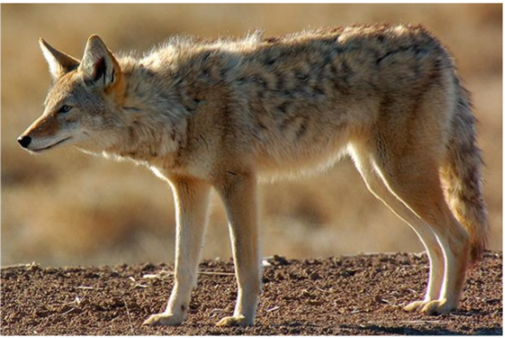
unguligrade
hooved animals
only the 3rd phalanx is on the ground
proximal carpal bones
accessory carpal bone
named for adjacent long bones
ulnar carpal bone lateral
radial carpal bone medial
distal carpal bone
numbered from medial to lateral
clavicle
not found in dogs only cats
stabilizes thoracic limb allows for abduction
antebrachium in ruminant
complete ulna
partial fusion to the radius
carpus of ruminant
proximal carpal bones (radial, intermediate, ulnar accessory)
distal carpal bones (fused II and III) and IV (lateral)

metacarpals in ruminant
III and IV (III is medial IV is lateral)
II and V vestigial

equine carpus osteology
only 1 metacarpal bearing weight (metacarpal III)
II and IV act as splint bones

equine antebrachium
vestigial ulna
fused radius
thoracic sling
Trapiezius (superficial)
Rhomboid (deep)
serratus ventrails
pectoralis
passive stabilization
requires no muscular activity and is provided by bony anatomy
active stabilization
muscles actively contract to prevent movement
shoulder stablizers
supraspinatus (cranial to spine of scapula)
infraspinatus (caudal to spine of scapula)
subscapularis (medial aspect of the scapula)
Equine biceps brachii
contains bicipital bursa to lubricate the joint
olecranon
formed from apophysis

anconeal process
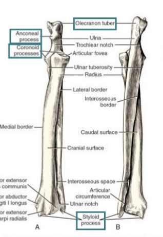
coranoid processes

styloid process

cranial elbow flexors
biceps
brachialis

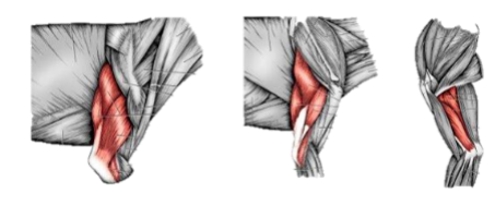
caudal elbow flexors
triceps brachii (4 heads in dogs)
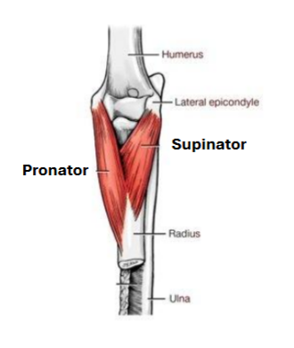
elbow rotations
pronator teres
supinator
Small animal carpal bones
Proximal intermedioradial, ulnar, and accessory
Distal I, II, III, IV
fetlock
metacarpophalangeal joint
pastern
proximal interphalangeal joint
Coffin
distal interphalangeal joint
Carpal and digital extenders
Extersor carpi radialis
Common digital extensor
Later digital extensor
ulnaris lateralis
Carpal and digital flexors
Flexor carpi radialis
flexor carpi ulnararis
superficial digital flexor tendon
deep digital flexor tendon
digital flexors and extenders in the horse
no muscle fibers in distal limb
accessory ligaments for superficial and deep digital flexors
Feline digit
digit III retracts
Fibers of ECM
collagenous or fibrilis
reticular
Elastic
Ground substance of ECP
contains GAG’s and PG
structural glycoproteins
why does collagen quarter stagger
in bone the first mineral accumulates in those spaces
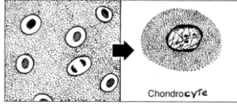
chondrocyte
cell pertaining to cartilage
how are fibrils aligned in cartilage
crossed over each other
why is a type 2 collagen molecule made of
trimeric protein
positively charged
what is the backbone of ground substance made of
GAG’s (chondroitin sulfate or Keratan sulfate)
where do sugars attach in cartilage
The PG (very negatively charged) attach to the GAG’s
what is the downside of PG’s being negativly charged
PG’s try to maximize the distance between each other due to their charge
PG’s are hydrophilic meaning that thy bind lots of water leaving the matrix to be squishy
causes stain to be very purple
Type 2 collagen
positively charged
trimeric molecule
PG/PAG’s
negativly charged
chondroitin and keratan sulfated side chains
Hyaline cartilage
avascular
nutrient and gas exchange in ECM
releases water when compressed but can reabsorb when compression alleviated
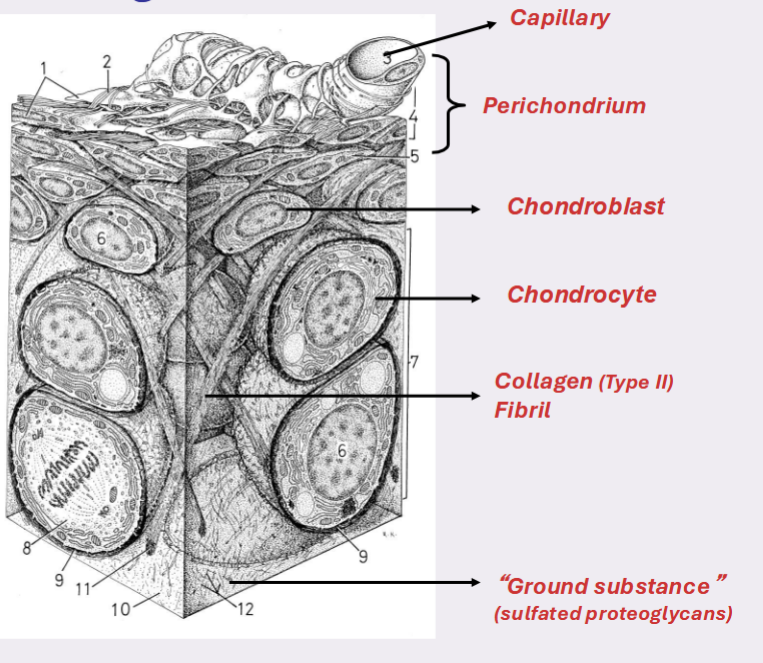
mesenchymal cell
precursor of all connective tissue cells
chondroblasts
divide and secrete ECM
distance themselves from neighbors

chondrocytes
cell completely surround by ECM
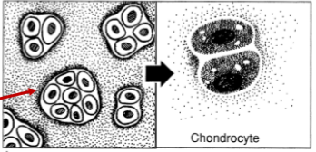
Isogenic cells
group of daughter cells in close proximity to one another
apositional growth
differentiation of chondroprogenitor cells into chondroblasts
interstitial growth
division of per-existing chondricytes in cartilage matrix
hyaline cartilage
structural support, rapid growth withstanding compression
found in fetal skeleton and respiratory passage