HP- Lec 2 Intro to Nervous System
1/27
There's no tags or description
Looks like no tags are added yet.
Name | Mastery | Learn | Test | Matching | Spaced | Call with Kai |
|---|
No analytics yet
Send a link to your students to track their progress
28 Terms
Nervous System Terminology- CNS,PNS, Inter, snesory, motor neuron
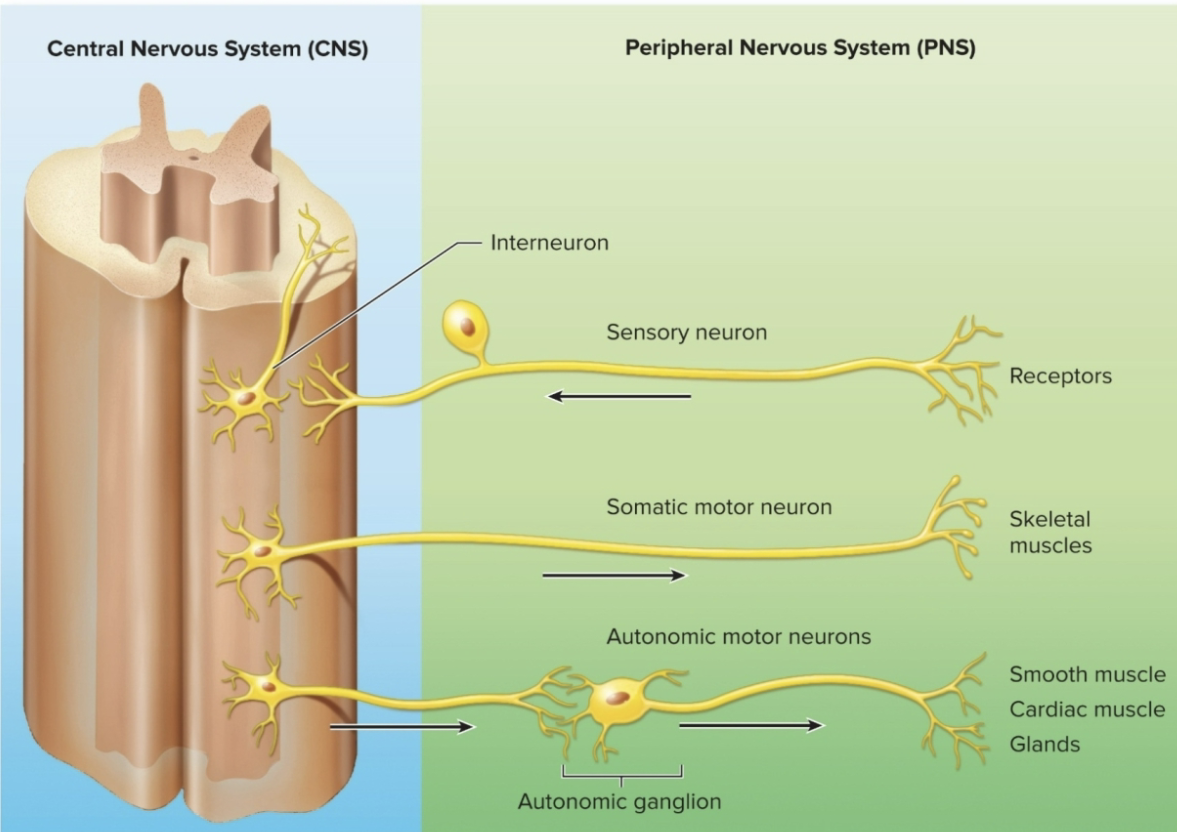
CNS: Brain and Spinal Cord
PNS: Nerves, ganglia, and nerve plexuses (outisde of CNS)
Interneuron: multipolar neuron located entirely within the CNS
Sensory Neuron (afferent Neuron): neuron that transmits impulses from a asensory receptor into the CNS
Motor Neuron (efferent neuron): neuron that transmits impulses from the CNS to an effector organ (ex: muscle)
Nervous system Division & Tissues
Divided into:
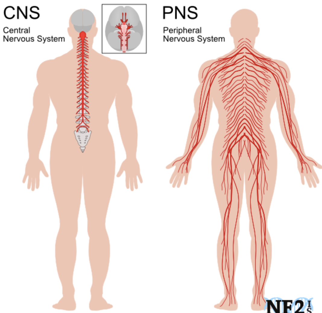
Central nervous system (CNS): Organs- brain and spinal cord
Peripheral nervous system(PNS): Organs- cranial and spinal nerves, and ganglia
Tissue is composed of two types of cells
Neurons: conduct electrical activity (impulses), but, in adults, typically lack the ability to divide
Glial cells (neuroglia): support neurons, do not generate electrical impulses, but retain the ability to divide
Neurons
Structural & Functional units of the nervous system
Features & General Functions
generates and conducts electrical activity
release neurotransmitters, which are chemical regulators used for neuronal communication through chemical synapses
depending on their role, neurons can sense external sensory info (sensory neuron), send motor inputs (motor neuron) or be an interneuron
neurons enable perception of sensory stimuli from both the external environment and the internal body as well as memory and control of muscles and glands (IMAGE)
Neurons Features
there are approx 100 billion neurons in the human brain, they vary in size and shape
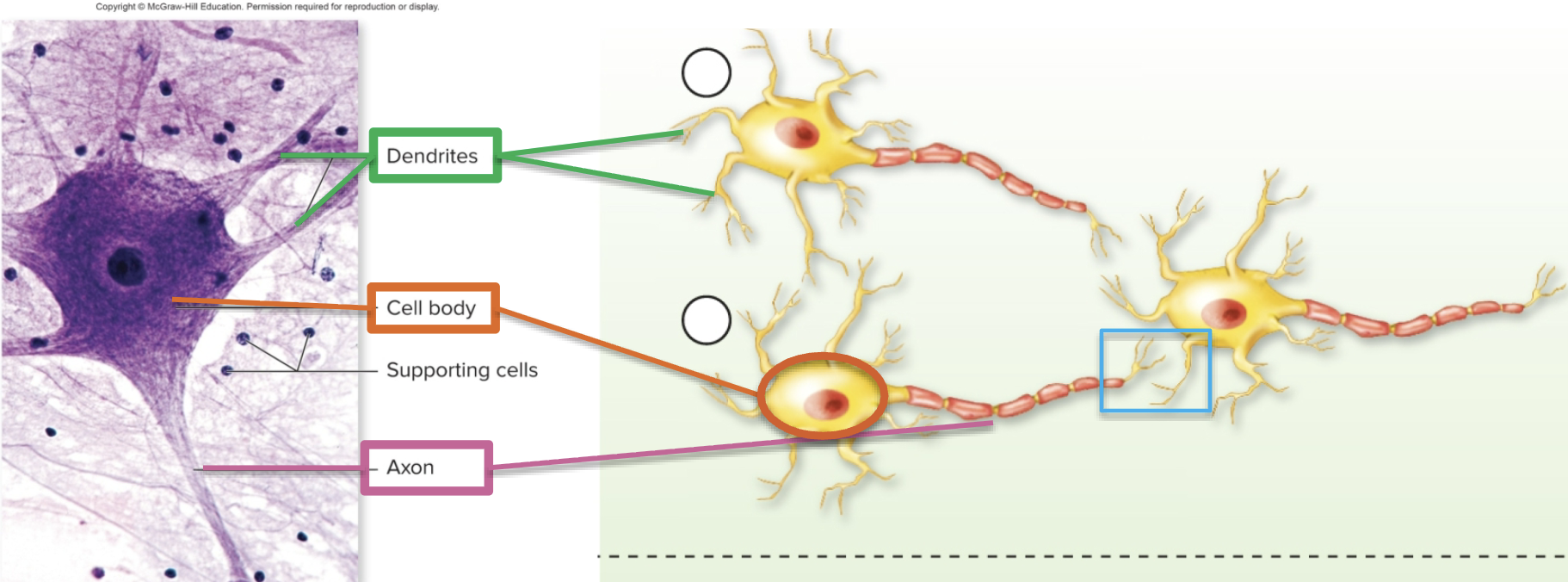
Dendrites (input): receive signals and conducts a graded impulse towards the cell body
Cell Body: contains the nucleus and other organelles; after integrating all the graded impulses from the dendrites, it may generate action potential.
Cluster in groups= Nuclei in CNS; ganglia in PNS
Axon (output): conducts action potentials AWAY from the cell body
Synapse: place at which the axon of one neuron comes in close contact to the dendrite of another neuron
Functional Classification of Neurons
Neurons can be classified based on the direction in which they conduct electrical impulses
Interneurons: located entirely within the CNS, these neurons integrate the functions of the nervous system
Sensory Neurons: conduct impulses from sensory receptors to the central nervous system (afferent)
Motor neurons: conduct impulses from the CNS to target organs (efferent)
Somatic motor neurons: control voluntary movements
Autonomic motor neurons: regulate involuntary functions
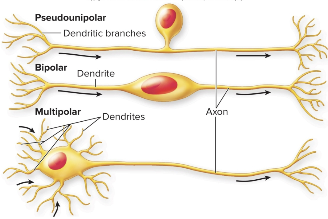
Structural Classification of Neurons (3)
based on their morphology
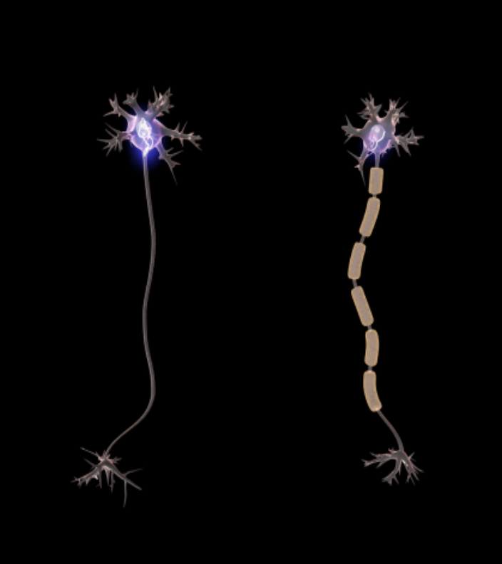
Pseudounipolar: single short process that branches like a T to form 2 longer processes; sensory neurons
Bipolar Neurons: have two processes, one on either end, found in retina of eye
Multipolar neurons: several dendrites and one axon; most common type
Axons(output)
conducts action potentials AWAY from the cell body
vary in length from a few millimeters to a meter
connected to the cell body by the axon hillock, where action potentials are generated at the initial segment of the axon
can form many branches called axon collaterals
covered in myelin with open spots called nodes of ranvier
Classification of bundle of axons
Nerves are bundle of axons located in the PNS
Tracts are bundles of axons located in the CNS
most are composed of both sensory an motor neurons and are called mixed nerves
some of the cranial nerves have sensory fibers only
Neuroglial cells and their functions
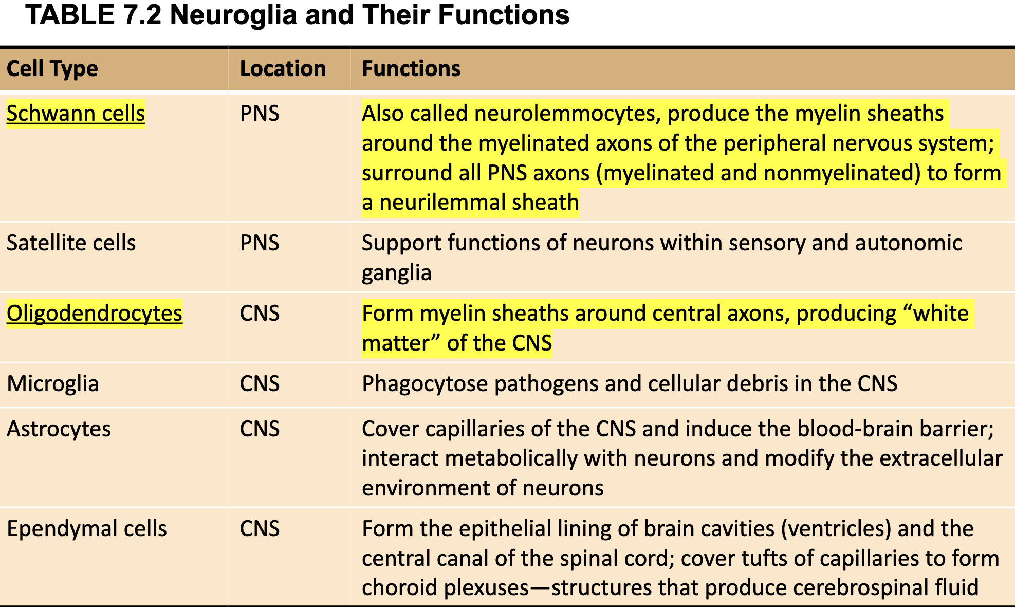
Schwann Cells: PNS; produce the myelin sheaths around the MYELINATED axons of the PNS; surrounded all PNS (myelinated/nonmyelinated) to form a neurilemmal sheath
Oligodendrocytes: CNS; form myelin sheaths around CENTRAL axons, producing “white matter” of the CNS
Myelin Sheath
in the CNS: the myelin sheath is produced by oligodendrocytes
in the PNS: the myelin sheath is produced by Schwann cells
One oligodendrocyte sends extensions to several axons and each wraps around a section of an axon (like insulation)
axon is like a power cord, wrapped by insulation cord
can have unmyelinated fiber
Demyelinating Diseases
are those in which the myelin sheaths are specifically attacked
Guillain-Barre syndrome: the T cells of the immune system attack the myelin sheaths of the PNS, this produces rapid onset of symptoms that include muscle weakness
Multiple Sclerosis: produced by an autoimmune attack by T lymphotcytes causing lymphocytes and monocyte-derived macrophages to enter the brain and target the myelin sheaths of the CNS causing demyelination
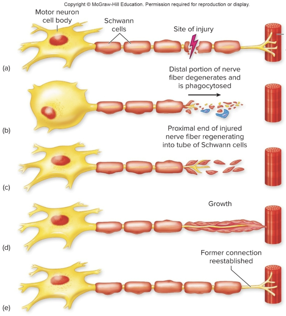
Neuroregeneration in the PNS
when an axon in the PNS is cut, the severed part degenerates, and a regeneration tube is formed by Schwann cells
growth factors (neurotrophic factors) are leased that stimulate growth of axon sprouts within the tube
new axon eventually connects to the undamaged axon or effector
Neurotrophic (growth) Factors or Neurotrophins
neurotrophins are secreted proteins that promote the survival, differentiation and growth of neurons
promote neuronal growth in the fetal brain both in CNS and PNS
Nerve Growth Factor (NGF)
Brain-derived neurotrophic factor (BDNF)
Glial-derived neurotrophic factor (GDNF)
Neurotrophin-3, neurotropin-4/5
In adults, neurotrophins aid in the maintenance of sympathetic ganglia (PNS) and the regeneration of sensory neurons
CNS Regeneration
injury in the mature (adult) CNS triggers limited regeneration in central axons compared to peripheral axons
Nogo: proteins produced predominantly by oligodendrocytes, inhibit axon regeneration in the mature CNS
Glial scars form from astrocytes also prevent regeneration
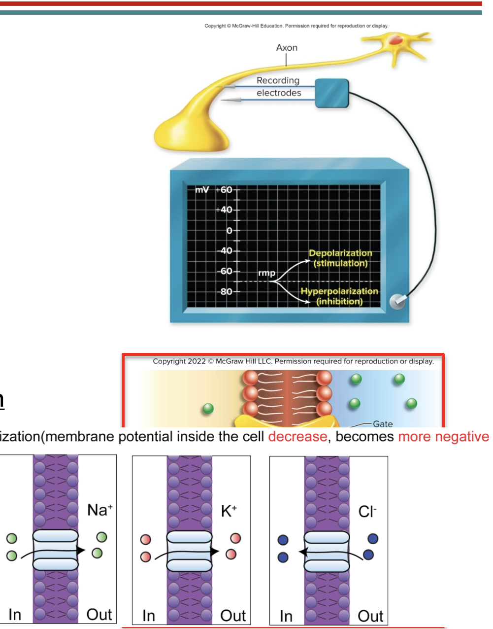
Electrical Activity in Neurons
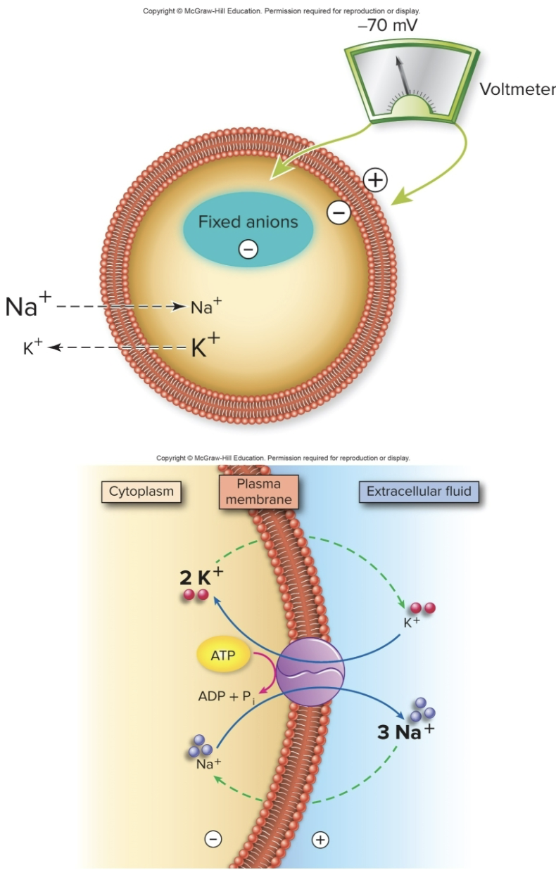
Resting Membrane Potential
Neurons have a resting potential of -70mV
why? bc of the imbalance of charged ions across the membrane, the inside of the resting neuron is negative relative to the outside
Mechanism responsible?
established by large negative molecules inside the cell
Na+/K+ pumps (moves 3 Na+ positive charges OUT and 2K+ in)
At Rest, we do have:
electrical gradient: more negative inside
concentration gradient: K+ inside the cell, Na+ outside the cell
Altering Membrane Potential
neurons and muscle cells can change their membrane potentials
excitability: the property of a neuron to produce electrical activity (change in membrane potential)
caused by changes in the permeability to certain ions
ions will follow their electrochemical gradient: combination of concentration gradient and attraction to opposite charges
ion currents: flow of ions which occur where ion channels are located
Two main types of electrical activity in neurons
Graded Potential
Action Potential
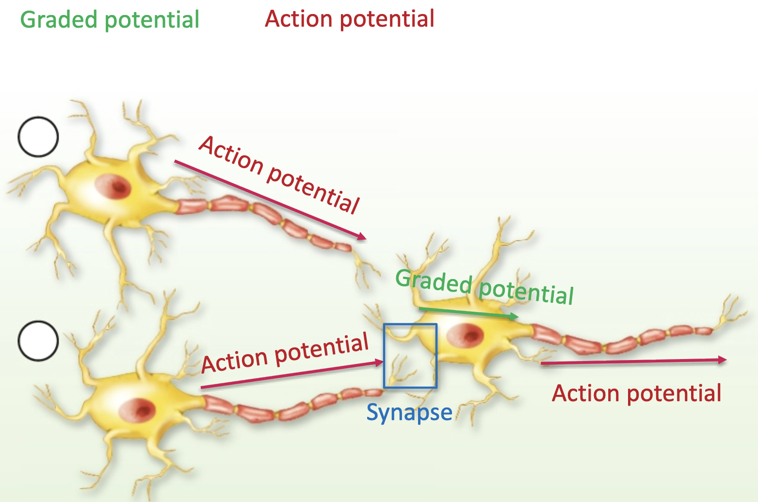
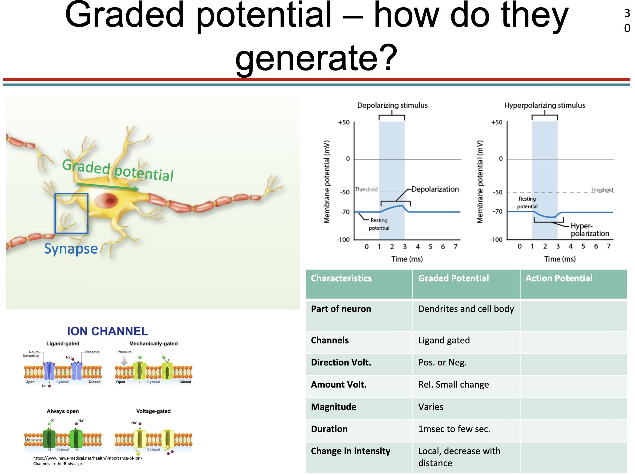
Graded Potential
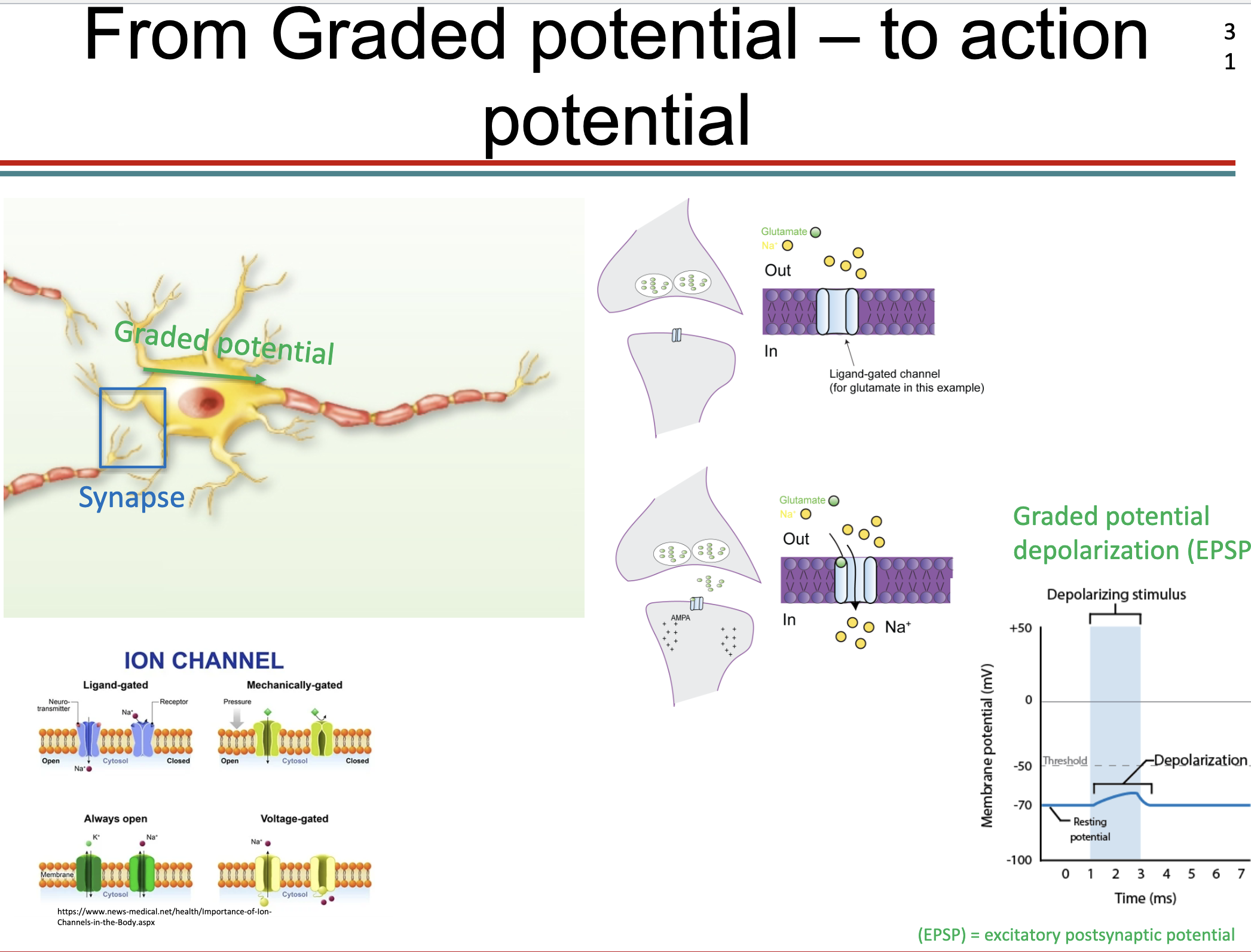
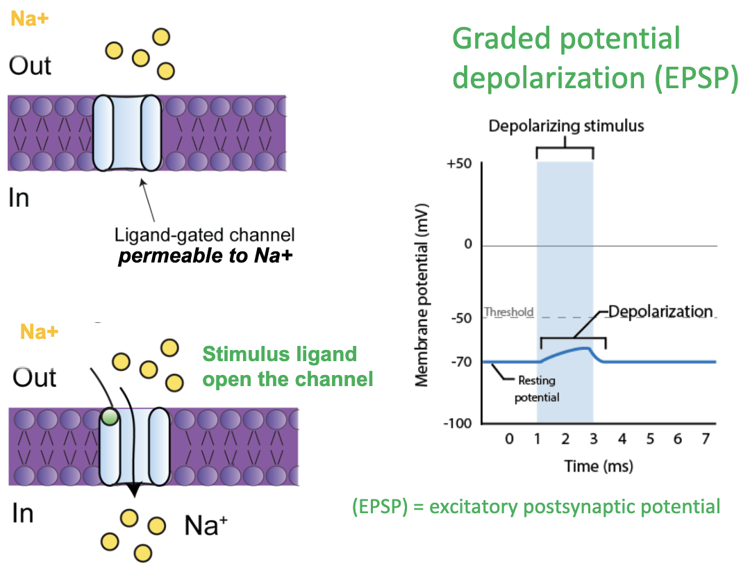
Graded Potential (ex: Depolarization)
Graded potential: is a small local change in the membrane voltage
when a “stimulus” (ex:ligand) reaches the neuron membrane certain channels (like Na) open, positively charged ions (sodium) flow into the cell
this makes the inside of the neuron slightly less negative (a small depolarization, also called excitatory postsynaptic potential (EPSP)
EPSPs are additive: if enough occur close together, they can bring the neuron to threshold for an action potential
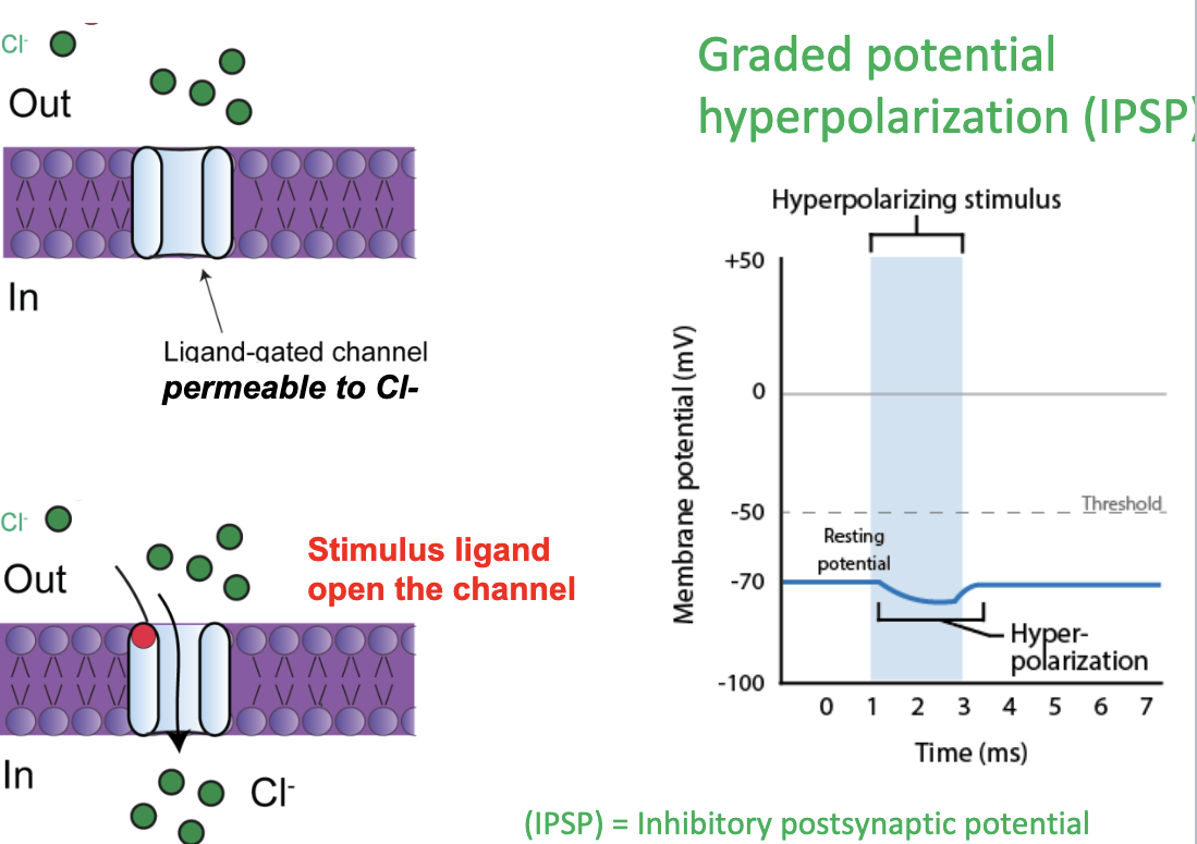
Graded Potential-How do they generate? (ex: Hyperpolarization)
Graded Potential: small local change in membrane voltage
when a stimulus (ligand) reaches the neuron membrane certain channels (Cl channel) open, negatively charged ions (Chloride) flow into the cell
making the inside of the neuron slightly more negative ( a small hyper polarization, also called inhibitory postsynaptic potential or IPSP)
IPSPs are additive- if enough occur close together, they can bring the neuron to FAR AWAY from the threshold, REDUCING the chance for an action potential
EPSP, IPSP
EPSPs are additive: if enough occur close together, they can bring the neuron to threshold for an action potential
IPSPs are additive: if enough occur close together, they can bring the neuron to FAR AWAY from the threshold, REDUCING the chance for an action potential
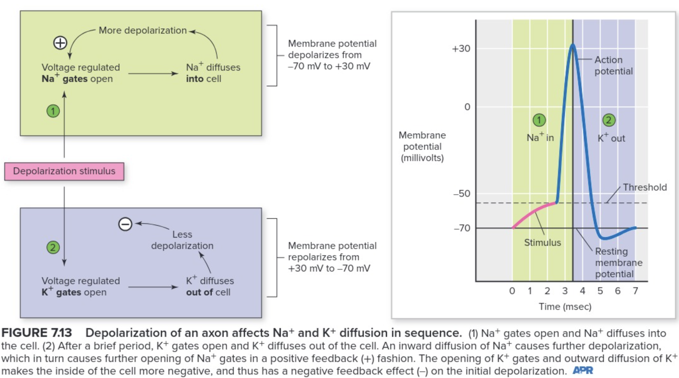
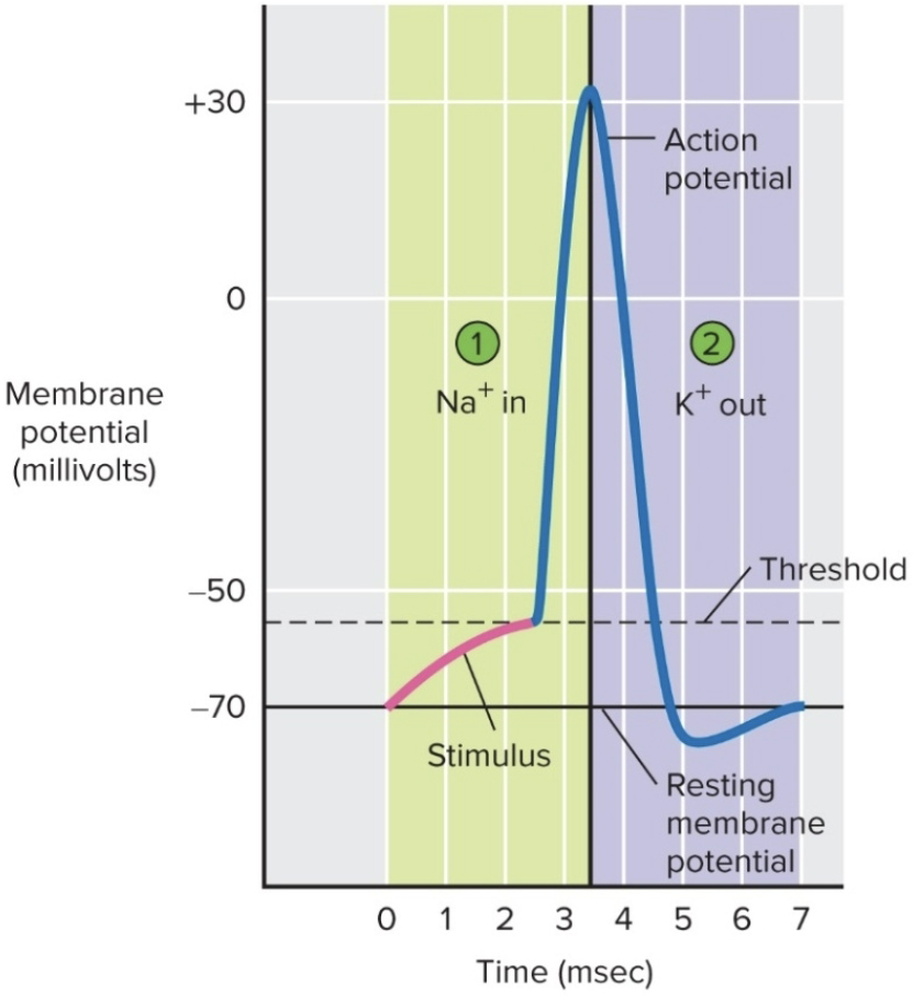
Action Potential Generation
Excitatory Post Synaptic Potential (EPSP): as graded stimulus, depolarize the membrane
if the depolarization reaches the threshold (-55mV) an action potential is generated
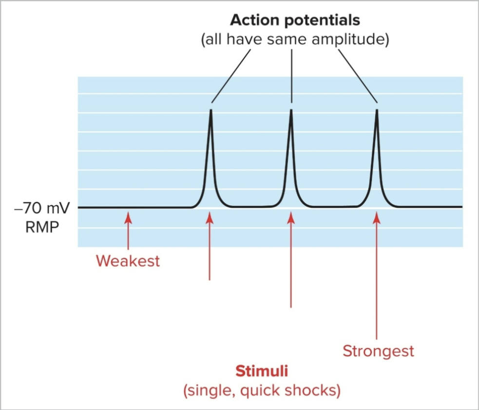
All or None Law:
once the threshold has been reached an action potential will happen
stimulus will not affect the size of the action potential; it will always reach +30mV
the size of the stimulus (EPSP) will not affect action potential duration
All or None Law
Once threshold has been reached, an action potential WILL happen
The size of the stimulus (EPSP) will NOT effect the size of the Action Potential; it will always reach +30mV
The size of the stimulus (EPSP) will NOT affect the Action potential duration
Generation of Action Potential Cont
changes in membrane potentials and the generation of action potentials are controlled by changes in the flow of ions (Na+ and K+) through channels
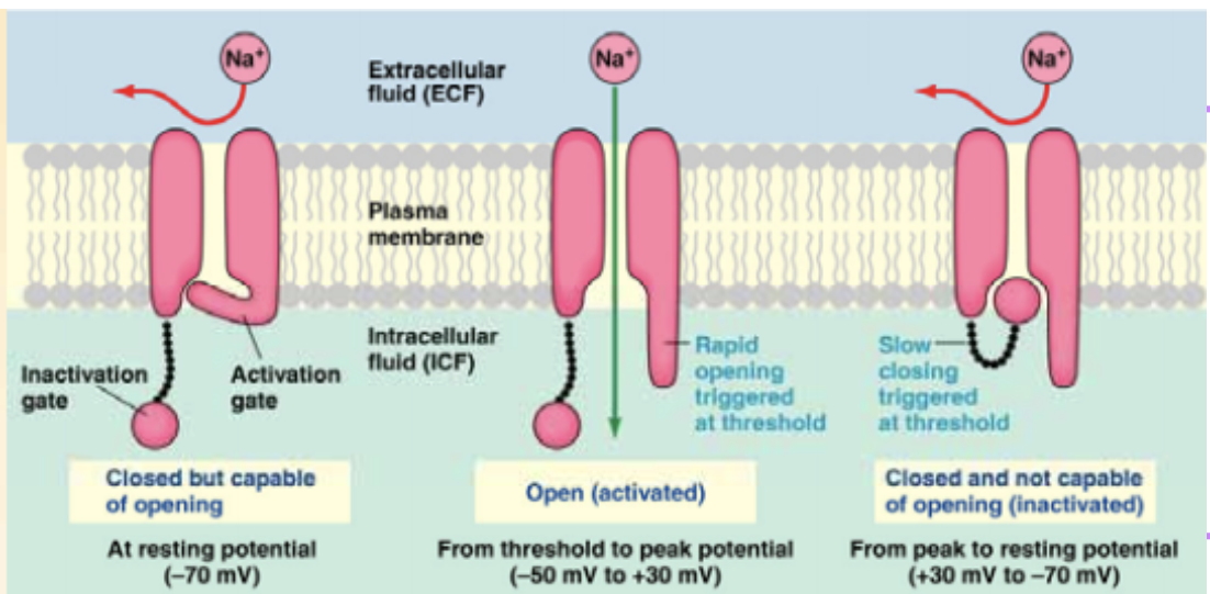
Na+ Channels
Na+ has voltage-gated channels that are closed at rest (they have an inactivation and an activation “gate”)
the membrane is less permeable to Na+ at rest, normally they open at -55mV (threshold)
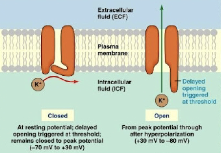
K+ Channels
has 2 types:
Not gated (always open): sometimes called K+ leakage channels
Voltage gated K+ channels: open when a particular membrane potential is reached; closed at resting potential (open at +30mV to -80mV)