Sacrum, Coccyx, and SI Joints
 Call Kai
 Learn
Learn Practice Test
 Spaced Repetition
 Match
Match Flashcards
 Knowt Play
1/129

There's no tags or description
Looks like no tags are added yet.
Name | Mastery | Learn | Test | Matching | Spaced | Call with Kai | Chat |
|---|
No analytics yet
Send a link to your students to track their progress
130 Terms
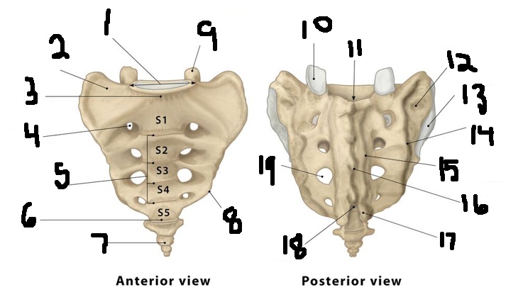
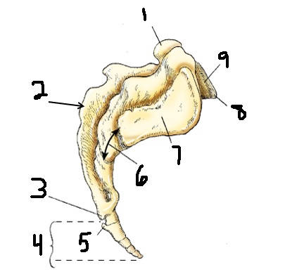
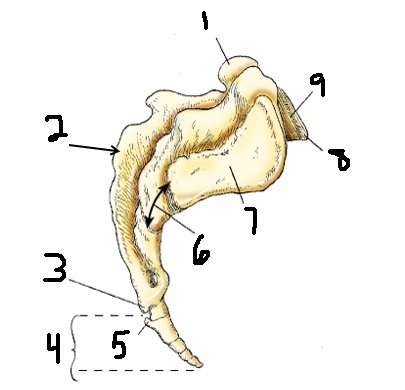
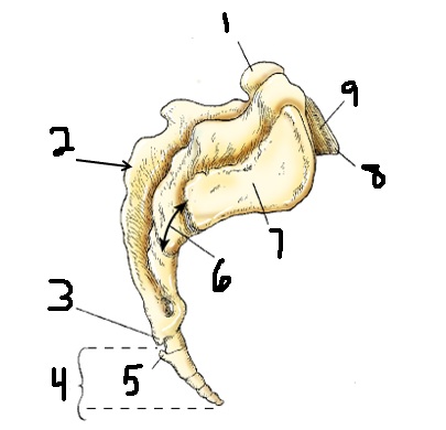
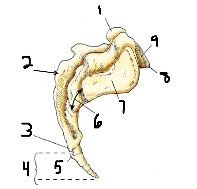
base
1
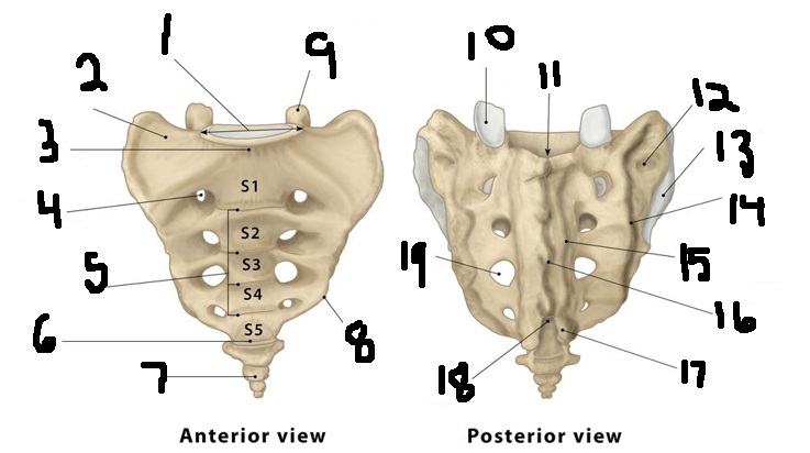
sacral ala
2
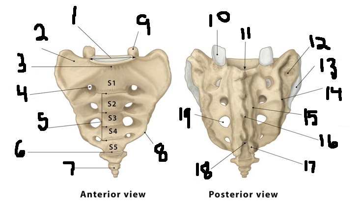
sacral promontory
3
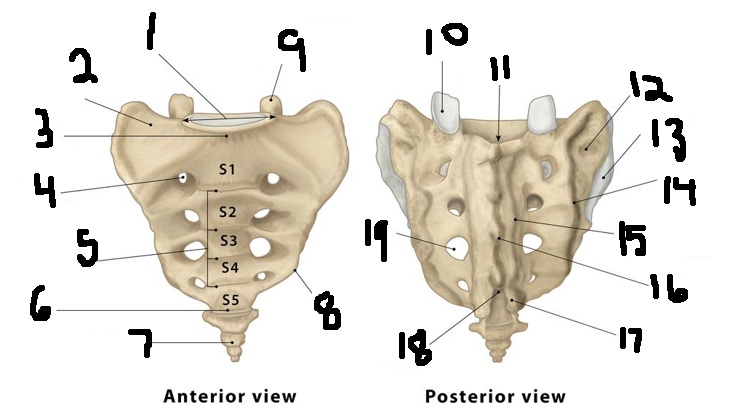
sacral foramen
4
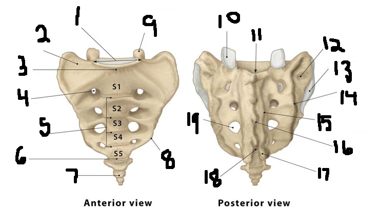
transverse lines
5
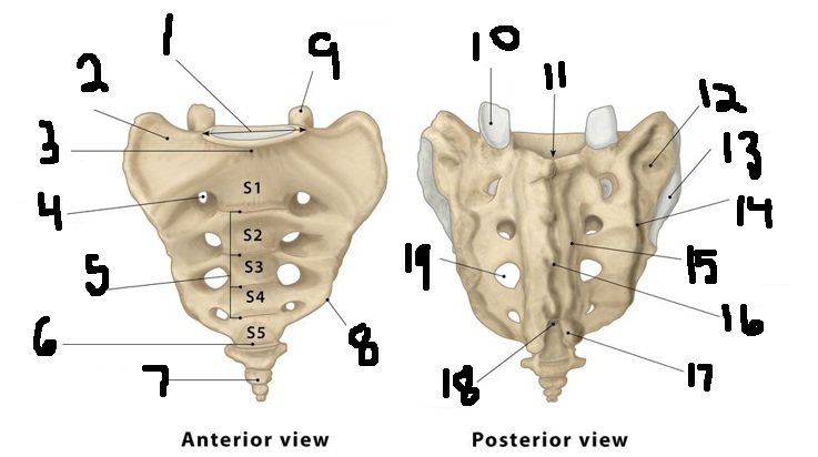
apex
6
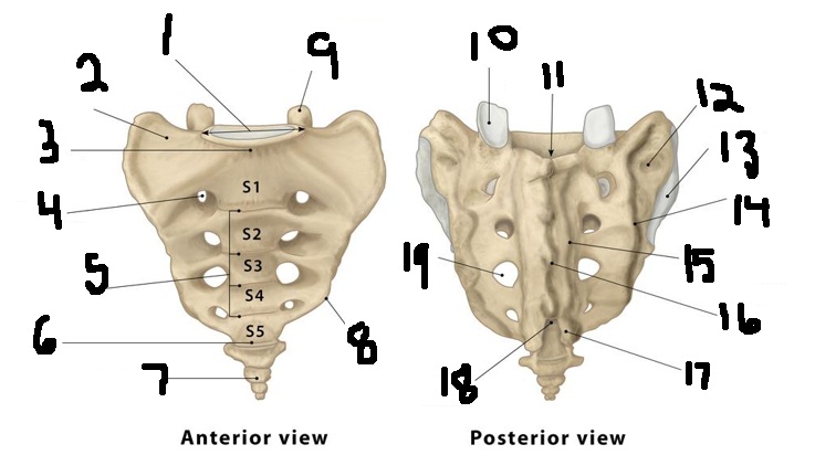
coccyx
7
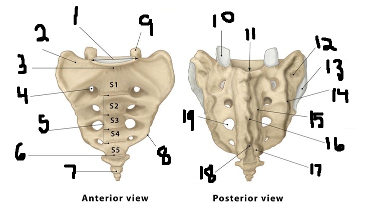
inferior lateral angle
8
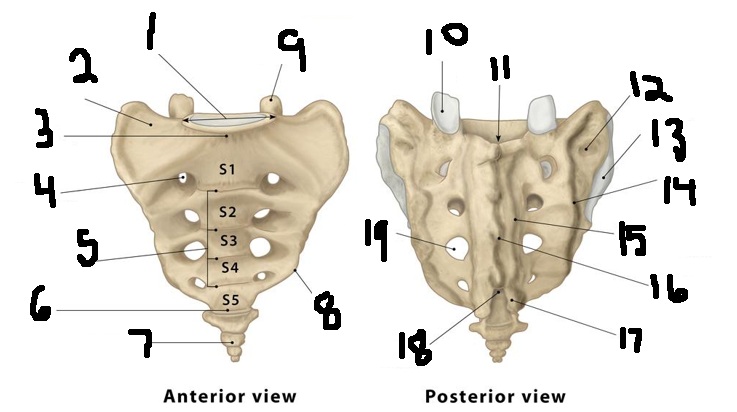
superior articular process
9
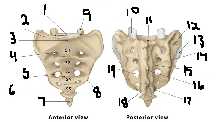
superior articular facet
10
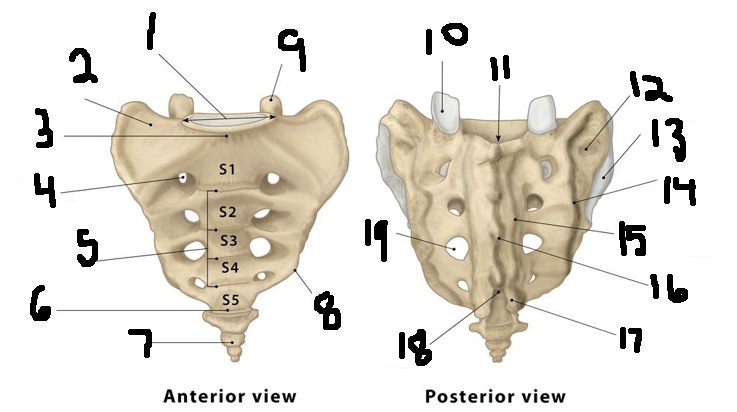
sacral canal
11
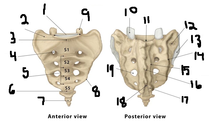
sacral tuberosity
12
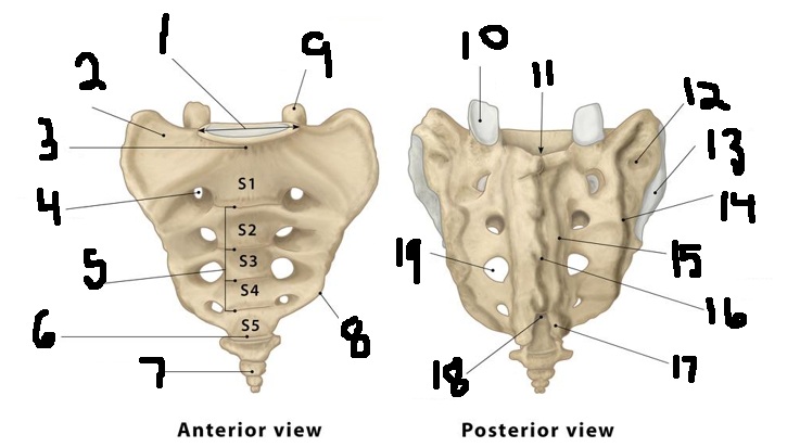
articular surface
13
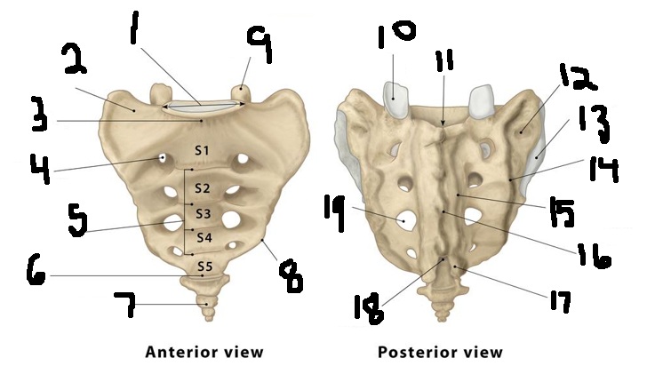
lateral sacral crest
14
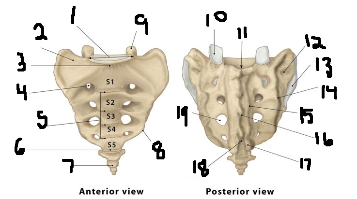
Intermediate sacral c
15
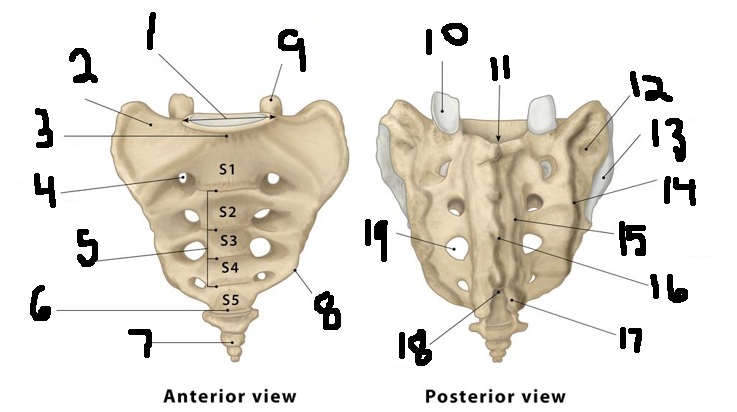
Median sacral crest
16
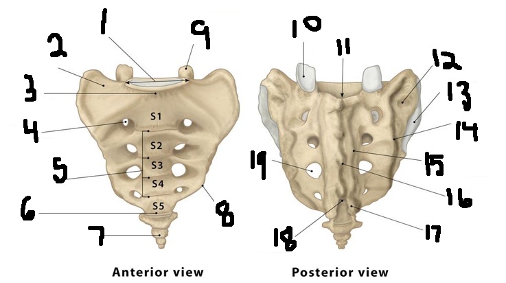
Sacral cornu
17
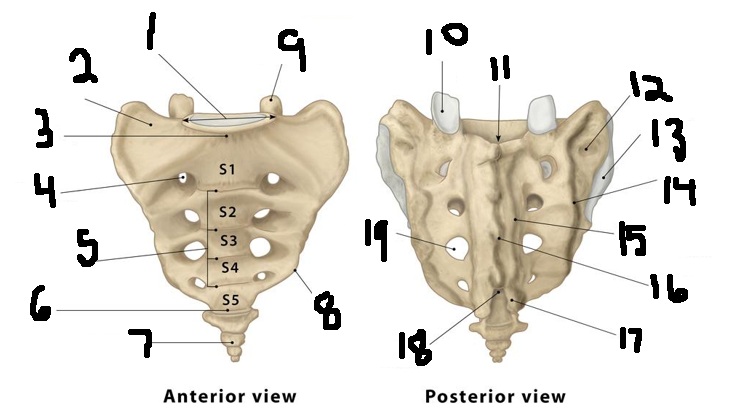
Sacral hiatus
18
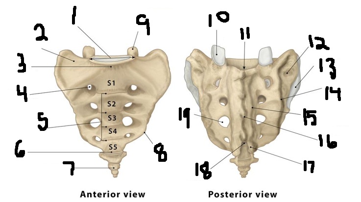
Sacral foramen
19
5
sacrum is made of how many vertebral segments?
sacral nerves and blood vessels
foramen carry what through sacrum?
superior articular process
1
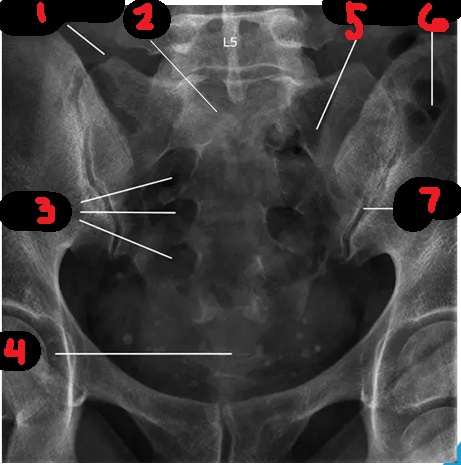
base
2
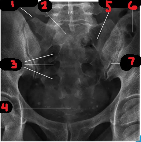
sacral foramina
3
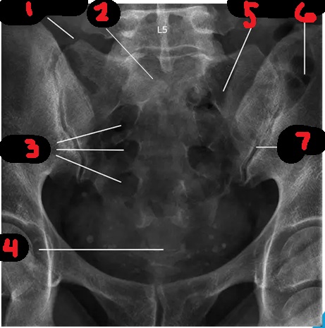
apex
4
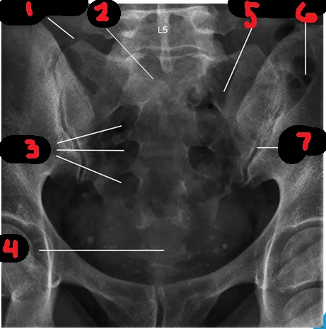
sacrum
5
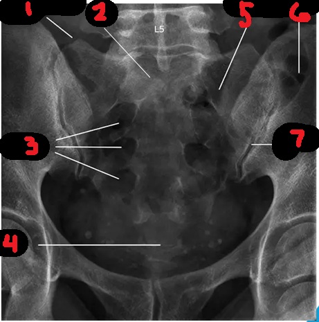
ilium
6
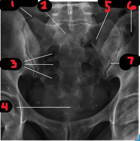
SI joint
7
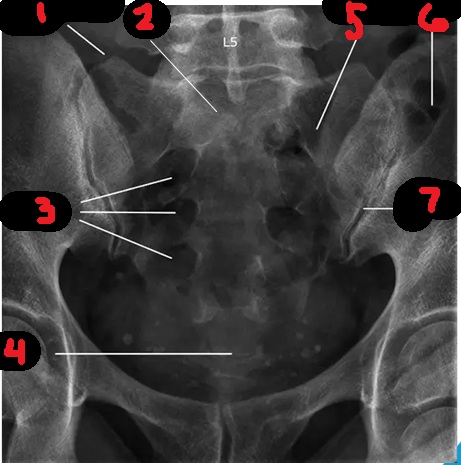
base
1
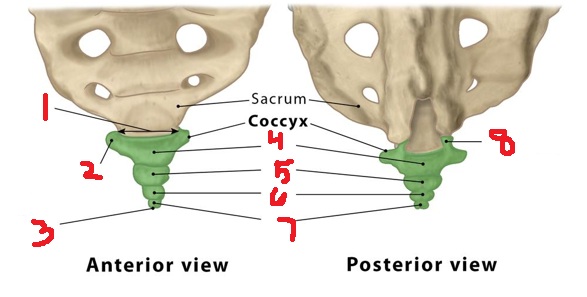
transverse process
2
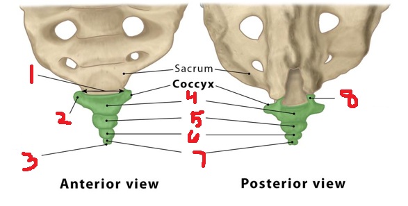
apex
3
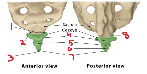
Co1
4
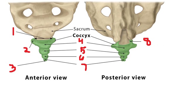
Co2
5
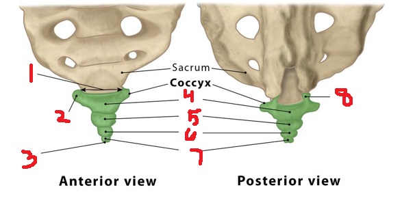
Co3
6
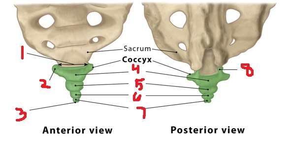
Co4
7
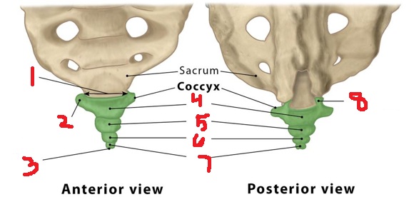
coccygeal cornua
8
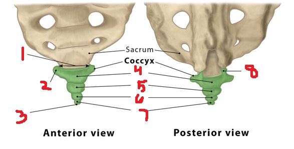
3-5
how many segments make up the coccyx?
coccyx
gives stabilization while sitting
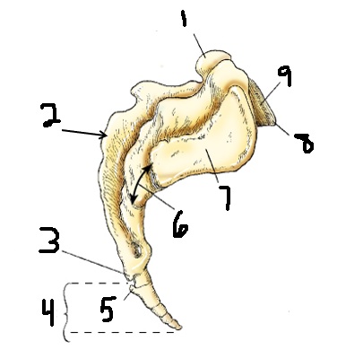
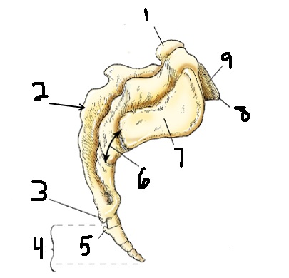
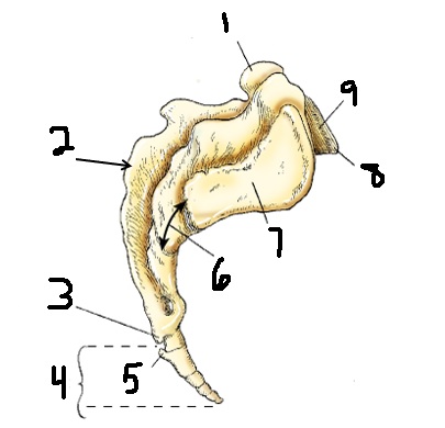
superior articular process
1
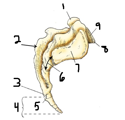
median sacral crest
2
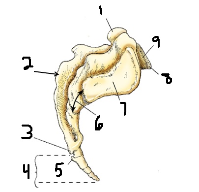
sacral cornu
3
coccyx
4
coccygeal cornu
5
sacrum
6
articular surface
7
promontory
8
base
9
sacral canal
1
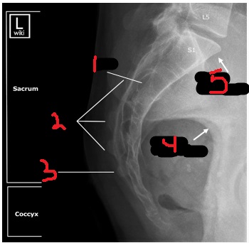
median sacral crest
2
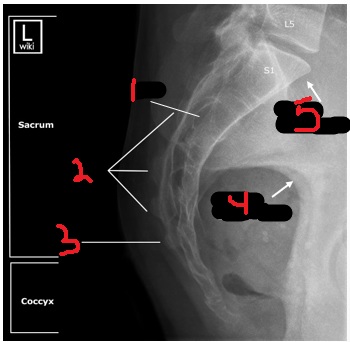
sacral horn
3
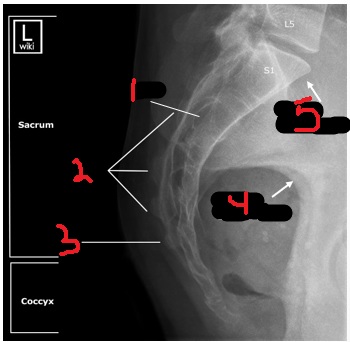
greater sciatic notch
4
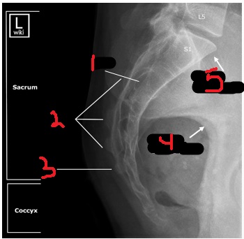
promontory
5
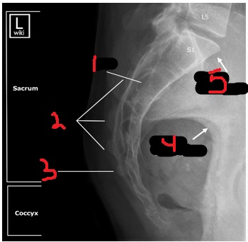
sacroiliac joints
junction of iliac ala and sacral ala
concave
sacral surface shape for SI joints
convex
iliac surface shape for SI joints
fibrous part of SI joint
1
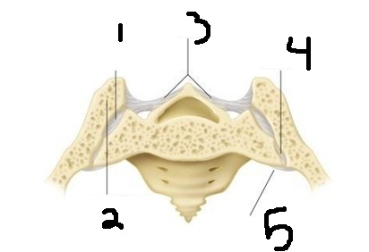
PSIS
2
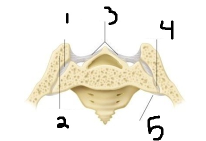
interosseous SI ligaments
3
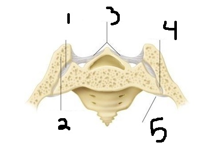
synovial part of SI joint
4
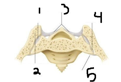
anterior SI ligament
5
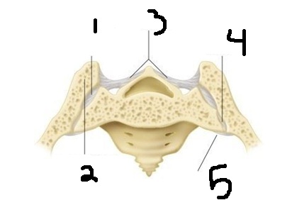
synovial amphiarthrodial joint
SI joint type
ligaments
superior portion of SI joints are connected by what?
synovial joint
inferior portion of SI joint is?
posteriorly and anteriorly
SI joint is angled?
40
S1 degree
25
S2 degree
-10
S3 degree
radiographic appearance of dislocated coccyx
transverse orientation or displacement of fracture fragment
dislocated coccyx
what pathology is this?
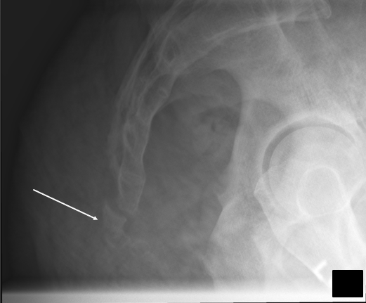
radiographic appearance of dislocated SI joint
misalignment of SI joints
dislocated SI joint
what pathology is this?
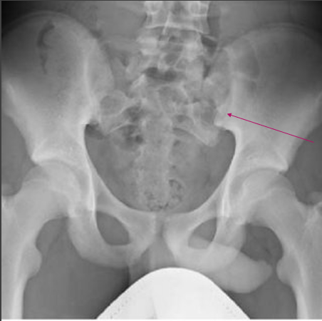
sacrum AP axial
Sacrum centered and seen in its entirety
Sacrum free of foreshortening, with the sacral curve straightened
Pubic bones not overlapping the sacrum
No rotation of the sacrum, as demonstrated by symmetric alae
repeat → no tube angle
critique sacrum AP axial
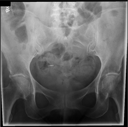
repeat → too much tube angle
apex of sacrum covered by pubic bone
critique sacrum AP axial
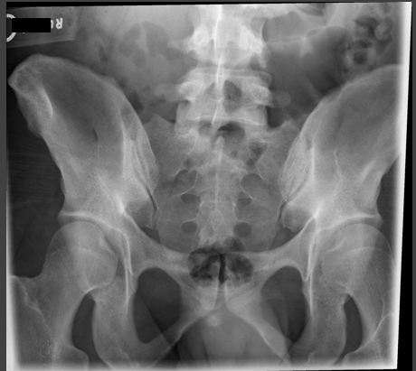
good
critique sacrum AP axial
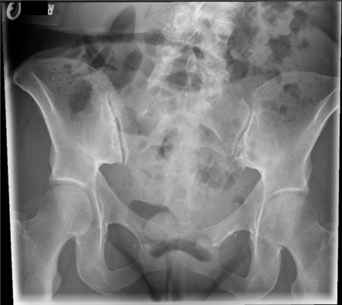
coccyx AP axial
Coccyx centered and seen in its entirety
Coccyx free of foreshortening, with the sacral curve straightened
Pubic bones not overlapping the Coccyx
No rotation of the Coccyx , as demonstrated by symmetric pelvis
good even though slight rotation
critique coccyx AP axial
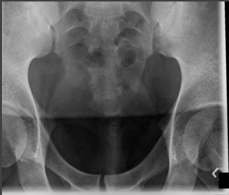
repeat → no tube angle
pubic bone over coccyx (pubic bone should look thin when there is an angle)
critique coccyx AP axial
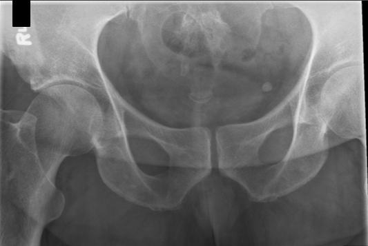
good
critique for coccyx AP axial
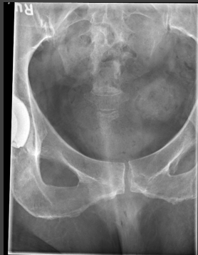
sacrum lateral
Sacrum
Closely superimposed posterior margins of the ischia and ilia, demonstrating no rotation
good
centering is too superior and anterior
critique for sacrum lateral
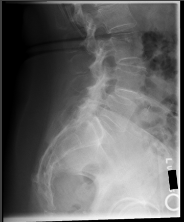
repeat → rotation
acetabulums are off, sacral canal not open
critique for sacrum lateral
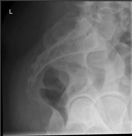
good
slight rotation
critique for sacrum lateral
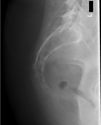
coccyx lateral
Coccyx
Closely superimposed posterior margins of the ischia and ilia, demonstrating no rotation
Bony trabecular detail and surrounding soft tissues
good
critique for coccyx lateral
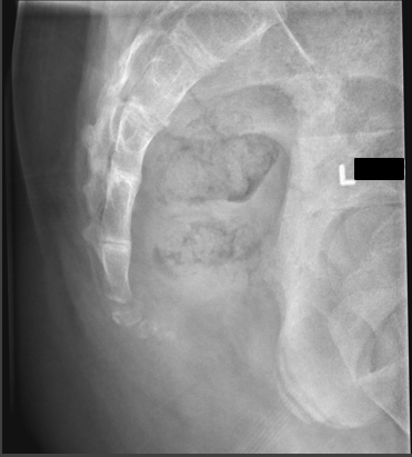
good
critique for coccyx lateral
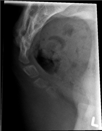
repeat → rotation
starting to superimpose coccyx
critique for coccyx lateral
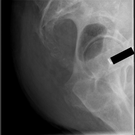
SI joints AP axial
Lumbosacral junction and sacrum
Open intervertebral disk space between L5 and S1
Both sacroiliac joints
Central Ray angled
30° cephalad angle for males
35° cephalad angle for females
repeat → too much angle
covering bottom of left SI joint
critique for SI joint AP axial
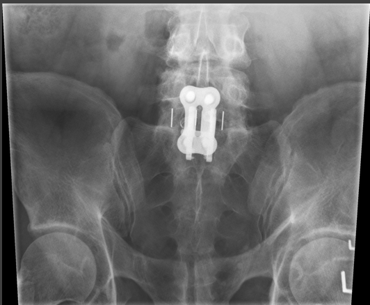
good
critique for SI joint AP axial
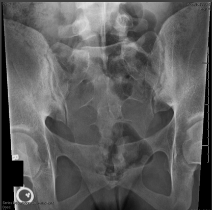
repeat → no tube angle
SI joints are seen but not elongated indicating no angle
critique for SI joint AP axial
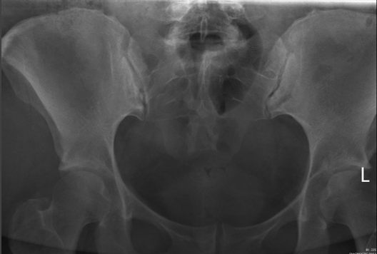
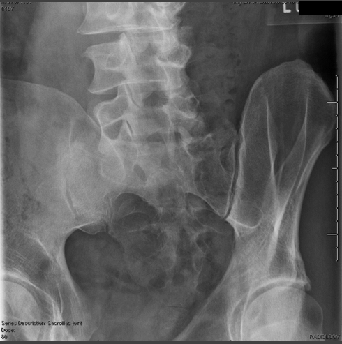
SI joint - RPO/ LPO
Open sacroiliac joint space furthest from the IR with minimal overlap of the ilium and sacrum
Joint centered on the radiograph
Appropriate right or left marker should be used to mark laterally on the elevated side.
elevated
what side do you mark for RPO/LPO SI joints?
good
critique for SI obliques
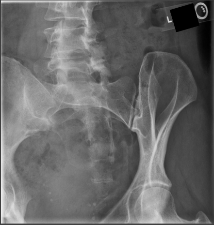
repeat → over rotated
joint closed/ covered
critique for SI obliques
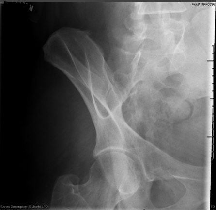
repeat → under rotated
joint covered by ala
critique for SI obliques
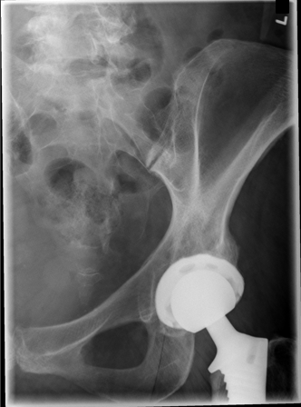
good
critique for SI obliques
90 kVp 14 mAs
tech for sacrum AP
center
cell for sacrum AP
10×12
collimation for sacrum AP