Medical microbiology module 1
 Call Kai
 Learn
Learn Practice Test
 Spaced Repetition
 Match
Match Flashcards
 Knowt Play
1/46

There's no tags or description
Looks like no tags are added yet.
Name | Mastery | Learn | Test | Matching | Spaced | Call with Kai | Chat |
|---|
No analytics yet
Send a link to your students to track their progress
47 Terms
Colonization
Bacteria are on surface of body and do not cause harm to host
Media used for Staphylococcus
Blood Agar, Chocolate agar, Thioglycollate broth
Mannitol Salt Agar or MRSA ChromAgar as additional primary plating media
Infection
Bacteria break through barriers and multiple in deep tissue leading to an immune response or inflammation
Sir Alex Ogston
1800’s Scottish surgeon
Discovered greek staphyle (bunch of grapes) and kokkos (berry)
Fredrich Julius Rosenbach
1884 German physician
differentiated the bacteria by colony color: S.aureus (latin aurum gold) and S. albus
(latin for white)
Pathogenic Staphylococcus
staphylococcus aureus
staphylococcus epidermidis
staphylococcus saprophyticus
Staphylococcus lugdunensis
Staphylococcus pseudintermedius
Rarely pathogenic Staphylococcus
staphylococcus haemolyticus
staphylococcus intermedius
staphylococcus simulans
Staphylococcus capitis
Staphylococcus hyicus
Staphylococcus saccharolyticus
Micrococcus
Other name: M. luteus
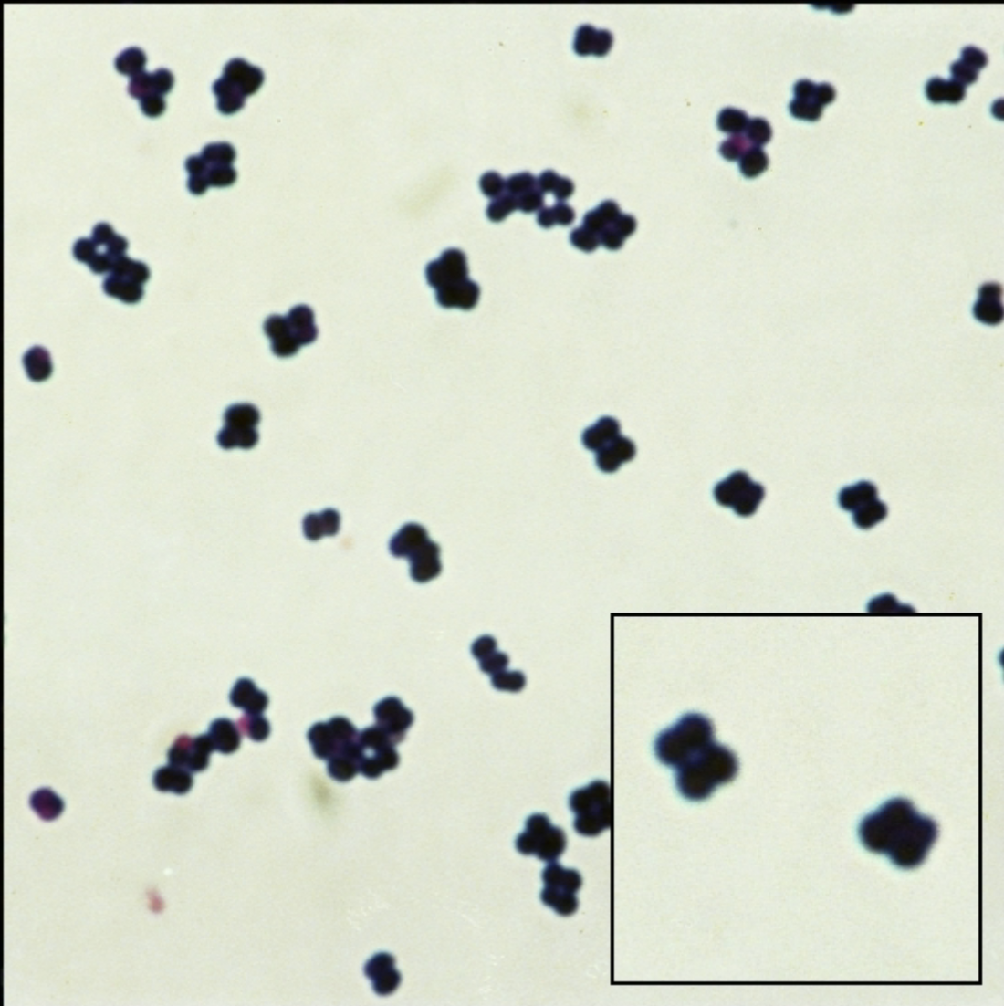
Gram stain: Gram Positive: clusters, single, tetrads
Catalase: positive
Coagulase: negative
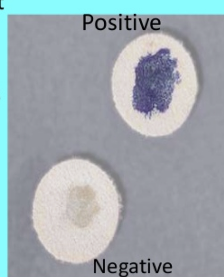
Test: modified oxidase is positive
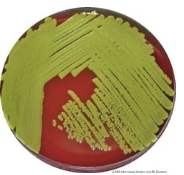
Micrococcus
Located: Normal skin flora
Appearance: Yellow pigment on BA
Associated with: abscesses, septic arthritis, endocarditis, pneumonia & meningitis
Other tests: Microdase (modified oxidase) it detects cytochrome enzyme
Micrococcus is blue (+) Staphylococcus has no color (-)
Micrococcus gram stain
GPC in pairs, tetrads, and no fermenting glucose
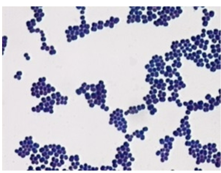
Staphylococcus aureus
Located: Nares, axillae, vagina, pharynx, and skin surfaces
Gram stain: Gram Positive Cocci
Morphology: yellowish white, Beta hemolytic
Virulence factors: enterotoxins, toxic shock syndrome (toxin-1), Exfoliative toxin, cytolytic toxins, Enzymes, lipase, hyaluronidase, and Pro A
Catalase: positive
Plate growth: Blood agar, Chocolate agar, Mannitol Salt Agar, MRSA chromagar as additional culturing media
Abscess
Caused by: Staphylococcus Aureus
pocket of infection / tender mass that forms at the site of injury
-Area surrounding is red, painful and swollen
– Skin warm to the touch
– Usually filled with pus
Cellulitis
Caused by: Staphylococcus Aureus
Usually results from a scrape or cut in the skin which allows bacteria to enter
– Can occur anywhere in the body, but most often on the legs or arms.
– Symptoms include redness, swelling, and pain at the site of infection
Folliculitis
Caused by: Staphylococcus Aureus
inflammation of hair follicles due to an
infection, injury, or irritation
Impetigo
AKA School sores
Caused by: Staphylococcus Aureus
highly contagious skin
infection forming pustules and yellow, crusty sores
-Very common in children
– Most common skin infection in Northern Europe, U.S.A &
Canada
– > 3million cases per year in U.S.
– First sign is a patch of red, itchy skin
– Sores rapidly burst -> leaving yellow, brown crus
Boils / Furuncles
Caused by: Staphylococcus Aureus
Deep folliculitis
-painful swollen area on skin
– an accumulation of pus and dead tissue
Carbuncles
Caused by: Staphylococcus Aureus
Collection of furuncles
Called Skin and soft tissue infections SSTIs
Scalded Skin Syndrome
Caused by: Staphylococcus Aureus
Caused by toxin
New borns & young healthy children under 5
Adults: renal disease (high mortality)
Profuse peeling of epidermal layer (2- 4 days)
Toxic Shock Syndrome (TSS)
Caused by: Staphylococcus Aureus
Caused by toxin - Toxin 1 (super antigen → strong immune response)
production of cytokines → cytotoxic
Rare sudden high fever, a rash (like a sunburn on palms and soles), dehydration, shock
Risk factors: skin wounds, surgery, and tampons
Food poisoning
Caused by: Staphylococcus Aureus
Caused by toxin - enterotoxin A or D → GI problems with fever 2-8 hrs after ingestion
Resolved in 24-48 hours
Cramping, diarrhea, Headache
come from: mayo, meats, poultry, creamed filled pastries, dairy
DIAGNOSED BY LOCAL PUBLIC HEALTH LABS NOT HOSPITAL LABS
Exfoliative Toxin or epidermolytic toxin
Caused by: Staphylococcus Aureus
(strain) produced by phage group II
causes scalded skin syndrome aka Ritter’s disease
Bullous impetigo (fluid filled blisters)
Osteomyelitis
Caused by: Staphylococcus Aureus
an infection and inflammation of the bone (usually caused by bacteria). Can occur from an infection in bloodstream or post surgery
wound → Blood → bones
Pneumonia
Caused by: Staphylococcus Aureus
a serious lung infection & 2nd to viral / high mortality rate
Bacteremia
Caused by: Staphylococcus Aureus
common and serious condition in hospitalized patients and IV drug users
Septic Arthritis
Caused by: Staphylococcus Aureus
painful infection in a joint. It can come from bacteria that travel through the bloodstream from another part of the body
Necrotizing fasciitis
Caused by: Staphylococcus Aureus
“flesh eating disease”
Staphylococcus aureus DISEASES
Abscess
Cellulitis
Folliculitis
impetigo
Boils/ furuncles
carbuncles
Caused by toxin:
scaled skin syndrome aka ritter’s disease
Toxic shock syndrome
Food poisoning
Exfoliative Toxin
Virulence Factors of Staphylococcus Aureus: Cytolytic Toxin
affects RBC & Leukocytes (leukocidins)
4 hemolysis: α, β, delta, and gamma
α hemolysin lyzes platelets, macroph → severe tissue damage
β hemolysin → hot / cold lysin
delta hemolysin: found in more staph but less toxic
Gamma hemolysin - kills wbcs aka Panton valentine leukocidin
Severe cutaneous infections, necrotizing pneumonia
Virulence Factors of Staphylococcus Aureus: Enzymes
Enzymes: coagulase, protease, hyaluronidase, lipase
hyaluronidase- hyaluronic acic of conenctive tissue
Lipase- act on lipids on skin surface
facilitate spread of infection to adjoining tissue
PROTEIN A- found in cell wall of Staphylococcus Aureus
binds Fc portion of IgG to block phagocytosis
CNS
Coagulase Negative Staphylococcus
do not produce the enzyme coagulase
Staphylococcus epidermidis
Staphylococcus saprophyticus
Staphylococcus haemolytic
Staphylococcus hominis
Staphylococcus capitis
CNS is C H H E S!
staphylococcus epidermis
Located: Normal skin flora & mucous membranes
Diseases caused: UTI’s and endocarditis (prosthetic valve)
Virulence factors: Slime, Biofilm, contaminate catheters, shunts, and prosthetic devices
staphylococcus epidermis
Morphology: white, smooth, creamy
Hemolysis: non hemolytic
number one cause of blood culture contamination

Staphylococcus lugdunensis
Antibiotics: pan susceptible (vulnerable to most antibiotics)
Tests: PYR on sterile body site , Ornithine positive, Rapid staph (may be positive)
Morphology: Colonial morphology not definitive + may resemble s. aureus and s. epidermidis
Diseases caused: causes SSTI and endocarditis
Other tests: Rapid panel ID or MALDI
More virulent than other CNS
Staphylococcus saprophyticus
Located: Skin, Mucous membranes (genitourinary tract), rare on mucous membrane
Antibiotics: Novobiocin resistant
Diseases caused: UTIs in young sexually active women
Morphology: smooth, creamy + may produce a light yellow pigment
Catalase: positive
Coagulase: negative
Catalase test
Tests for enzyme catalase
2H2O2 breaks into 2H2O + O2
Differentiates catalase (+) Staphylococcus & micrococcus from catalase negative streptococcus
(+) bubbles present & catalase
(-) absent bubbles
Coagulase test
3 methods: Slide test, Tube test, and several commercial brand (Bacti staph)
Differentiates: Staphylococcus aureus (+) & Catalase Negative Streptococcus
(+) Clumping (fibrinogen → fibrin)
(-) clear background
Selective media
Mannitol Salt Agar (MSA): high salt content
Selective and differential: Staphylococcus aureus from Catalase Negative Streptococcus
(+) Staphylococcus aureus ferment mannitol and turns plate yellow
(-) Staphylococcus epidermidis remains pink
Antimicrobial susceptibility
Staphylococcus aureus is methicillin susceptible
Penicillinase resistance: Resistant to all beta lactam antibiotics (Methicillin, Penicillin, nafcillin, oxacillin, and amoxicillin)
Ex.) Methicillin Resistant Staphylococci Aureus is detected with the “Spectra” plate
(+) MRSA appears blue
MRSA & MRSE use vancomycin combined with rifampin or gentamicin
Expensive: Biomerieux Vitek 2, Microscan Walk-Away, BD Phoenix
MRSA
Methicillin Resistant Staphylococci Aureus
Located: colonizes patients nasal, axilla, and groin
Morphology: Weakly beta hemolytic or Non hemolytic
Antibiotics: Resistant to certain, commonly used antibiotics
Treated with vancomycin IV
Other tests: Diff / selective media (MRSA chromagar) 24 hrs + PCR test 1 hr
Cepheid GeneXpert (polymerase chain reaction)
HAI (Hospital acquired infection)
Infections that patients get while or soon after receiving health care
Causes: unclean hands, improper use or reuse of equipment between health care workers and patients
48 hours or more after patient is admitted
CAI (community acquired infection)
Infection contracted outside of a healthcare setting or an infection present on admission
1st case: Boston 1999
MRSA prevention
Regular handwashing
cover cuts and scrapes with a bandage until healed
Avoid contact with other people’s wounds / bandages
Avoid sharing personal items
Keep skin healthy and moisturized
Shower after sports
MSSA
Methicillin Susceptible Staphylococci Aureus
Susceptible to commonly used antibiotics
Treat with cefazolin IV
Resistance & Surveillance
mecA gene encodes for new penicillin binding protein
Passive: Isolate infected patients who are infected / colonized
Active: Screen patient before surgery to temporarily eradicate pathogen before it causes infection
Patient care impact
Patients can get hospital acquired MRSA → longer visit + more pain
Hospital acquired infection is 8th leading death in USA
Financial impact
MRSA cost $$$
long treatment + more equipment used (IV)
Drugs are expensive
Biofilm
Community of microbes attached to a solid surface
Ex.) plaque, biofilms on catheters
particularly resistant to antibiotics