Week 1 & 2 Integ & Lymph
 Call Kai
 Learn
Learn Practice Test
 Spaced Repetition
 Match
Match Flashcards
 Knowt Play
1/81

There's no tags or description
Looks like no tags are added yet.
Name | Mastery | Learn | Test | Matching | Spaced | Call with Kai | Chat |
|---|
No analytics yet
Send a link to your students to track their progress
82 Terms
What are the primary phases of wound healing?
1. Hemostasis
2. Inflammatory
3. Proliferative
4. Remodeling
________: first phase of wound healing; involves immediate vasoconstriction + formation of a platelet clot = stop bleeding; main cell type = platelets
Hemostasis
_______: second phase of wound healing that can be broken down into 3 subphases (killing and containing invaders, wound debridement, neo-angiogenesis); main cell types = neutrophils, monocytes & macrophages
Inflammatory
_______: third phase of wound healing that involves new tissue (made of collagen) formation as the wound contracts; main cell types = monocytes & macrophages, fibroblasts
Proliferative
_______: fourth and final phase of wound healing that involves organization of different variations of collagen; main cell types = myofibroblasts, lymphocytes
Remodeling (some texts also consider this the maturation phase)
Describe the maturation of tissue healing. What's the maximum tensile strength the tissue obtains?
Skin is only able to obtain 80% of the original tensile strength
How is scar tissue different from original tissue?
Primarily composed of collagen fibers in a disorganized and parallel fashion, lacks elastin, may appear raised, less strong, less resistant to UV damage, more susceptible to infection
What are the three primary types of wound closure?
1. Primary intention
2. Secondary intention
3. Tertiary intention
_______: type of wound closure that occurs immediately. Clean incised wound/ surgical wound. Wound closed by suturing, skin grafting, or flap closure.
Primary intention
_______: type of wound closure that involves leaving the wound open to heal from the inside-out. Highly contaminated wound or tissue loss. Closed by reepithelialization.
Secondary intention
_______: type of wound closure that involves delayed closure. Contaminated wound --> treated by repeated debridement --> wound is closed by suturing skin grafting or flap.
Tertiary intention (also called delayed primary closure)
Describe the primary general factors affecting wound healing.
Local factors (at the wound site): oxygenation & blood supply, infection/contamination, moisture balance, edema/pressure, foreign bodies/necrotic tissue, wound size & location
Systemic factors (whole body): age, nutrition, comorbidities, medications, immune status, lifestyle factors, systemic infection/illness
In summary: adequate blood flow, oxygen, nutrition, infection control, and balanced inflammation are KEY to wound healing.
Describe the primary local factors (intrinsic) affecting wound healing.
Edema, hx of injury, hypoxia (pressure, limited blood flow, etc.)
Describe the primary systemic factors (intrinsic) affecting wound healing.
Age, nutritional status, arterial disease, smoking, medications (steroids, chemo, overused anti-inflammatories), obesity, stress
Describe the extrinsic factors affecting wound healing.
Treatment, foreign bodies, infection, mechanical pressure, too much moisture
List pertinent lab values: complete blood count (CBC)
WBC 3.8-11.8 K/ul (infection), hemoglobin 11.7-15.3 g/dl (oxygen), RBC count 3.63-4.92 M/ul (oxygen), hematocrit 34.1-43.9% (oxygen), platelet count 150-450 K/ul (clotting)
List pertinent lab values: basic (BMP) or comprehensive (CMP) metabolic panel
Glucose 70-100 mg/dL (blood sugar), creatinine 0.51-0.95 mg/dL (kidney fx, protein metabolism, edema), urea nitrogen 7-22 mg/dL (hydration status), albumin 3.4-5.0 gm/dL (protein), total protein 6.4-8.2 gm/dL
What is a normal HgbA1C value? What does it indicate?
What is the difference b/w ESR and CRP?
CRP is a more specific marker for acute inflammation, rises rapidly within hours of an inflammatory stimulus, and is preferred for detecting ACUTE inflammation
ESR is a non-specific indicator, takes longer to show changes and peaks after a day or more, often used as a screen test for CHRONIC inflammation
Why are prothrombin time (PTT), international normalized ratio (INR), and platelet count important laboratory values?
Each indicate coagulation profiles and clotthing abilities
What is the difference b/w aerobic and anaerobic wound cultures?
Aerobic = creature needs oxygen, anaerobic = creature does NOT need oxygen
What is the best collection method for a wound culture?
Levine technique
1. Cleanse the wound w/ sterile saline (avoid antiseptic use)
2. Locate an area within the wound which has health tissue (red)
3. Rotate the cotton-tipped applicator, stay within 1 cm2 area
4. During the rotation, press into the wound bed to extract tissue
What are the key components of the history for a patient with a wound?
Subjective history, past medical history, past surgical history, social history
What questions within the history are different for a patient with a wound?
Remember the "OLD CARTS" acronym for onset, location, duration, characteristics, aggs, relieving factors, treatment, and severity
Can you describe the rationale for each of the questions asked within the history?
Onset: MOI? Insidious/gradual vs. traumatic condition?
Location: where is it?
Duration: how long has the patient had this wound? which stage of healing SHOULD it be in?
Characteristics: what type of wound is it?
Aggs: what is bothersome?
Relieving factors: what is easing?
Treatment: what has the patient already done?
Severity: how bad is it?
How are the tests and measures similar or different for a patient with a wound?
General assessment consists of skin (ABCDE's, color, turgor, temperature, drainage, palpation, texture, moisture), edema, and wound analysis
How do you grade edema of the skin?
0 = 0 mm deep
1+ = 2 mm (immediate rebound effect)
2+ = 4 mm (10-15 seconds to rebound)
3+ = 6 mm (>1 min to rebound)
4+ = 8 mm (2-5 min to rebound)
What specific tests and measures are used to document a wound assessment?
Location, tissue type within the wound bed, drainage, odor, dimensions (size/depth), wound edges, periwound skin
Explain in further detail what it means to assess the location of a wound(s).
Apply correct terminology to describe location (medial/lateral, left/right, proximal/distal, etc.), utilize a body chart, take photos, assign numbers to multiple wounds
________: overgrowth; exuberant granulation tissue. Granulation tissue is protruding past the epithelial ridge.
Hypergranulation tissue (note: precursor to keloid scar; may be related to excess moisture or bacteria)

________: appearance of a wound (color) that may indicate poor blood supply or nutritional status
Pale pink
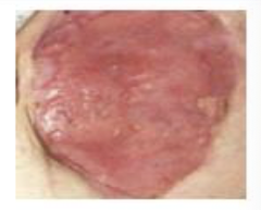
________: denatured protein, fiber strands, and dead skin cells that naturally collect in the wound. May appear yellow, white, or gray in color.
Slough (note: precursor to scabbing over; similar to the appearance of skin when getting out of a bath)
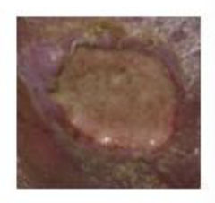
________: appearance of a wound (structure) that involves exposure of healthy ________ that are shiny and white
Tendon exposure

________: collection of dead tissue within the wound bed. It is important to differentiate this from a scab, which is dried blood over the wound.
Eschar (REALLY thick, leathery scab that affects the healing process)
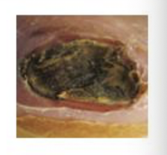
________: appearance of a wound (structure) that involves exposure of pink to dark red, highly vascularized, striated (striped, grooved, or ridged) tissue
Muscle

________: new connective tissue and microscopic blood vessels that form on the surfaces of a wound during the healing process. It typically grows from the base of wounds. Appears beefy and red.
Granulation tissue ("where's the beef?")

________: area of redness that persists (does not blanch/become pale) when pressure is applied to the area
Non-blanchable erythema
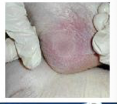
________: reddened area that turns pale when light pressure is applied
Blanchable erythema

Explain in further detail what it means to assess the drainage of a wound(s).
Type, color, consistency, amount (none, scant, small, moderate, large, copious)
What are the types of drainage and how would you document appropriately within the medical record?
Serous, sanguineous, serosanguinous, purulent -- document each based on type, color, consistency, and amount
________: type of drainage; contains protein rich fluids w/ WBCs; clear-pale yellow, watery
Serous (helps w/ healing)
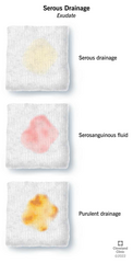
________: type of drainage; contains blood or drying blood; red-dark brown, consistency of blood or slightly thickened water
Sanguineous
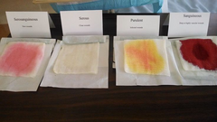
________: type of drainage; contains RBCs within the serum; light red or pink (pale-yellow in some cases), consistency is thin and watery
Serosanguinous (mix of serous and sanguineous -- it is the MOST common)

________: type of drainage; indicator of infection; white-pale yellow, viscous or creamy consistency (note: could also be green in some cases)
Purulent (pus)

Explain in further detail what it means to assess the odor of a wound(s).
Assess AFTER cleaning to avoid confusion w/ dressing or exudate --> document presence or absence of odor, description of odor quality, changes over time or w/ treatment
________: type of odor; may indicate infection
Foul
________: type of odor; may be associated w/ specific bacteria (e.g., pseudomonas)
Sweet or fruity
________: type of odor; may suggest proteolytic activity
Ammonia-like
Explain in further detail what it means to assess the dimensions of a wound(s).
Multiple methods available to determine the SIZE of the wound
Can you describe how to measure a wound using the perpendicular (ruler) technique?
Length (longest dimension cephalad to caudal), width (perpendicular to length at the wound's widest dimension), depth (deepest point - use a sterile Q-tip)
**NOTE: undermining always measured using clock positions**

Can you describe how to measure a wound using the clock method (note: this is the MOST common method of measuring dimensions)?
12-o'clock ALWAYS the area of wound closest to head; length (12-6), width (9-3), depth (various clock positions, 2, 4, 8, 10, or others)
**NOTE: undermining always measured using clock positions**

________ (special measurement consideration): tissue under wound edge is eroded, similar to cave under skin
Undermining

How is undermining best measured and documented?
Clock method --> "undermining of 4 cm from 10-12 o'clock"

________: narrow passageway, tube like extension of wound, no exit
Sinus tract (ex: tract at 5 o'clock 7 cm)
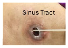
________: entrance and exit, truly a ________
Tunnel (document location and length)

What is the difference between undermining, tracts and tunnels? Are they a positive or negative prognostic healing factor and why?
Undermining is much more superficial to the surface than sinus tracts or tunnels; tracts do NOT have an exit vs. tunnels DO have an exit
**note: they are negative prognostic healing factors because they indicate tissue destruction and separation (undermining), infection or abscess extending deep into the wound (sinus tract), and deep tissue involvement and loss of tissue continuity (tunnel)**
Explain in further detail what it means to assess the wound edges of a wound(s).
Determine well defined (demarcated) or diffuse, attached or detached to wound base (i.e., undermining), raised (hypergranulation) or flush to wound bed, rolled (epibole), callus (hyperkeratosis), evidence of epithelialization
What is the difference b/w a rolled edge and callus?
Rolled edge is vascularized and WILL bleed vs. callus consists of epithelial tissue and will NOT bleed
What characteristics of wound edges offer insight into slow or delayed healing?
In general, the following characteristics indicate problematic/delayed healing:
- appearance: rolled, undermined, irregular
- color: pale/white (ischemic), black/brown (necrotic), yellow/gray (slough), red/inflamed
- texture: hard, thickened (callus), macerated (wet, white), indurated (firm, stiff)
- progressing: non-advancing
- moisture: excessively wet
ID and describe the primary characteristics of epibole vs. no epibole.
Rolled (epibole) ("lip-like") indicates that epithelial cells have stopped migrating = chronicity; elevated above wound bed; pale, shiny, or white (epithelial cells stopped migrating); firm, sometimes calloused; common in pressure injuries, diabetic ulcers, venous ulcers
No epibole indicates that epithelial cells are migrating normally to close the wound; flat, thin, continuous w/ surrounding skin; pink or red; soft, fragile, moist
**ROLLED = BAD**
ID and describe the primary characteristics of well-defined vs. diffuse wound edges.
Diffuse edges are poorly demarcated and blend gradually into periwound tissue (even in terms of coloration (redness, inflammation, maceration)); common w/ venous ulcers, pressure injuries, inflammatory or infected wounds; suggest ongoing inflammation, infection, edema, or moisture imbalance
Well-defined edges are clear w/ sharp margins in a round, oval, or straight shape; abrupt transition b/w wound bed and intact skin, making color distinct (pink, red, or dark); common w/ arterial ulcers, surgical/incisional wounds, neuropathic ulcers; suggest more localized pathology (may be ischemic and slow to heal)
*DIFFUSE = BAD*
ID and describe the primary characteristics of attached vs. detached wound edges.
Detached (undermining) means the edge is separated from wound bed; may feel loose, rolled, or firm above an empty pocket; indicates stalled healing, as epithelial cells cannot bridge wound effectively; common w/ pressure injuries, infection, chronic ulcers
Attached means the edge is flush w/ the wound bed and anchored to granulation tissue; contour is flat or gently sloping; texture is soft, fragile, moist; indicates good epithelial migration and wound progression
**DETACHED = BAD**
ID and describe the primary characteristics of raised vs. flushed wound edges.
Raised means the edge is elevated above the wound bed; may be firm, fibrotic, or rolled; can be pale/while, dark, or hyperpigmented; often indicates stalled epithelial migration in chronic ulcers, hypertrophic scarring, or excessive pressure
Flushed means the edge is level w/ the wound bed and continuous w/ granulation tissue; soft, fragile, moist; pink or red; suggests active epithelial advancement
**RAISED = BAD**
ID and describe the primary characteristics of callus vs. no callus wound edges.
Callus is thickened, yellowish, hard rim; dry, rough, firm w/ a yellow, tan, or brown color; acts as a physical barrier to prevent epithelial migration, trap bacteria, and increase pressure; common w/ diabetic foot ulcers, neuropathic plantar ulcers, and areas of repeated friction/pressure
No callus is thin, soft, and continuous; fragile, pliable, moist w/ a pink or red color; allows epithelial cells to migrate; common w/ surgical wounds, venous ulcers, and traumatic wounds
**CALLUS = BAD**
Explain in further detail what it means to assess the periwound skin of a wound(s).
Inspect skin surrounding the wound (4 cm margin), assess color (erythema, pallor, bruising, discoloration), evaluate texture (induration (hardness), softness, bogginess), check moisture level (dryness, maceration (overhydration), desiccation), look for signs of infection (warmth, swelling, pain, or purulent drainage), note presence of callus, scaling, or peeling
What could an induration indicate vs. softness or bogginess?
Induration ("woody") = lymphedema, cellulitis
Softness or bogginess = fluctuating wound, abscess w/ fluid
________: technique that reduces the # of microorganisms to minimize risk of infection; used for chronic wounds (e.g., venous ulcers, pressure injuries, home care, and OP settings)
Clean technique
Describe the environment, supplies, hand hygiene, and cost & efficiency for the clean technique.
Clean, non-sterile setting; clean gloves + dressing packs; hand hygiene before and after care; most cost-effective & faster
________: technique that eliminates ALL microorganisms to maintain a sterile field; used for surgical wounds, acute or invasive procedures, and wounds in immunocompromised patients
Aseptic technique
Describe the environment, supplies, hand hygiene, and cost & efficiency for the aseptic technique.
Sterile field maintained t/o procedure; sterile gloves + instruments + dressing; hand hygiene performed before sterile gloving w/ strict protocol; more resource-intensive and time-consuming
Describe the steps of the clean technique.
1. Prepare the area
2. Hand hygiene
3. Glove up (non-sterile)
4. Remove old dressing
5. Cleanse the wound
6. Re-glove if needed
7. Apply new dressing
8. Dispose of waste
9. Final hand hygiene
Why does maintaining a clean field matter?
Prevents intro of pathogens into the wound, protects immunocompromised and high-risk patients, supports proper wound healing and reduces complications
What are clean field basics? (hint: 5x)
Perform hand hygiene before and after, use clean gloves for setup and wound cleaning, place a clean barrier (e.g., sterile drape or blue pad) on surface, keep all supplies on clean barrier, open dressings carefully and avoid touching inner surfaces
________ (do vs. don't) change gloves if they become contaminated
DO
________ (do vs. don't) discard used gauze and tools immediately
DO!!
________ (do vs. don't) return used gauze or tools to clean field
DON'T!!
________ (do vs. don't) touch clean supplies w/ used gloves
DON'T!!
Describe the steps of the aseptic technique.
1. Prepare the area
2. Hand hygiene
3. Remove old dressing (if necessary, remove w/ clean gloves first --> re-perform hand hygiene and don new sterile gloves)
4. Open supplies
5. Apply sterile gloves
6. Perform wound care or procedure
7. Apply new dressing
8. Dispose of waste
9. Final hand hygiene
What are the key components of a patient history for a basic vascular assessment?
Claudication (Pain in calf w/ activity? What about sleeping in bed?), rest pain, hx of comorbidities (diabetes, smoking, HTN, HLD, CVD), previous vascular surgery or wounds
What are the key components of a visual inspection for a basic vascular assessment?
Skin color, skin temp, hair on LE, shiny + thin skin, nail changes, presence of edema or varicosities, wound location and characteristics
What are the key components of a palpation of pulses for a basic vascular assessment?
Check bilaterally and compare dorsalis pedis artery & posterior tibial artery; if unable to palpate, use Doppler
What are the key components of a capillary refill for a basic vascular assessment?
Press nailbed or skin for 2 seconds and release (normal =