🌱 Anatomy of Flowering Plants – Quick Intro 🔍 What is Anatomy?
1/130
There's no tags or description
Looks like no tags are added yet.
Name | Mastery | Learn | Test | Matching | Spaced | Call with Kai |
|---|
No analytics yet
Send a link to your students to track their progress
131 Terms
🌱 Anatomy of Flowering Plants – Quick Intro 🔍 What is Anatomy?
Anatomy = Study of internal structure and functional organization of organisms.
In plants, it involves tissues, cells, and organs.
🧬 Hierarchy of Plant Structure
Level | Example |
|---|---|
Cell | Parenchyma, collenchyma, etc. |
Tissue | Meristematic, Permanent |
Organ | Root, Stem, Leaf |
🌾 Monocots vs Dicots (Anatomy Level)
Feature | Dicot | Monocot |
|---|---|---|
Vascular bundles | Few, in a ring | Many, scattered |
Secondary growth | Present | Absent |
Pith | Prominent | Reduced/Absent |
Vascular bundle type | Open (has cambium) | Closed (no cambium) |
🌿 Adaptations in Internal Structures
Hydrophytes: Aerenchyma for buoyancy.
Xerophytes: Thick cuticle, sunken stomata.
Halophytes: Special salt glands for excretion.
Plant Tissues 📌 1. Meristematic Tissue
Definition: Tissues with actively dividing cells.
Features:
Thin cell walls
Dense cytoplasm
Large nucleus
No intercellular spaces
Types:
Apical Meristem – at root and shoot tips (growth in length)
Intercalary Meristem – base of leaves or internodes (growth at specific points)
Lateral Meristem – along the sides (growth in thickness, e.g., vascular cambium)
📌 2. Permanent Tissue
Definition: Tissues with cells that have lost the power of division.
Formed from: Meristematic tissues.
Types:
(A) Simple Permanent Tissue
🔹 Parenchyma – Living, thin walls, large vacuole (storage & photosynthesis)
🔹 Collenchyma – Living, thick corners, flexible (support in dicot stems)
🔹 Sclerenchyma – Dead, thick lignified walls (mechanical strength)
(B) Complex Permanent Tissue
👉 Made of different types of cells working together.
Xylem – Conduction of water
Tracheids, vessels, xylem fibres, xylem parenchyma
Phloem – Transport of food
Sieve tubes, companion cells, phloem parenchyma, phloem fibres
🌿 Tissues in Plants – NEET Focus 🧩 What is a Tissue?
Tissue = Group of cells with a common origin and usually performing a common function.
🧪 Types of Plant Tissues 🌱 1. Meristematic Tissues (Actively dividing cells)
Feature | Description |
|---|---|
💡 Function | Cell division (growth) |
🌍 Location | Growing regions – tips of roots and shoots |
💠 Characteristics | Small cells, dense cytoplasm, large nucleus, no vacuoles |
🧠 Types:
Type | Location |
|---|---|
Apical Meristem | Tips of root & shoot (↑ growth) |
Intercalary Meristem | Base of leaves/internodes (length) |
Lateral Meristem | Sides of stem/root (thickness – secondary growth) |
🌿 2. Permanent Tissues (Non-dividing cells)
🪑 Formed from meristematic tissue, but lose ability to divide.
🍃 Types of Permanent Tissue: A. Simple Permanent Tissues (Made of one type of cell)
Tissue | Function | Key Points |
|---|
Parenchyma | Storage, photosynthesis | Living cells, thin walls |
Collenchyma | Flexibility | Thick corners, living |
Sclerenchyma | Strength | Dead, thick lignified walls |
B. Complex Permanent Tissues (More than one cell type)
Tissue | Function |
|---|---|
Xylem | Water conduction (🧊) |
Phloem | Food transport (🍬) |
🌱 Meristematic Tissues – NEET Notes
Meristems are regions in plants with active cell division, responsible for growth.
(Greek: meristos = divisible)
📚 Types of Meristematic Tissue
Type | Location | Function | Special Notes |
|---|---|---|---|
Apical Meristem | Tips of roots & shoots | Primary growth – Increases length | Produces primary tissues. Forms axillary buds that can grow into branches/flowers. |
Intercalary Meristem | Between mature tissues, like at base of leaves or internodes | Regrowth in length | Found in grasses; helps in regeneration after grazing. |
Lateral Meristem (Secondary Meristem) | Mature regions of stem/root (in woody plants) | Secondary growth – Increases girth | Includes: |
Vascular cambium
Interfascicular cambium
Cork cambium |
🌿 Primary vs Secondary Meristem
Feature | Primary Meristem | Secondary Meristem |
|---|---|---|
Appearance | Early in plant’s life | Later stage (woody plants) |
Growth Type | Primary growth (↑ length) | Secondary growth (↑ thickness) |
Examples | Apical, Intercalary | Lateral (vascular & cork cambium) |
🔄 What Happens After Cell Division?
New cells from meristems become:
Specialised
Mature
Lose ability to divide → Form Permanent Tissues
🌈 Bonus: Apical Meristem Produces
Dermal tissue → Outer protective layer
Ground tissue → Basic plant support/storage
Vascular tissue → Xylem & Phloem
🌱 Permanent Tissues – NEET Notes
Permanent tissues are made of mature, non-dividing cells that are specialised for a specific function.
🧬 Types of Permanent Tissues
Simple Tissues → Made of one type of cell
Complex Tissues → Made of more than one type of cell (like xylem, phloem)
1⃣ SIMPLE TISSUES 📘 1. Parenchyma
Feature | Description |
|---|---|
Structure | Thin-walled, isodiametric (round, oval, polygonal etc.) |
Cell Wall | Made of cellulose |
Arrangement | Loose with small intercellular spaces |
Functions | ✅ Photosynthesis (if chloroplasts present – called chlorenchyma) |
✅ Storage, Secretion | |
✅ Wound healing | |
Location | Soft parts of plants – pith, cortex, mesophyll, etc. |
📗 2. Collenchyma
📗 2. Collenchyma
Feature | Description |
|---|---|
Structure | Thickened at corners due to cellulose, hemicellulose & pectin |
Cell Shape | Oval, spherical or polygonal |
Special Feature | No intercellular spaces, sometimes contain chloroplasts |
Functions | ✅ Mechanical support to growing parts |
✅ Photosynthesis (when chloroplasts are present) | |
Location | Below epidermis in dicot stems, petioles |
📙 3. Sclerenchyma
📙 3. Sclerenchyma
Feature | Description |
|---|---|
Structure | Thick, lignified walls, dead cells, no protoplast |
Types | 🔹 Fibres – long, pointed, thick-walled, in bundles |
🔹 Sclereids – round, oval, short, very thick-walled | |
Functions | ✅ Mechanical strength |
Location | |
🔸 Sclereids – found in nutshells, pulp of fruits (guava, pear), seed coats, tea leaves | |
🔸 Fibres – present in stems, bark, vascular bundles |
🧠 Quick Memory Trick
PCS for simple tissues
:
P → Parenchyma (Photosynthesis + Packing)
C → Collenchyma (Corners + Chloroplast)
S → Sclerenchyma (Strength + Support)
🌱 Complex Tissues – NEET Notes
Complex tissues are made of more than one type of cell, all working together for a common function.
Xylem – Water & Mineral Transport 🚰
🔹 Component | 🔸 Description |
|---|---|
Function | Conducts water & minerals upward from roots → stems → leaves + provides mechanical strength |
Made of | 🔹 Tracheids |
🔹 Vessels | |
🔹 Xylem fibres | |
🔹 Xylem parenchyma |
🌾 Xylem Elements
🌾 Xylem Elements
Element | Type | Function & Structure |
|---|---|---|
Tracheids | Dead | 🔸 Long, tapered ends |
🔸 Lignified thick walls | ||
🔸 Found in gymnosperms & angiosperms | ||
🔸 Main conducting tissue in gymnosperms | ||
Vessels | Dead | 🔸 Long tube-like, made of vessel elements |
🔸 Have perforations → continuous column | ||
🔸 Found only in angiosperms | ||
Xylem fibres | Dead | 🔸 Thick-walled, narrow |
🔸 Give mechanical support | ||
Xylem parenchyma | Living | 🔸 Store food (starch, fats, tannins) |
🔸 Help in radial conduction of water (sideways movement) |
🔄 Types of Primary Xylem
🔄 Types of Primary Xylem
Type | Description |
|---|---|
Protoxylem | First-formed xylem (small lumen, few thickenings) |
Metaxylem | Later-formed xylem (large lumen, more thickening) |
Endarch: Protoxylem towards pith (stem)
Exarch: Protoxylem towards periphery (root)
Phloem – Food Transport 🍞
2⃣ Phloem – Food Transport 🍞
🔹 Component | 🔸 Description |
|---|---|
Function | Transports food (sucrose) from leaves to other parts (translocation) |
Made of | 🔹 Sieve tube elements |
🔹 Companion cells | |
🔹 Phloem parenchyma | |
🔹 Phloem fibres |
🌿 Phloem Elements
🌿 Phloem Elements
Element | Type | Function & Structure |
|---|---|---|
Sieve Tube Elements | Living (without nucleus) | 🔸 Main conducting cells |
🔸 Have sieve plates (with pores) | ||
🔸 No nucleus, but active | ||
Companion Cells | Living | 🔸 Connected to sieve tubes via plasmodesmata |
🔸 Help sieve tubes in functioning | ||
Phloem Parenchyma | Living | 🔸 Store food and resins, latex etc. |
Phloem Fibres (Bast fibres) | Dead | 🔸 Provide mechanical strength |
🔸 Only dead component of phloem |
📌 In monocots, phloem parenchyma is absent.
💡 Quick Tip to Remember
📚 Xylem = Xtra Water → dead elements except parenchyma
📚 Phloem = Food Phone Line → living (except fibres)
🌿 Phloem – Detailed Notes (NEET Ready)
📦 Function: Transports food (mainly sucrose) from leaves → other parts of plant = Translocation
🧬 Phloem Components (Angiosperms)
🔹 Tissue | 🔸 Type | 🧠 Key Functions & Features |
|---|---|---|
Sieve Tube Elements | Living (no nucleus) | 🔸 Long tube-like, arranged longitudinally |
🔸 Sieve plates at end walls (perforated) | ||
🔸 No nucleus, has cytoplasm + large vacuole | ||
🔸 Function regulated by Companion Cell | ||
Companion Cells | Living | 🔸 Nucleated, thin-walled |
🔸 Connected to sieve tubes via pit fields | ||
🔸 Maintain pressure gradient | ||
🔸 Control sieve tube activity | ||
Phloem Parenchyma | Living | 🔸 Cylindrical, tapering, nucleated |
🔸 Store food, resins, mucilage, latex | ||
🔸 Absent in monocots | ||
Phloem Fibres (Bast Fibres) | Dead | 🔸 Thick-walled, sclerenchymatous |
🔸 Found in secondary phloem | ||
🔸 Used in industry: jute, flax, hemp | ||
🔸 Absent in primary phloem |
🌲 Phloem in Gymnosperms
🧾 Tissue | 🧠 Info |
|---|---|
Sieve Cells | Present instead of sieve tubes |
Albuminous Cells | Replace companion cells |
🔸 Sieve Tubes + Companion Cells | ❌ Absent |
🕰 Primary Phloem Types
Type | Features |
|---|---|
Protophloem | 🔸 First formed |
🔸 Narrow sieve tubes | |
Metaphloem | 🔸 Formed later |
🔸 Wider sieve tubes |
🧠 Quick Visual Association
Phloem Tissue | Memory Trick |
|---|---|
Sieve Tube | 🍝 Like pasta tubes – conduct food |
Companion Cell | 💁♀ Helper cell (with nucleus) |
Phloem Parenchyma | 🧃 Stores juices (resin, latex) |
Phloem Fibre | 🪢 Tough like rope – Jute |
🌿 TISSUE SYSTEM IN PLANTS (NEET Notes)
🧩 Based on location and structure, tissues are grouped into three main systems:
1⃣ Epidermal Tissue System
📍 Location: Outer protective covering of entire plant body (young stems, roots, leaves, flowers)
🔹 Key Components:
Component | Function |
|---|---|
Epidermis | Single layer, protective barrier |
Cuticle | Waxy layer (made of cutin), reduces water loss |
Stomata | Regulate gas exchange & transpiration |
Trichomes (shoot) | Hair-like, prevent water loss, defense |
Root Hairs | Increase surface area for water absorption |
2⃣ Ground (Fundamental) Tissue System
📍 Location: All tissue except epidermis and vascular tissue
🔹 Key Components:
Region | Tissue | Function |
|---|---|---|
Cortex | Mostly parenchyma | Storage |
Endodermis | Innermost cortex layer | Controls water movement |
Pericycle | Just outside vascular tissue | Lateral root initiation |
Pith/Medulla | Center part (stem) | Storage, sometimes support |
3⃣ Vascular (Conducting) Tissue System
📍 Location: In vascular bundles (roots, stems, leaves)
🔹 Key Components:
Tissue | Function |
|---|---|
Xylem | Transports water + minerals (roots → shoots) |
Phloem | Transports food (leaves → other parts) |
📌 Vascular Bundles:
Conjoint (xylem + phloem together)
Radial (xylem & phloem separate, alternate) – common in roots
Open (with cambium – can do secondary growth) / Closed (no cambium – no secondary growth)
🧠 NEET QUICK RECALL TRICKS:
Epidermis = Skin of plant 🌿
Ground Tissue = Filler & support 🧱
Vascular Tissue = Transport system 🚚
🌿 ANATOMY OF DICOT ROOT (e.g., Sunflower Root) 📸 Transverse Section (T.S.) – What You'll See:
From outside to inside ⬇
1⃣ Epidermis (Rhizodermis / Epiblema)
Outermost layer
Unicellular root hairs present 🌱 (absorb water)
No cuticle
2⃣ Cortex
Several layers of parenchyma cells (thin-walled, loosely packed)
Intercellular spaces for gas exchange
3⃣ Endodermis
Innermost layer of cortex
Barrel-shaped cells, no intercellular spaces
Casparian Strips on radial and tangential walls (suberin deposition – water-impermeable 🚫💧)
4⃣ Pericycle
Just inside endodermis
Thick-walled parenchyma
Origin of:
Lateral roots 🌱
Vascular cambium (in secondary growth) 🌳
5⃣ Vascular Bundles
Radial arrangement (xylem and phloem alternate)
Exarch xylem (protoxylem towards periphery, metaxylem towards centre) 🔁
Usually diarch to tetrarch (2–4 xylem patches)
Conjunctive tissue: Parenchyma between xylem & phloem
6⃣ Pith
Small or absent (inconspicuous) in dicot root
💡 STELE = All tissues inner to endodermis
🧩 Includes: Pericycle + Vascular bundles + Pith
🧠 NEET KEY POINTS TO REMEMBER:
Feature | Dicot Root |
|---|---|
Xylem & Phloem | Radial |
Xylem Type | Exarch |
Vascular Bundles | 2–4 (diarch to tetrarch) |
Pericycle | Forms lateral roots & vascular cambium |
Pith | Small or absent |
🌾 ANATOMY OF MONOCOT ROOT (e.g., Maize Root) 📸 Transverse Section (T.S.) – What You’ll See:
Just like dicot root, arranged from outside to inside, but with key differences! 👇
1⃣ Epidermis (Rhizodermis)
Outermost layer
Unicellular root hairs present
No cuticle
2⃣ Cortex
Multiple layers of parenchyma
Intercellular spaces for aeration
3⃣ Endodermis
Barrel-shaped cells
With Casparian strips (suberin)
Controls water flow into stele
4⃣ Pericycle
Thin layer just inside endodermis
No lateral roots or vascular cambium initiation here (because NO secondary growth!)
5⃣ Vascular Bundles
Radial and Polyarch (more than 6 xylem bundles 🔟+)
Exarch xylem (protoxylem towards outside)
Xylem and phloem alternate
No cambium ➡ no secondary growth
6⃣ Pith
Large and well-developed (unlike dicot root) 💡
🌟 KEY DIFFERENCES: Dicot Root vs Monocot Root
Feature | Dicot Root | Monocot Root |
|---|---|---|
Xylem Bundles | 2–4 (Diarch–Tetrarch) | >6 (Polyarch) |
Pith | Small or absent | Large & well developed |
Secondary Growth | Present 🌳 | Absent 🚫🌳 |
Cambium | Forms during growth | Absent |
🔍 NEET Exam Tip:
"Polyarch + No secondary growth + Big pith = Monocot Root" – lock it in! 🔒🧠
🌿 ANATOMY OF DICOT STEM
(Example: Sunflower Stem)
🔬 Transverse Section (T.S.) shows these layers (Outside to Inside):
🌿 ANATOMY OF DICOT STEM
(Example: Sunflower Stem)
🔬 Transverse Section (T.S.) shows these layers (Outside to Inside):
2⃣ Cortex
👉 Divided into 3 zones:
Sub-zone | Tissue Type | Function |
|---|---|---|
Hypodermis | Collenchyma | Mechanical support to young stem |
General cortex | Parenchyma | Storage + Intercellular spaces |
Endodermis | Parenchyma rich in starch | Called Starch sheath 🍚 |
3⃣ Pericycle
Sclerenchyma patches (semi-lunar)
Located just inside endodermis,
🌱 ANATOMY OF MONOCOT STEM
(Example: Maize stem)
🔬 Key Features (T.S. View):
1⃣ Epidermis
Outermost single cell layer
Covered by cuticle
Protective function 🛡
2⃣ Hypodermis
Sclerenchymatous (not collenchyma like dicots)
Provides mechanical strength 💪
3⃣ Ground Tissue
Large, parenchymatous, no clear cortex/pith separation
Fills most of the stem 🧱
Storage and basic support
4⃣ Vascular Bundles
Scattered throughout ground tissue (NO ring formation❌)
Conjoint & Closed (no cambium = no secondary growth)
Surrounded by a sclerenchymatous bundle sheath 🔒
Peripheral bundles smaller, central bundles larger
5⃣ Phloem
Lacks phloem parenchyma ❌
Sieve tubes + Companion cells present
6⃣ Xylem
Contains water-filled cavities 💧 inside vascular bundles
Helps in water conduction & pressure regulation
🍃 ANATOMY OF DORSIVENTRAL (DICOTYLEDONOUS) LEAF
(e.g., sunflower leaf)
🔍 1. Epidermis
Two layers:
🔸 Adaxial (upper) epidermis – thick cuticle, fewer or no stomata
🔸 Abaxial (lower) epidermis – more stomata, thinner cuticleFunction: Protection + gas exchange 🌬
🌿 2. Mesophyll (photosynthetic layer)
Made of parenchyma cells containing chloroplasts 🌞
📏 2 Types:
🔹 Palisade parenchyma (upper side)
Elongated, vertical, tightly packed
Rich in chloroplasts
Main site of photosynthesis 🧪
🔸 Spongy parenchyma (lower side)
Loosely arranged, round/oval cells
Air spaces → helps in gas exchange
Fewer chloroplasts
💧 3. Vascular System (veins & midrib)
Made of vascular bundles:
Surrounded by bundle sheath (thick-walled)
Xylem: towards adaxial (upper) side 🌊
Phloem: towards abaxial (lower) side 🍭
Veins show reticulate venation – network-like
QUICK TIPS:
Mesophyll is differentiated (palisade + spongy)
Xylem up, Phloem down (think: XP → xylem-phloem!)
More stomata on lower surface
🌿 ANATOMY OF ISOBILATERAL (MONOCOT) LEAF
(e.g., grass leaf)
🔍 1. Epidermis
Present on both surfaces:
🔸 Adaxial (upper) & 🔹 Abaxial (lower) epidermisStomata on both sides (unlike dicot)
Cuticle present on both sides
Bulliform cells (special feature!):
Found in adaxial epidermis
Large, empty, colourless cells
Help in rolling/unrolling of leaves during water stress 💧➡🍃
🌱 2. Mesophyll
Not differentiated into palisade & spongy parenchyma ❌
All cells are parenchymatous and contain chloroplasts for photosynthesis ☀
📌 QUICK TIPS:
x
Vascular bundles are conjoint and closed
Arranged in parallel venation
Similar size bundles (except larger ones in midrib)
Bundle sheath present
📌 QUICK TIPS:
Stomata on both sides
Bulliform cells → key monocot feature!
Parallel venation
Mesophyll is undifferentiated
Adapted for water conservation and sunlight capture 🌞
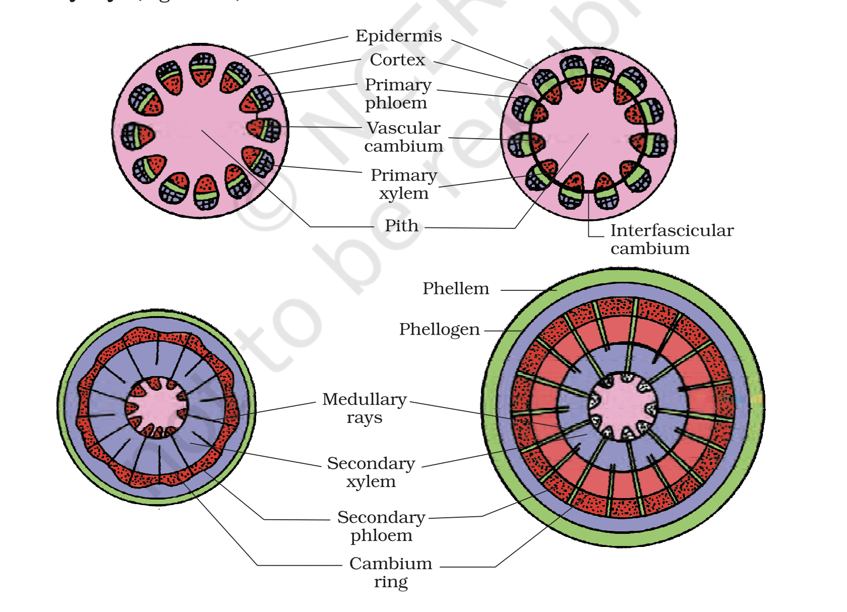
🌱 Vascular Cambium & Cambial Ring Formation 🧠 What is Vascular Cambium?
A lateral meristem that produces secondary xylem (wood) & secondary phloem
Responsible for secondary growth (increase in thickness)
In a young dicot stem, vascular cambium is initially:
A) Absent
B) Present as a continuous ring
C) Present as patches between xylem and phloem
D) Present in the medullary rays only
✅ Correct Answer:
C) Present as patches between xylem and phloem
💡Explanation:
As per NCERT, in the young dicot stem, the vascular cambium is not initially a complete ring. It exists in small patches between xylem and phloem of vascular bundles. Later, it develops into a continuous cambial ring during secondary growth.
Which of the following best describes the role of vascular cambium?
A) Helps in absorption of water
B) Produces new epidermal cells
C) Cuts off new xylem and phloem tissues
D) Causes elongation of root tips
✅ Answer: C
Explanation: Vascular cambium is a lateral meristem that cuts off secondary xylem and phloem during secondary growth.
In the young dicot stem, the vascular cambium is:
A) Present as a complete ring
B) Found in medullary rays only
C) Found as scattered patches between xylem and phloem
D) Present in phloem only
✅ Answer: C
Explanation: Initially, it appears as patches between xylem and phloem, which later connect to form a continuous ring.
The vascular cambium ring is completed by:
A) Activity of cork cambium
B) Fusion of intrafascicular and interfascicular cambium
C) Lenticel formation
D) Fusion of pericycle and endodermis
✅ Answer: B
Explanation: Intrafascicular cambium (within vascular bundles) and interfascicular cambium (between bundles) fuse to form the ring.
Which of the following tissues originates from vascular cambium?
A) Parenchyma
B) Sclerenchyma
C) Secondary xylem
D) Primary phloem
✅ Answer: C
Explanation: Vascular cambium gives rise to secondary vascular tissues, especially secondary xylem and phloem.
Which meristem is responsible for the increase in girth of the plant stem?
A) Apical meristem
B) Lateral meristem (vascular cambium)
C) Intercalary meristem
D) None of the above
✅ Answer: B
Explanation: Lateral meristem, i.e., vascular cambium, causes secondary growth, increasing stem thickness.
Assertion (A): In a dicot stem, vascular cambium ring is formed from both intrafascicular and interfascicular cambium.
Reason (R): Cells of medullary rays become meristematic and form the interfascicular cambium.
A) Both A and R are true, and R is the correct explanation of A
B) Both A and R are true, but R is not the correct explanation of A
C) A is true, but R is false
D) A is false, but R is true
✅ Answer: A
Explanation: Both statements are correct, and R explains how interfascicular cambium is formed.
🌿 Diagram-based MCQ: Q7.
In the transverse section of a young dicot stem, where would you find the interfascicular cambium developing?
A) Between the phloem and cortex
B) In the center of xylem
C) Between two vascular bundles, in medullary rays
D) Inside pith region
✅ Answer: C
Explanation: Interfascicular cambium arises from medullary ray cells between vascular bundles
What triggers the formation of a continuous cambium ring in a dicot stem?
A) Formation of root hairs
B) Thickening of the cortex
C) Meristematic activity of medullary ray cells
D) Activity of cork cambium
✅ Answer: C
Explanation: Medullary ray cells between vascular bundles become meristematic, forming interfascicular cambium that completes the cambial ring.
🔄 Types of Cambium in Dicot Stem
Cambium Type | Location |
|---|---|
Intrafascicular Cambium | Between primary xylem & phloem (within vascular bundles) |
Interfascicular Cambium | Formed from medullary ray cells (between vascular bundles) |
🌳 Formation of Cambial Ring (Step-by-Step)
1⃣ Young Dicot Stem
Intrafascicular cambium exists within vascular bundles
2⃣ Medullary Ray Activation
Medullary ray cells become meristematic
3⃣ Interfascicular Cambium Formation
These meristematic ray cells form interfascicular cambium
4⃣ Continuous Cambial Ring
Intra + Interfascicular cambium unite to form a complete ring of vascular cambium
Interfascicular cambium is formed from:
A) Apical meristem
B) Xylem parenchyma
C) Medullary ray cells
D) Pith cells
✅ Answer: C
📘 Explanation: The medullary ray cells (which are parenchymatous) become meristematic to form interfascicular cambium.
The continuous cambial ring in dicot stems is formed by the fusion of:
A) Phloem and xylem
B) Pith and cortex
C) Intrafascicular and interfascicular cambium
D) Apical and intercalary meristem
✅ Answer: C
📘 Explanation: Intrafascicular + Interfascicular cambium = Cambial ring ➡ responsible for secondary growth.
Which of the following statements is correct?
A) Cambial ring formation occurs in monocots
B) Interfascicular cambium originates from vascular bundles
C) Intrafascicular cambium is formed from medullary rays
D) Interfascicular cambium forms outside the vascular bundles
✅ Answer: D
📘 Explanation: Interfascicular cambium develops between vascular bundles, outside the bundle, from medullary rays.
What does the cambial ring cut off towards the inner side?
A) Secondary phloem
B) Secondary cortex
C) Secondary xylem
D) Cork cells
✅ Answer: C
📘 Explanation: Cambium cuts cells towards the pith (inner side), which mature into secondary xylem.
Which of the following is TRUE about cambial activity?
A) Cambium is equally active on both sides
B) More secondary phloem is formed than secondary xylem
C) Cambium is more active on inner side, forming more secondary xylem
D) Primary phloem becomes secondary phloem
✅ Answer: C
📘 Explanation: Cambium shows more activity on the inner side, leading to more secondary xylem than phloem.
What happens to the primary and secondary phloem during secondary growth?
A) They increase in size
B) They form cork
C) They get gradually crushed
D) They turn into secondary xylem
✅ Answer: C
📘 Explanation: The accumulation of secondary xylem compresses and crushes the phloem layers.
What remains intact near the center of the stem even after secondary growth?
A) Secondary phloem
B) Secondary cortex
C) Primary xylem
D) Epidermis
✅ Answer: C
📘 Explanation: Despite secondary growth, the primary xylem remains more or less intact, near the pith.
What are secondary medullary rays, and where do they form?
A) Narrow bands of xylem near the epidermis
B) Bands of sclerenchyma formed outside the phloem
C) Bands of parenchyma passing radially through secondary xylem and phloem
D) Compressed regions of primary xylem
✅ Answer: C
📘 Explanation: Secondary medullary rays are parenchymatous bands that run radially, allowing lateral transport and storage.
🌳 Spring Wood vs Autumn Wood – Annual Rings
Feature | 🌸 Spring Wood (Early Wood) | 🍂 Autumn Wood (Late Wood) |
|---|---|---|
Season | Formed in spring | Formed in autumn/winter |
Cambium Activity | Highly active | Less active |
Xylary Elements | More numerous, with wider vessels | Fewer, with narrow vessels |
Appearance | Lighter in colour, lower density | Darker in colour, higher density |
Growth Rate | Fast growth | Slow growth |
Function | Facilitates rapid conduction of water | Provides mechanical strength |
Annual Ring | One spring wood + one autumn wood = 1 annual ring | |
Use in Age Determination | Used to estimate tree age | ✅ Yes, together with spring wood |
🔔 Key Point:
In temperate regions, trees form annual rings due to seasonal variation in cambial activity. Counting these rings reveals the tree’s age!
In which season is cambium most active, producing xylem with wider vessels?
A) Summer
B) Autumn
C) Winter
D) Spring
✅ Answer: D
📘 Explanation: In spring, cambium activity is high → more xylem → wider vessels → spring wood/early wood.
Annual rings are formed due to:
A) Formation of only spring wood
B) Alternate arrangement of primary xylem and phloem
C) Alternate appearance of spring wood and autumn wood
D) Continuous activity of cork cambium
✅ Answer: C
📘 Explanation: Spring wood + Autumn wood = 1 annual ring. These appear as concentric rings in stems.
How can the age of a tree be estimated?
A) By counting medullary rays
B) By counting lenticels
C) By counting annual rings in the stem
D) By measuring the diameter of xylem
✅ Answer: C
📘 Explanation: Each annual ring = 1 year. Count the rings in T.S. of stem to know the tree’s age.
Which of the following characteristics is true for autumn wood?
A) Lighter in color, low density
B) Vessels with wider cavities
C) Formed when cambium is highly active
D) Narrow vessels, darker wood, high density
✅ Answer: D
📘 Explanation: Autumn wood forms in winter, has narrow vessels, is darker and denser.
Why is spring wood lighter in color than autumn wood?
A) Due to higher tannin content
B) Due to wider vessels and lower density
C) Due to thicker cell walls
D) Because it is formed from primary xylem
✅ Answer: B
📘 Explanation: Spring wood has larger vessels, making it less dense and thus lighter in color.
Each combination of spring wood + autumn wood = ?
A) One vascular bundle
B) One medullary ray
C) One annual ring
D) One cambium ring
✅ Answer: C
Explanation: One spring + one autumn wood band = 1 annual ring, which helps estimate tree age.
If the diagram above has 4 spring wood + 3 autumn wood bands, what is the age of the tree?
A) 3 years
B) 4 years
C) 7 years
D) 1 year
✅ Answer: B
Explanation: Number of complete spring + autumn pairs = 3 → but we see a 4th spring forming → 4 years.
Why is heartwood darker in color?
A) Due to thickening of primary xylem
B) Due to deposition of organic compounds like tannins and resins
C) Because it conducts more water
D) Because it contains living parenchyma
✅ Answer: B
📘 Explanation: Heartwood is dark because of tannins, resins, oils, gums, and aromatic substances.
🌲 Q2.
Which of the following is true for heartwood?
A) Conducts water
B) Involved in mineral transport
C) Provides mechanical support
D) Contains active living cells
✅ Answer: C
📘 Explanation: Heartwood is dead, does not conduct water, but provides strength and support.