Thyroid Pathology
 Call Kai
 Learn
Learn Practice Test
 Spaced Repetition
 Match
Match Flashcards
 Knowt Play
1/41

There's no tags or description
Looks like no tags are added yet.
Name | Mastery | Learn | Test | Matching | Spaced | Call with Kai | Chat |
|---|
No analytics yet
Send a link to your students to track their progress
42 Terms
a goiter is defined as an
enlarged, hyperplastic thyroid gland
causes of a goiter
iodine deficiency, graves disease, and thyroiditis
thyroid enlargement can be diagnosed by calculating
volume measurements ot by obtaining an AP thickness of the thyroid isthmus
an isthmus that measures greater than — may be indicative of thyroid enlargement
1 cm
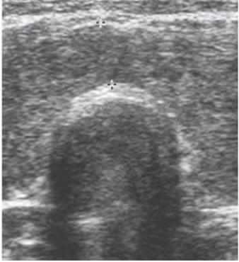
The thyroid isthmus measured 13 mm (between calipers), which is indicative of a goite
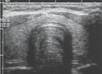
A normal thyroid gland and thyroid isthmus (2 mm) are shownclinical findings of a
clinical findings of a goiter
palpable (and possibly visually) enlarged thyroid gland
dyspnea
dysphagia
feeling of tightening in the throat
coughing
hoarseness
sonographic findingd of a goiter
enlarged thyroid gland (isthmus that exceeds 1 cm in the AP plane)
diffusely heterogeneous echotexture
multiple nodules with cystic and solid components
goiter is a general term for — that can result from inadequate iodine intake
thyroid enlargment
hyperthyroidismis a condition that results from the
overproduction of thyroid hormones
graves disease aka —
diffuse toxic goiter
the term — may be noted as hypervascularity within the thyroid gland
thyroid inferno
clinical findings of graves disease
buldging eyes
heat intolerance
nervousness
weight loss
hair loss
sonographic findings of graves disease
enlarged gland
heterogeneous or diffusely hypoechoic echotexutre
thyroid inferno
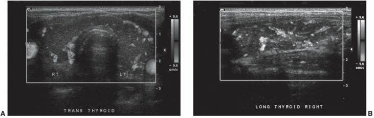
Hyperthyroidism. Transverse (A) and longitudinal (B) images with color Doppler in a patient with hyperthyroidism revealing hyperemia. (Color image provided online.)
— is the most common cause of hyperthyroidism.
Graves disease
— is the most common form of thyroid cancer
papillary carcinoma
other forms of thyroid malignancies
follicular carcinoma, medullary carcinoma, anaplastic carcinoma, lymphoma, and metastases of the thyroid
hypothyroidism is a condition that results from the
underproduction of thyroid hormones
Hashimoto thyroiditid is an
autoimmune disease
the most common cause of hypothyroidism in the United States
hashimotos thyroiditis
hashimoto thyroiditis may also be referred to as
chronic autoimmune lymphocytic thyroiditis
with hashimoto disease, the thyroid becomes inflamed, as, as a result, the thyroid produces —
smaller amounts of thyroid hormones
in order to compensate for hashimoto disease, the pituitary gland releases more —, which causes the thyroid to become enlarged
TSH
the end-stage of hashimoto disease may actually lead to —
fibrosis and atrophy of the gland
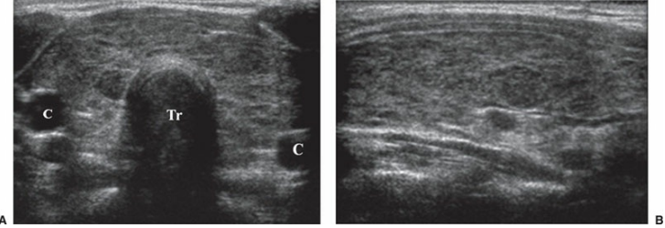
Hashimoto thyroiditis. A. Transverse image of the thyroid gland in a patient with hypothyroidism revealing a diffusely coarse, heterogeneous gland. The trachea (Tr) and carotid arteries (C) are also demonstrated. B. Longitudinal image of the same patient.
clinical findings of hashimoto thyroiditis
depression
increased cold sensitivity
elevated blood cholesterol levels
slight weight gain may occur
puffy face and puffiness under the eyes
sonographic findings of hashimoto thyroiditis
mild enlargement of the thyroid gland (initially)
heterogeneous echotexture
multiple, ill-defined hypoechoic regions seperated by fibrous hyperechoic tissue
hypervascular gland
hashimoto thyroiditis is the most common cause of —
hypothyroidism
— are the most common masses identified within the thyroid gland with sonography
benign thyroid nodules
follicular adenomas are most often —
small, round, and can have varying sonographic appearances, including completely anechoic, isoechoic, or hyperechoic.
follicular adenomas may also have a
surrounding halo
— is the most common cause of thyroid nodules
nodular hyperplasia
aka for hyperplastic nodules
adenomatous nodules
hyperplastic nodules are almost always — and have varying sonographic appearances
multiple
benign characteristics of thyroid nodules
extensive cystic components
cysts <5 mm
hyperechoic mass
“eggshell” calcifications
“hot” nodules (nuclear medicine)
malignant characteristics of thyroid nodules
hypoechoic mass
taller-than-wide
mass with internal microcalcifications (psammoma bodies)
solitary mass
marked vascularity within the central part of the nodule
interrupted peripheral calcification
extracapsular invasion
lobulated margins
enlargment of the cervical lymph nodes (metastasis)
“cold” nodule (nuclear medicine finding)
nuclear medicine utilizes scintigraphy to classify thyroid nodules as either
hyperfunctioning or hypofunctioning
hyperfunctioning or “hot nodules” yield — on a thyroid nuclear medicine scan
dark areas
hypofunctioning or “cold nodules” yield — on the nuc med scan
light or blank areas
most cancers are — nodules and will appear as — on nuc med scans, but thats not always the case
hypofunctioning; cold nodules
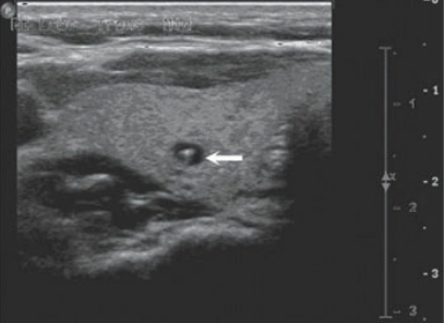
Colloid cyst. Transverse image of the thyroid revealing a small colloid cyst (arrow) with a small, hyperechoic focus contained within it that is producing comet-tail artifact.