Histology of bone
5.0(2)
Studied by 6 peopleCard Sorting
1/25
Last updated 4:22 AM on 10/11/22
Name | Mastery | Learn | Test | Matching | Spaced | Call with Kai |
|---|
No analytics yet
Send a link to your students to track their progress
26 Terms
1
New cards
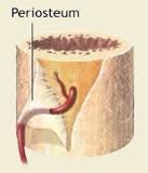
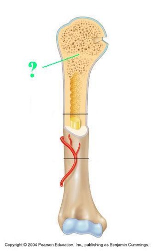
periosteum
thick, fibrous membrane that covers the outside of a bone; anchors tendons & ligaments; contains blood vessels, nerves, & lymph vessels
2
New cards
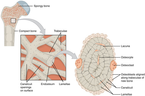
spongy bone
found in the ends of long bones; characterized by irregular spaces filled with red bone marrow that makes blood cells; helps keep bones light in weight
3
New cards
compact bone
dense, hard bone forming the shaft of long bones & the outer layer of other bones
4
New cards
epiphyseal plate
thin band of cartilage that is the growth zone between areas of ossification
5
New cards
Periosteum
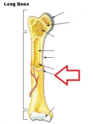
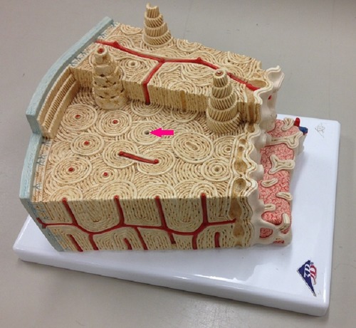
Identify the membrane that covers the bone seen at the pointer
6
New cards
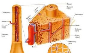
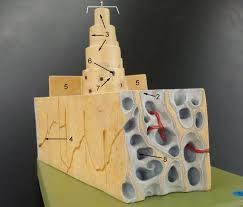
Osteon
Identify the structure at #1
7
New cards
Circumfrential lamellae
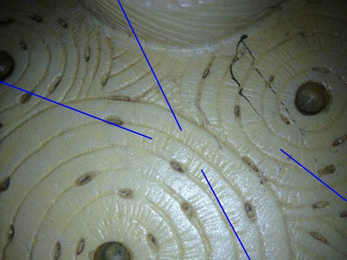
Identify the structure at #5

8
New cards
Concentric lamellae
Identify the structure at #3

9
New cards
Perforating canal
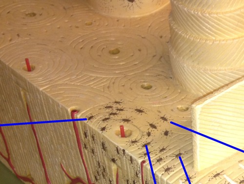
Identify the structure at #6
10
New cards
Central canal
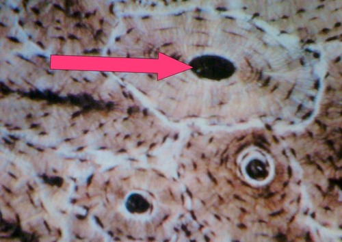
Identify the structure at the pointer
11
New cards
Canaliculi
Identify the structure at the blue pointers that are microscopic canals from lacuna to lacuna
12
New cards
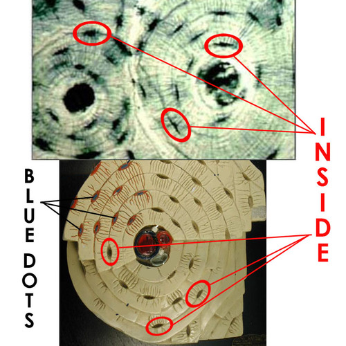
Osteocytes
Identify the dots at the blue pointers
13
New cards
Lacunae
Identify the spaces at the blue pointers

14
New cards
Interstitial lamellae
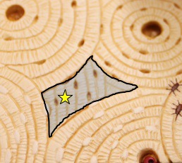
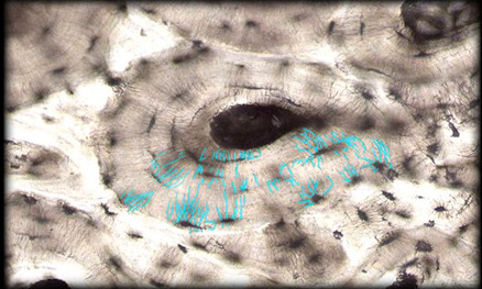
Identify the structure at the shadowed area with the star
15
New cards
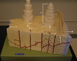
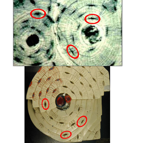
Osteon
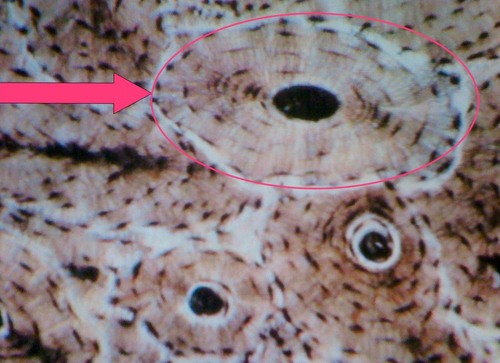
Identify the entire structure at the pink structure
16
New cards
Concentric lamellae
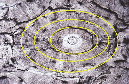
Identify the structure at the yellow circles
17
New cards
Central canal
Name the space at the pink pointer
18
New cards
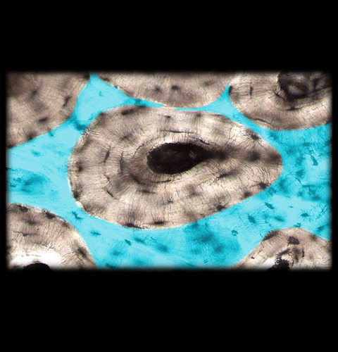
Canaliculi
Name the structure at the blue lines
19
New cards
Osteocyte
Name the dots inside the circled structures
20
New cards
Lacuna
Name the spaces that are circled
21
New cards
Interstitial lamellae
Name the structure in the highlighted area
22
New cards
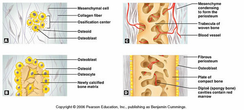
Intramembranous ossification timeline
occurs during fetal life
23
New cards
Intramembranous ossification location
mandible, clavicle, and cranial bones ( frontal, parietal, temporal, and occipital)
24
New cards
intamembranous ossification
bone tissue differentiates from fibrous membrane (mesenchyme become osteoblasts)
25
New cards
Intramembranous ossification process
ossification starts in the center of bone, moves towards edges
26
New cards
Intramembranous ossification image