Cardiac Embryology Phases
 Call Kai
 Learn
Learn Practice Test
 Spaced Repetition
 Match
Match Flashcards
 Knowt Play
1/13

There's no tags or description
Looks like no tags are added yet.
Name | Mastery | Learn | Test | Matching | Spaced | Call with Kai | Chat |
|---|
No analytics yet
Send a link to your students to track their progress
14 Terms
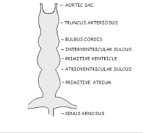
What does this image represent? Identify the week of development.
A single endocardial heart tube
The second phase of the hearts development
Week 3

What do the TRUNCUS ARTERIOSUS and the SINUS VENOSUS eventually become?
TRUNCUS ARTERIOSUS 🡪 the distal portion of bulbus cordis will form AO & main PA
SINUS VENOSUS 🡪 forms SVC, IVC, PV4, CS, oblique vein of LV, posterior portion of RA & LA
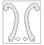
What does the image represent and what are the two layers present? Identify the week of development.
The first part of the heart development
inner, thinner layer - endocardium
outer, thicker layer - myocardium
Prior to week 3
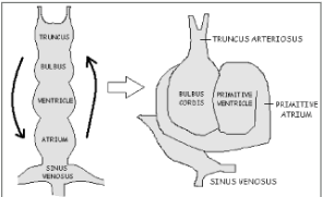
What does the image represent and what is occurring in this image? Identify the week of development.
The third phase of the heart’s development
S-shaped loop, normally folds to the right (known as D-looping)
Week 4

What does the image represent? Identify the week of development.
The fourth phase of the hearts development
Note the center of the endocardial cushion
Week 4.
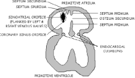
What does the image represent? What occurs during this phase? Identify the week of development.
The fifth phase of the heart’s development
Septum primum reaches endocardial cushions & closes ostium primum
Week 5
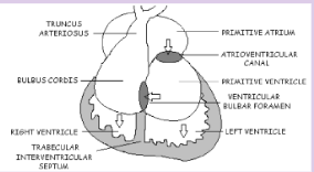
What does the image represent? What occurs during this phase? Identify the week of development.
The sixth phase of the hearts development
The conus cordis forms 🡪 RVOT & LVOT
Week 5
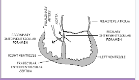
What does the image represent? What occurs during this phase? Identify the week of development.
The seventh phase of the heart’s development
LV communicates with RV via 2 routes
1) primary interventricular foramen
2) secondary interventricular foramen🡪 membranous IVS
Week 5

What does the image represent? What occurs during this phase? Identify the week of development.
The eighth phase of the heart’s development
At birth/first breath - LAP increases (doubles) forcing septum primum against septum secundum—closing the FO
Together, septum primum & septum secundum 🡪 form IAS
Week 7
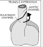
What does the image represent? What occurs during this phase? Identify the week of development.
The ninth and final phase of cardiac development
Bulbar ridges directly involved in rotation & division of truncus arteriosus into AO & main PA
Fetal circulation is in place! Final requirement is growth.
Week 7
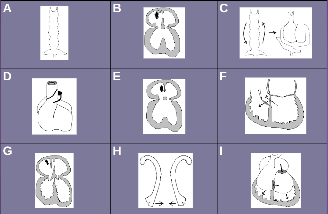
Put the images in order.
Order: H,A,C,E,B,I,F,G,D
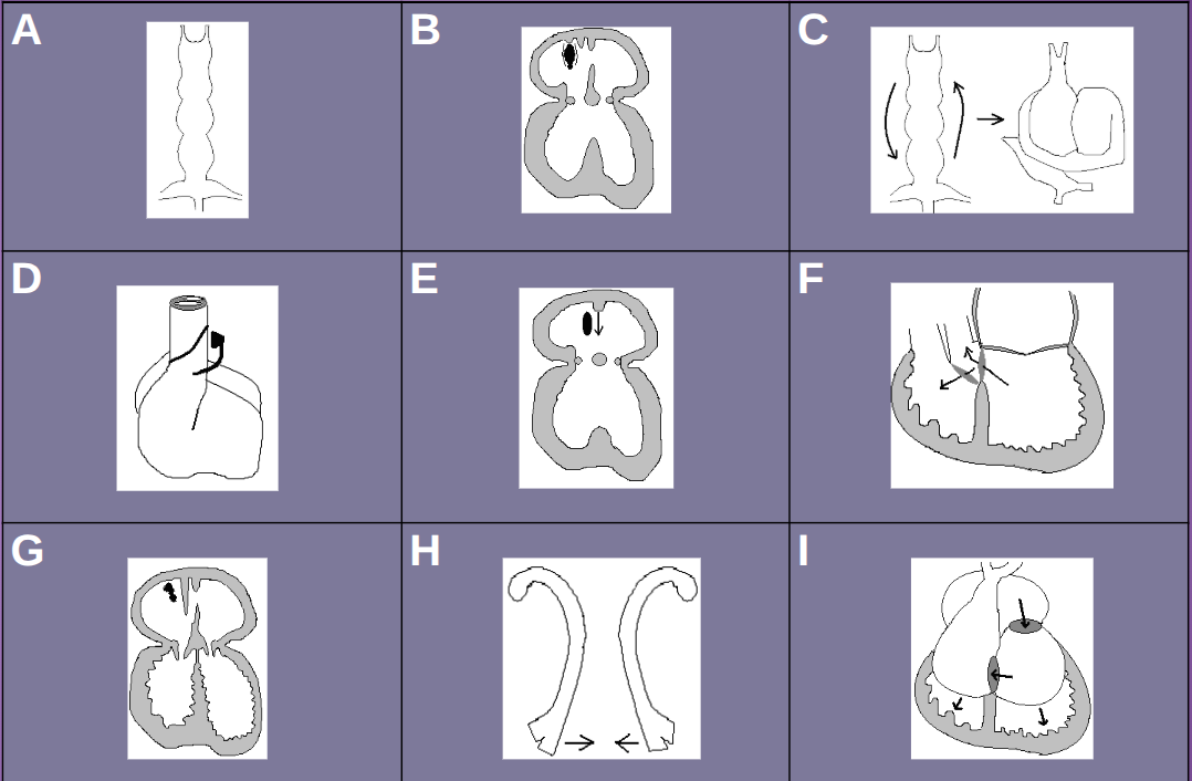
Identify the first three phases of development and the week.
H- Prior to week 3
A- Week 3
C- Week 4
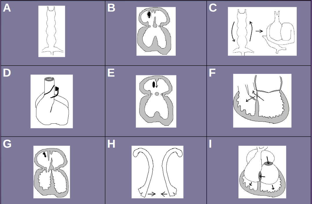
Identify the 4th, 5th, and 6th phases of development and the week.
E- Week 4
B- Week 5
I- Week 5
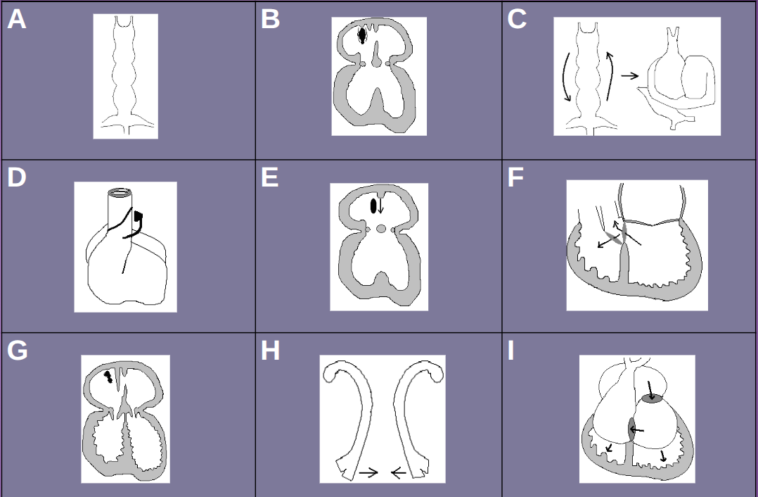
Identify the final three phases of development and the week.
F- Week 5
G- Week 7
D- Week 7