701 Larynx, Thyroid, Parathyroid Glands
1/100
There's no tags or description
Looks like no tags are added yet.
Name | Mastery | Learn | Test | Matching | Spaced |
|---|
No study sessions yet.
101 Terms
Endocrine Neck
Thyroid Gland & Parathyroid Gland
Anterior Jugular Veins
superficial veins of the neck that drain the mental triangle & empty into the subclavian veins &/or make connections w/ the external jugular veins
*overlies the investing fascia
Thyroid Gland (Overview)
-largest endocrine gland
-produces T3 & T4 hormones --> regulate metabolism
-produce calcitonin via parathyroid glands --> regulates level of Ca2+ in blood
-posterior to infrahyoid muscles
-anterior & lateral to trachea & esophagus
-medial to the carotid sheath (carotid arteries, Vagus Nerve, & Internal Jugular Vein)
-3 lobes --> right lobe, isthmus (middle), left lobe
-circoid & thyroid cartilage above thyroid that's apart of the larynx
*blood supply = superior thyroid artery (1st branch of external carotid) & inferior thyroid artery (branch of thyrocervical trunk of subclavian artery)
> can also have rare thyroid ima artery --> supplies the isthmus lobe of thyroid *
Thyroid Ima Artery
rare branch of brachiocephalic trunk that supplies the isthmus of the thyroid
*surgeons have to be careful not to cut this artery when removing the thyroid = hemorrhage
superior thyroid artery
1st branch of the external carotid artery that supplies the upper part of the thyroid
inferior thyroid artery
branch of thyrocervical trunk (of subclavian artery) that supplies the lower part of the thyroid artery
*runs perpendicularly across the recurrent laryngeal nerve --> thyroid removal can injure the laryngeal nerves & lead to laryngeal muscle paralysis (surgeons gotta be careful)
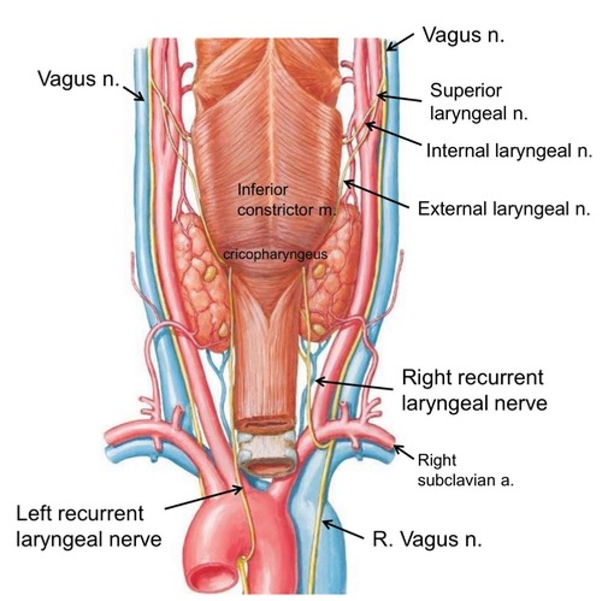
True or False: The left recurrent laryngeal nerve runs in a groove between the trachea & the esophagus
True
Venous Drainage of the Thyroid
-superior thyroid vein--> drains into the internal jugular vein
-middle thyroid vein --> drains into the internal jugular vein
-inferior thyroid vein --> drains into the brachiocephalic vein
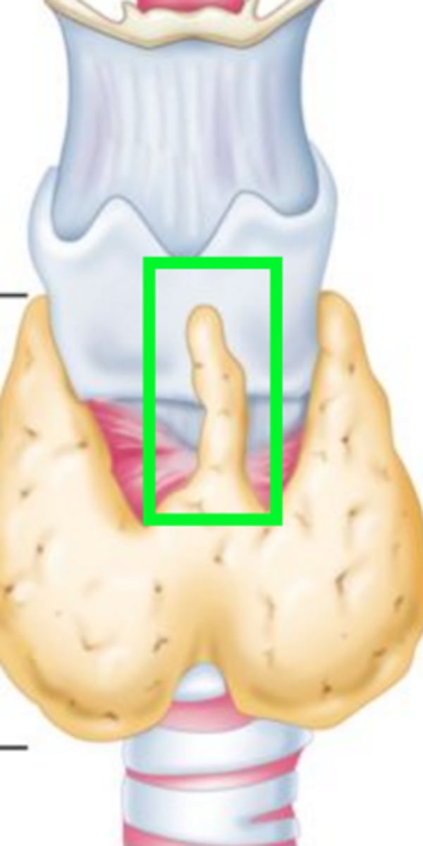
Pyramidal Lobe of Thyroid
remanent of thyroglossal duct from the development of the thyroid gland (only in 40% of ppl)
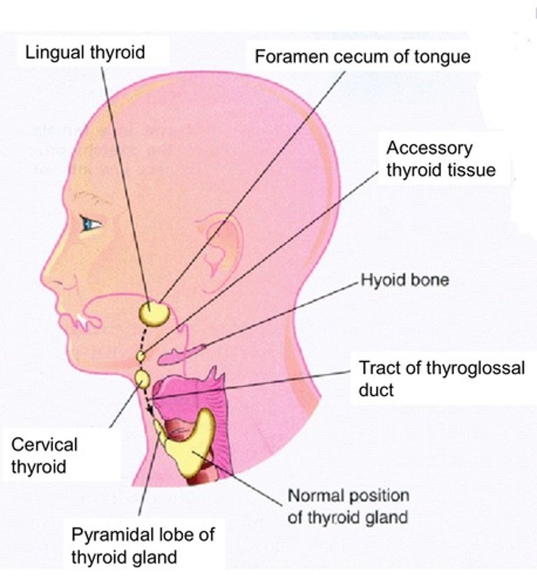
Development of the Thyroid Gland
thyroid begins development @ the base of the tongue during 3-4wks of development --> descends thru foramen cecum @ the back of the tongue --> travels down to the neck w/ thyroglossal duct fascia attachment bwtn the tongue & the pyramidal lobe of the thyroid --> thyroglossal duct degenerates at the end of the 5th wk--> thyroid makes it to it's final position during 6-8th wk (2 wks after descent)
*pyramidal lobe & thyroglossal duct can remain (thyroglossal duct can get infection = thyroglossal duct cyst)
foramen cecum
foramen between the anterior 2/3 & the posterior 1/3 of the tongue in which the thyroid travels through to descend into the neck during development
Ectopic Thyroid
thyroid can leave some tissue behind in its descent pathway anywhere from the tongue to the superior mediastinum
ex: lingual thyroid & cervical thyroid
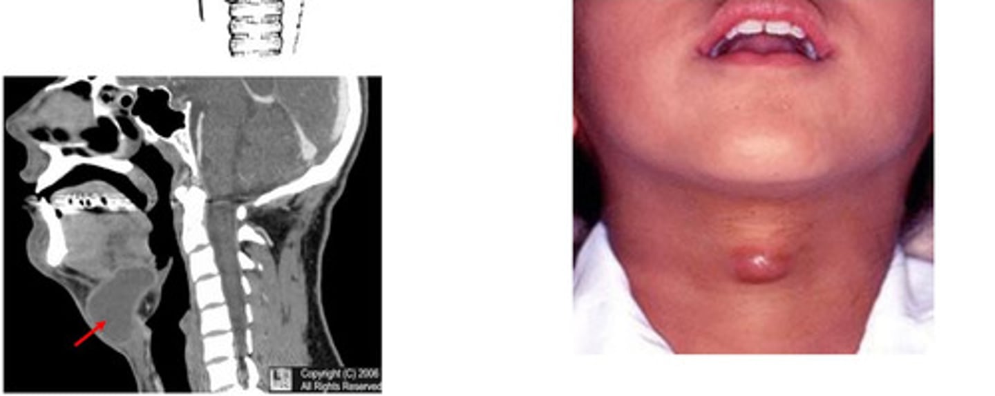
Thyroglossal Duct Cyst
thyroglossal duct from thyroid development can remain in the anterior neck & become infected==> presents as painless swelling in the midline near hyoid bone that moves when swallowing
*Tx= Sistrunk Procedure --> surgical resection of the duct to base of tongue & the hyoid bone to ensure complete removal of the duct
goiter
abnormal enlargement of the thyroid gland that's usually due to the lack of iodine
*no iodine = no T3 & T4 production = pituitary detect low hormone levels & increases TSH secretion -->>> overgrowth of thyroid follicles = hypertrophy=goiter
-symptoms
> swelling below the adam's apple
> tightness in the throat area
> hoarseness (scratchy voice) --> impinges the recurrent laryngeal nerves &/or the laryngeal aperature
>swelling of neck vein
*other causes of goiter --> Graves Disease, Thyroiditis, Thyroid Cancer
*Can compress other neck structures (ex: laryngeal aperature; the trachea)
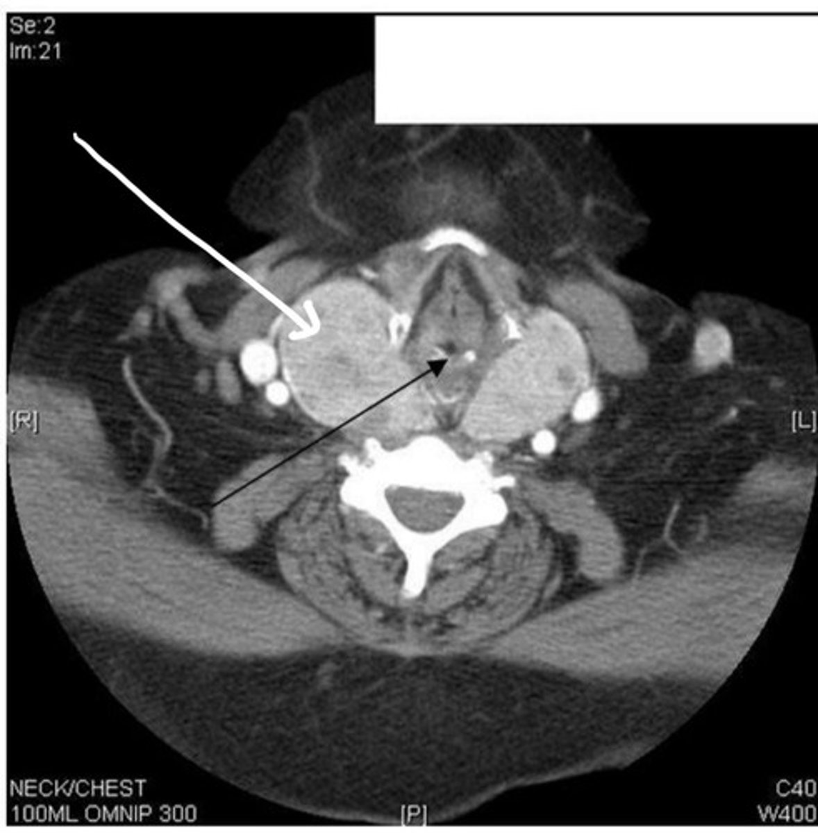
True or False: A goiter can get so big that it compresses and deviates the trachea
True
parathyroid glands
4 glands on the posterior aspect of the thyroid gland that produce calcitonin to regulate the level of Ca2+ in the blood, bones, & GI system
*blood supply = superior/inferior thyroid arteries
-removal of parathryoid glands --> not enough Ca2+ for muscles that leads to tetany
-cancer in parathyroid --> increased Ca2+ in blood due to overproduction of calcitonin from increased number of parathyroid cells
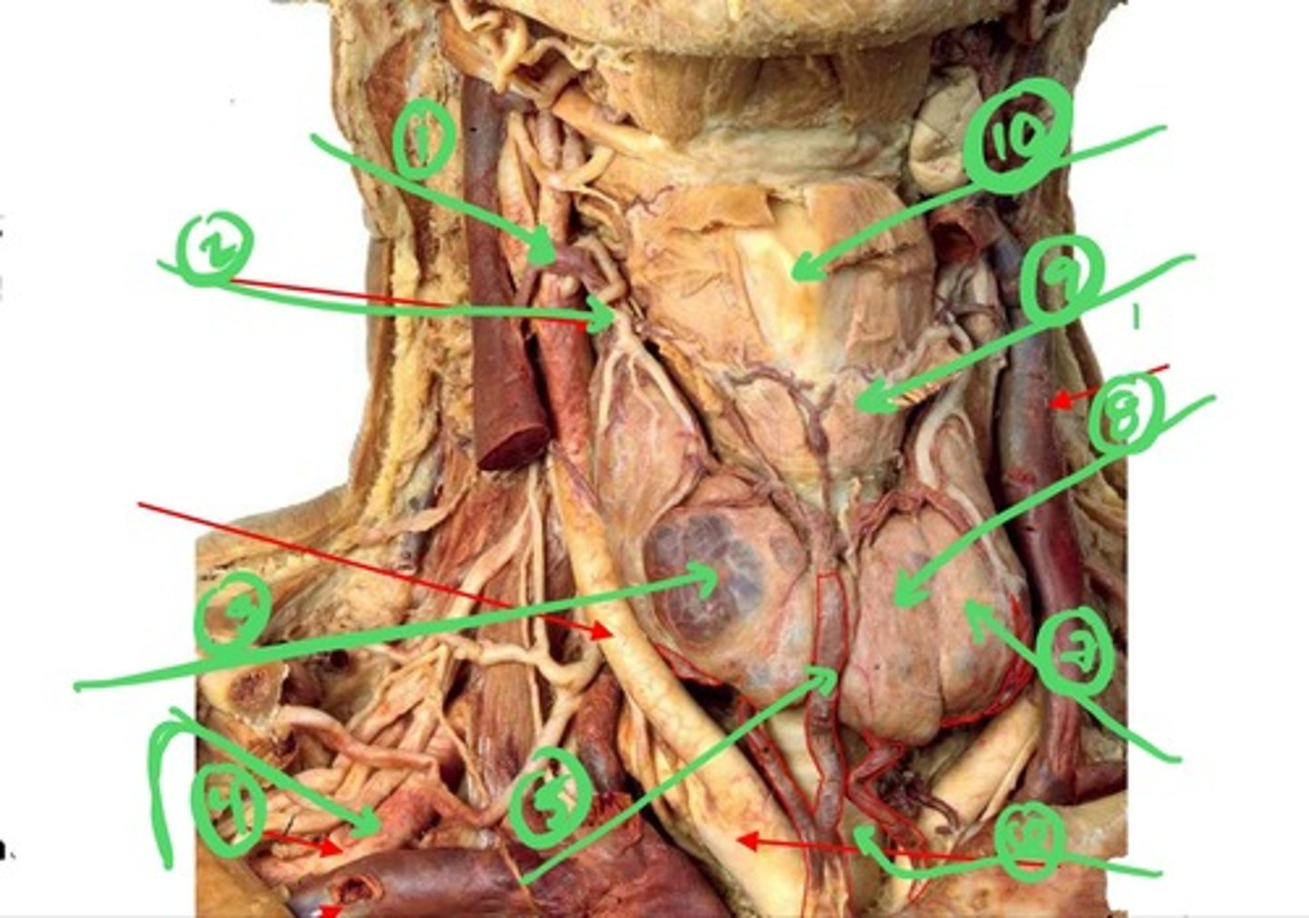
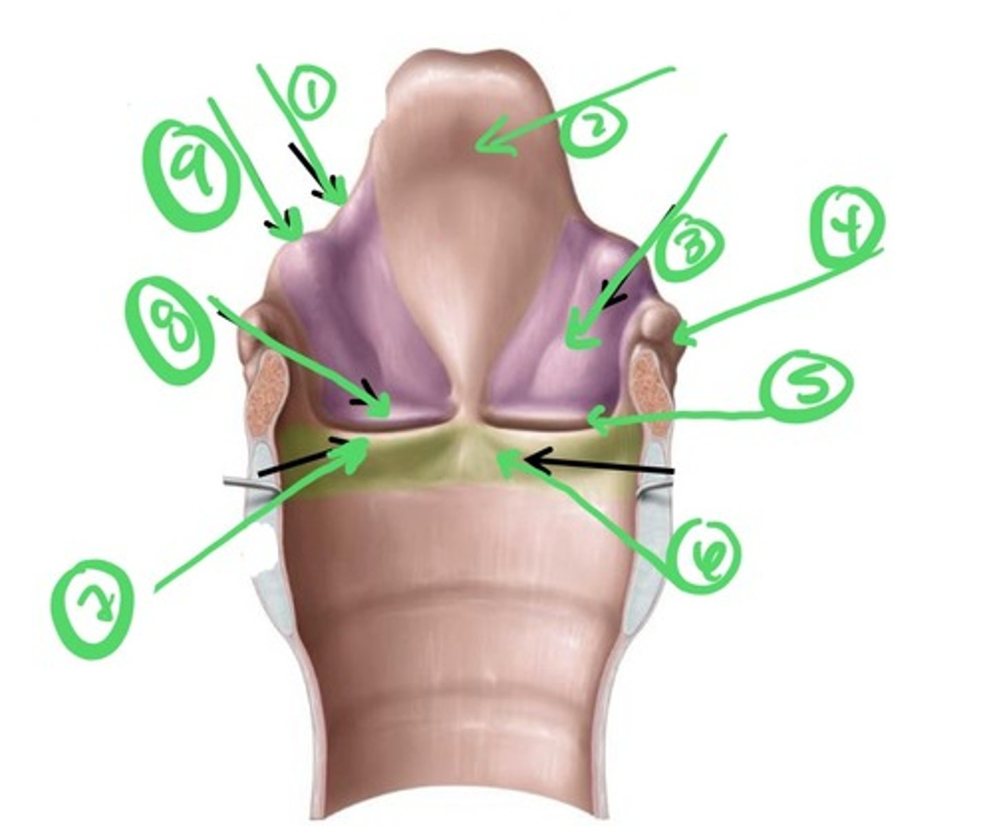
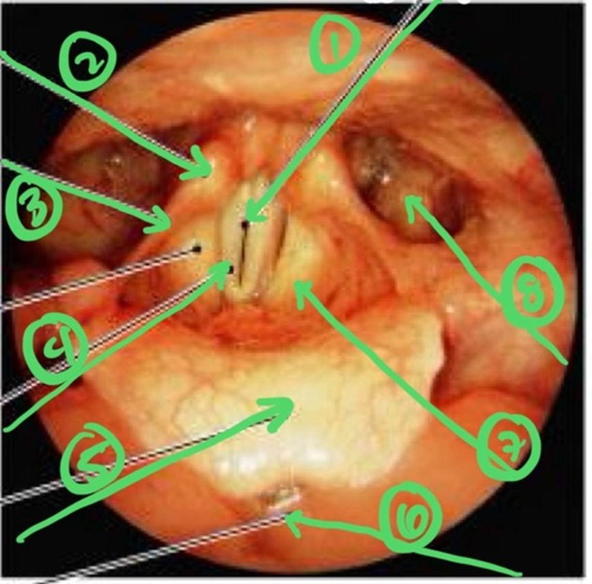
1.) superior thyroid vein
2.) superior thyroid artery
3.) right lobe of thyroid
4.) subclavian artery
5.) inferior thyroid vein
6.) trachea
7.) left thyroid lobe
8.) isthmus lobe of thyroid
9.) circothyroid muscle
10.) thyroid cartilage
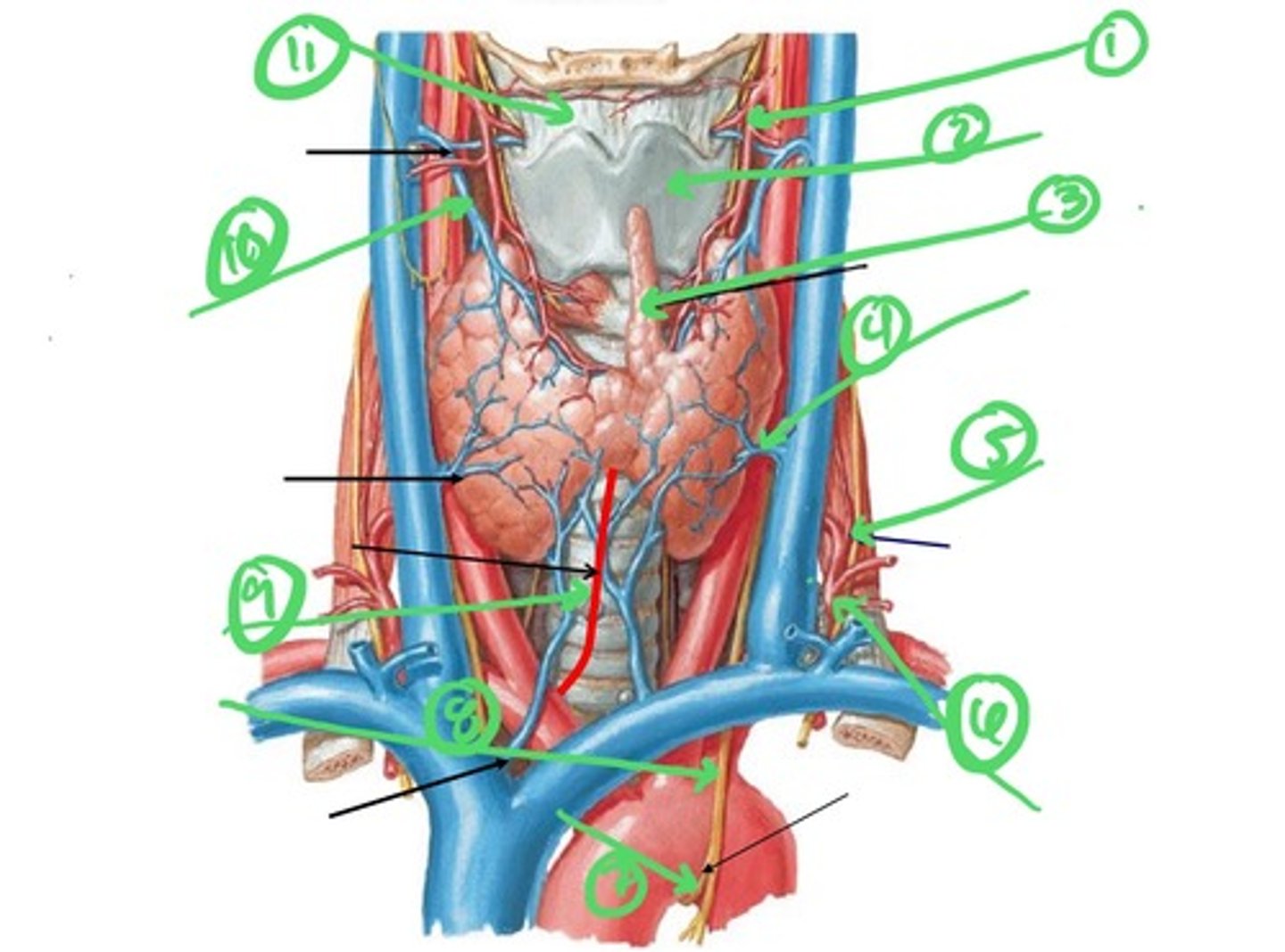
1.) superior thyroid artery
2.) thyroid cartilage
3.) pyramidal lobe of thyroid --> rare remanent of thyroglossal duct from thyroid development
4.) middle thyroid vein
5.) inferior thyroid artery
6.) thyrocervical trunk
7.) recurrent laryngeal nerve
8.) vagus nerve
9.) thyroid ima artery
10.) superior thyroid vein
11.) thyrohyoid membrane
True or False: Speech comes from the larynx
False ==> speech is from the oral cavity, but the larynx produces the tone of voice via the vocal cords
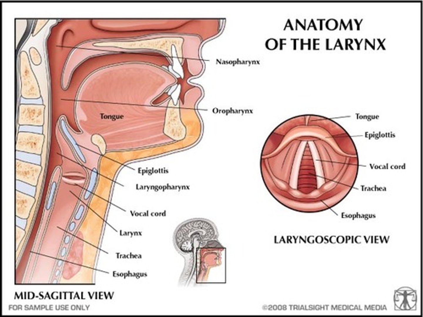
Larynx (Overview)
portion of the respiratory tract that contains the vocal cords (located right behind & below the tongue)
-about 5-7cm long
-upper boundary= tip of epiglottis, opposite of the 3-4th cervical vertebra
-lower end = lower border of the cricoid cartilage near the 6th cervical vertebra
-medial to the structures of the carotid sheath
-located above/medial to the inferior/superior thyroid arteries
-located below the hyoid bone
-located above the thyroid gland
*structure rigidity made from 3 unpaired cartilages --> epiglottis, thyroid cartilage, & cricoid cartilage
Function
-phonation = production of sound --> tone of voice produced when vocal cords vibrate & air moves past them
-sphincter for the respiratory system--> regulates the amount of air that comes in & out of lungs (can be used to increase abdominal pressure when pushing things out = Valsalva Maneuver)
-involved in action of swallowing (deglutition) --> acts w/ oral cavity & oropharynx
*innervated by the superior & recurrent laryngeal nerves (branches of Vagus Nerve CNX)
What forms the Adam's Apple?
the prominence created by the meeting of the 2 laminae that make up the thyroid cartilage at the midline
oblique line of thyroid cartilage
lines on lamina of thyroid cartilage that are the attachment points for thyrohyoid, sternohyoid, & inferior constrictor muscles (inferior circular muscle of the pharynx)
What attaches to the inferior border of the thyroid cartilage?
cricothyroid membrane
What attaches to the superior border of the thyroid cartilage?
thyrohyoid membrane
What structure is present at the rostral margin of the thyroid cartilage?
the superior thyroid notch
True or False: The superior & inferior horns of the thyroid cartilage lamina are located at its anterior border
False ==> the posterior border of each lamina forms the superior & inferior horns
superior horn of thyroid cartilage lamina
structure of the posterior part of thyroid cartilage lamina that attaches to the greater horn of the hyoid bone
inferior horn of the thyroid cartilage lamina
structure of the posterior part of thyroid cartilage lamina that forms a joint w/ cricoid cartilage to allow the thyroid cartilage to rock back & forth
True or False: Males have a wider angle of thyroid cartilage than females
False ==> Male thyroid angle = 90 deg; Female thyroid angle = 120 deg
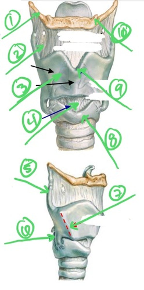
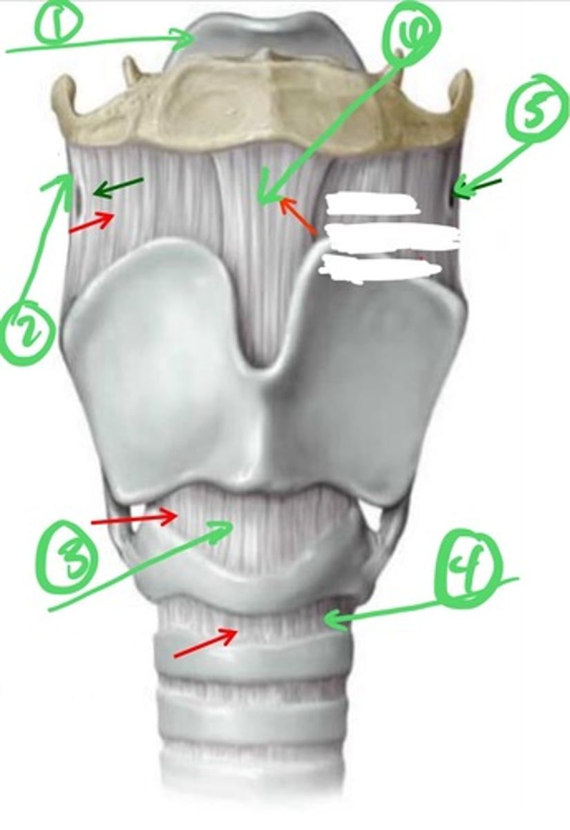
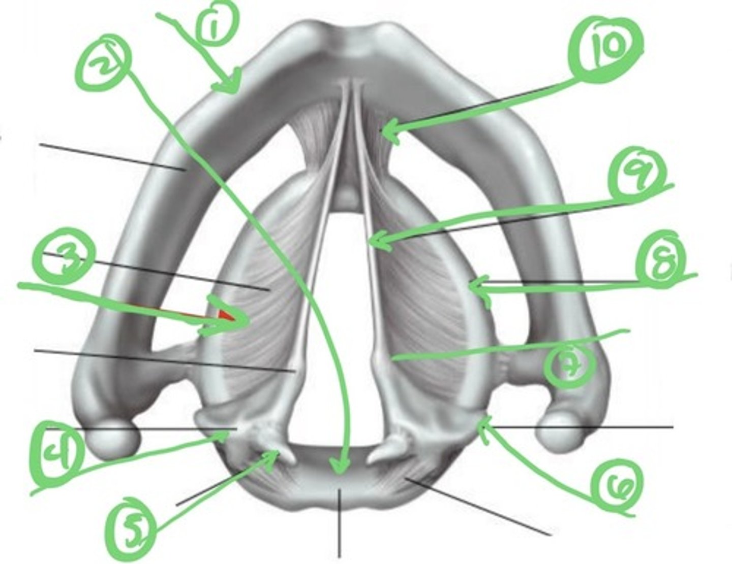
1.) greater horn of hyoid bone
2.) thyrohyoid cartilage
3.) laminae of thyroid cartilage
4.) cricothyroid membrane
5.) superior horn of thyroid cartilage laminae
6.) inferior horn of thyroid cartilage laminae
7.) oblique line of thyroid cartilage
8.) cricoid cartilage
9.) superior thyroid notch
10.) lesser horn of hyoid bone
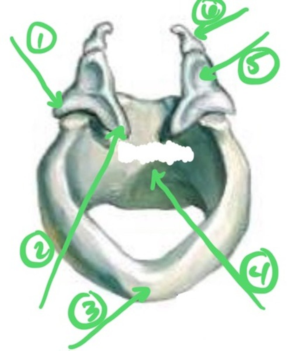
cricoid cartilage
ring shaped cartilage that is the 1st & only complete tracheal ring (narrow anteriorly & broad posteriorly)
-lies below the thyroid cartilage & its lateral surface articulates w/ the inferior horn of the thyroid cartilage
-upper border articulates with arytenoid cartilage (makes synovial joint)
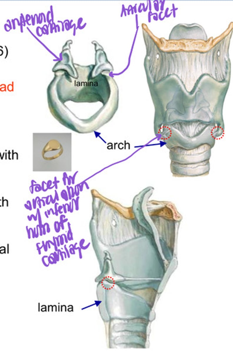
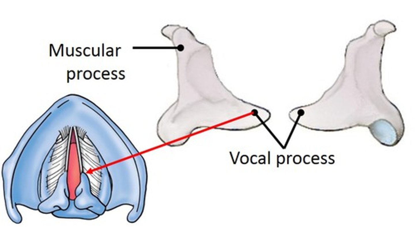
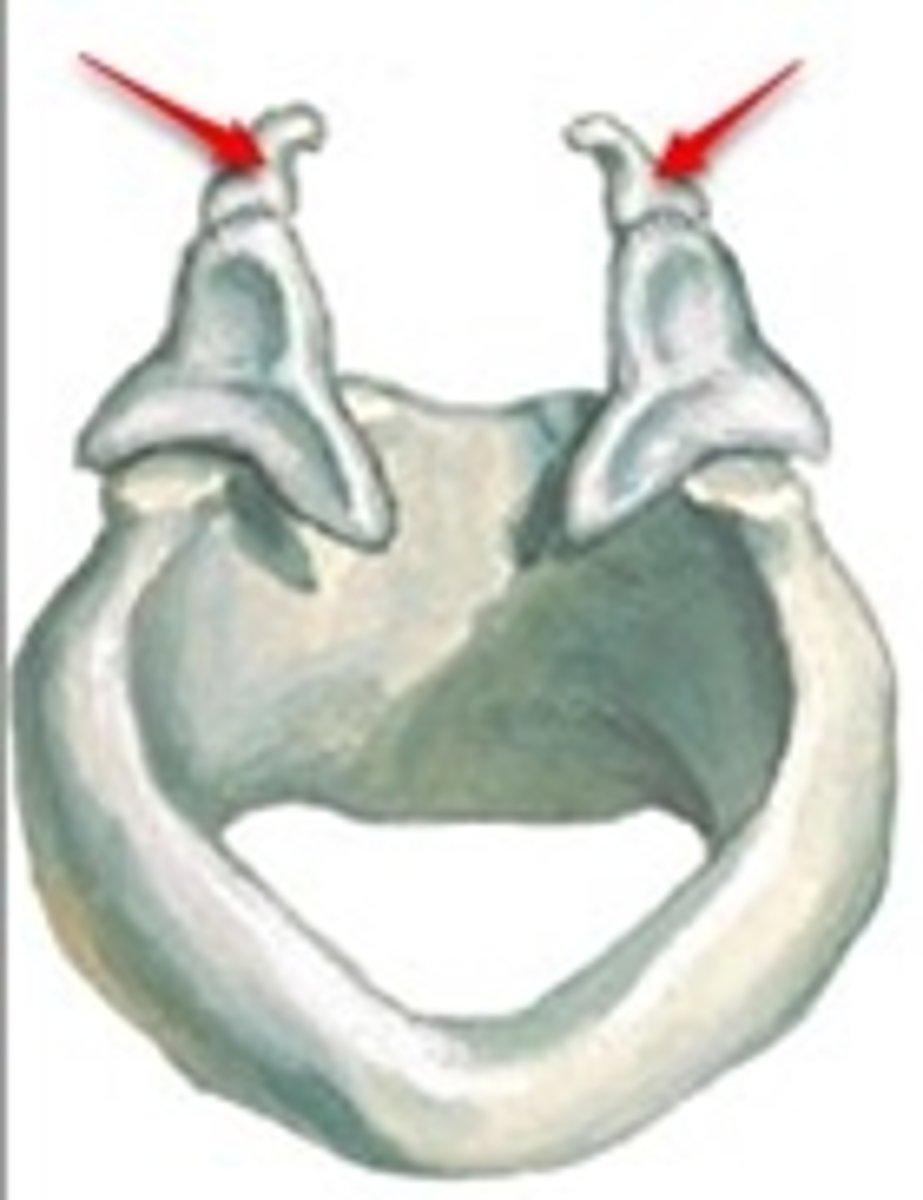
arytenoid cartilage
small pyramidal shaped cartilage that is rotated to tense the vocal cords for phonation
-base articulates w/ upper border of the cricoid cartilage
-apex supports the corniculate cartilage (cc sits on top of it)
-has medial vocal process that attaches to vocal ligments of vocal cords **
-has lateral muscular process that attaches to intrinsic muscles of larynx --> muscles pull on a. cartilage to rotate them on the face of the cricoid cartilage to abduct or adduct the vocal cords for respiration & phonation
intrinsic muscles of larynx
muscles of the larynx that pull on the muscular process of the arytenoid cartilage to move them on the face of the cricoid cartilage to tense the vocal cords for phonation
* attach to the muscular process of the aryntenoid cartilage
1.) muscular process of arytenoid cartilage
2.) vocal process of arytenoid cartilage
3.) arch of cricoid cartilage
4.) lamina of cricoid cartilage
5.) arytenoid cartilage
6.) corniculate cartilage
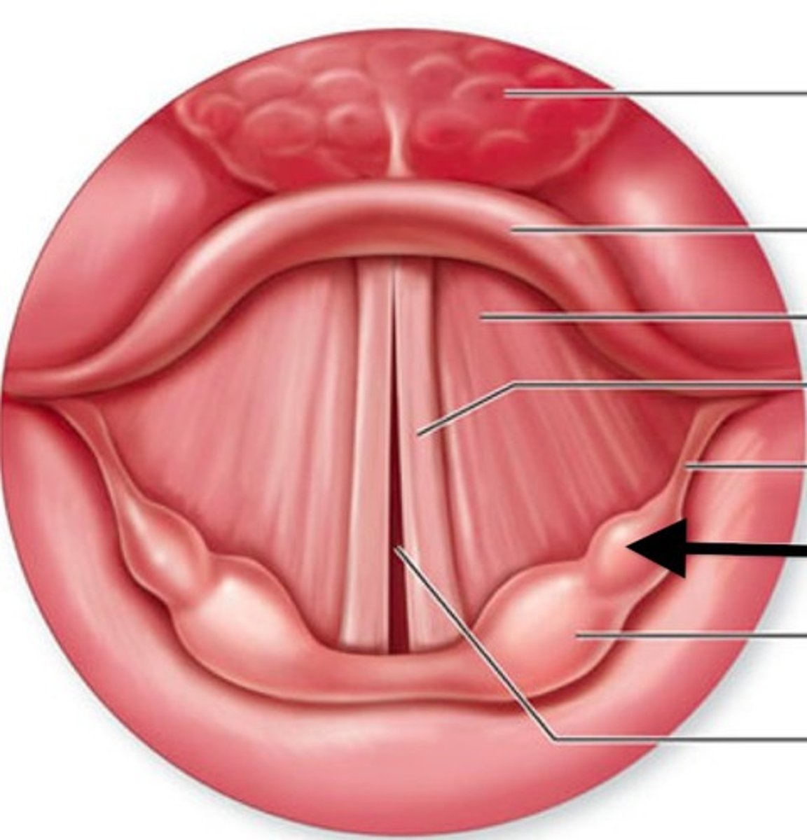
Rima Glottidis
the space bwtn the vocal cords ==> widens or narrows depending on the position of the vocal cords (abducted or adducted due to rotation of arytenoid cartilage by intrinsic muscles of the larynx)
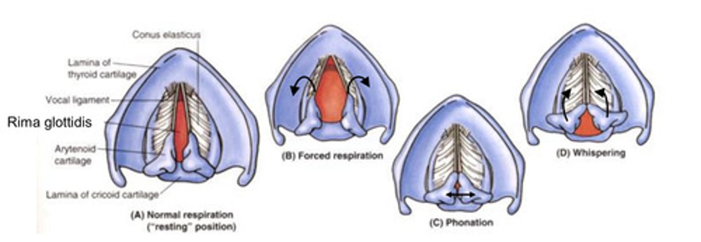
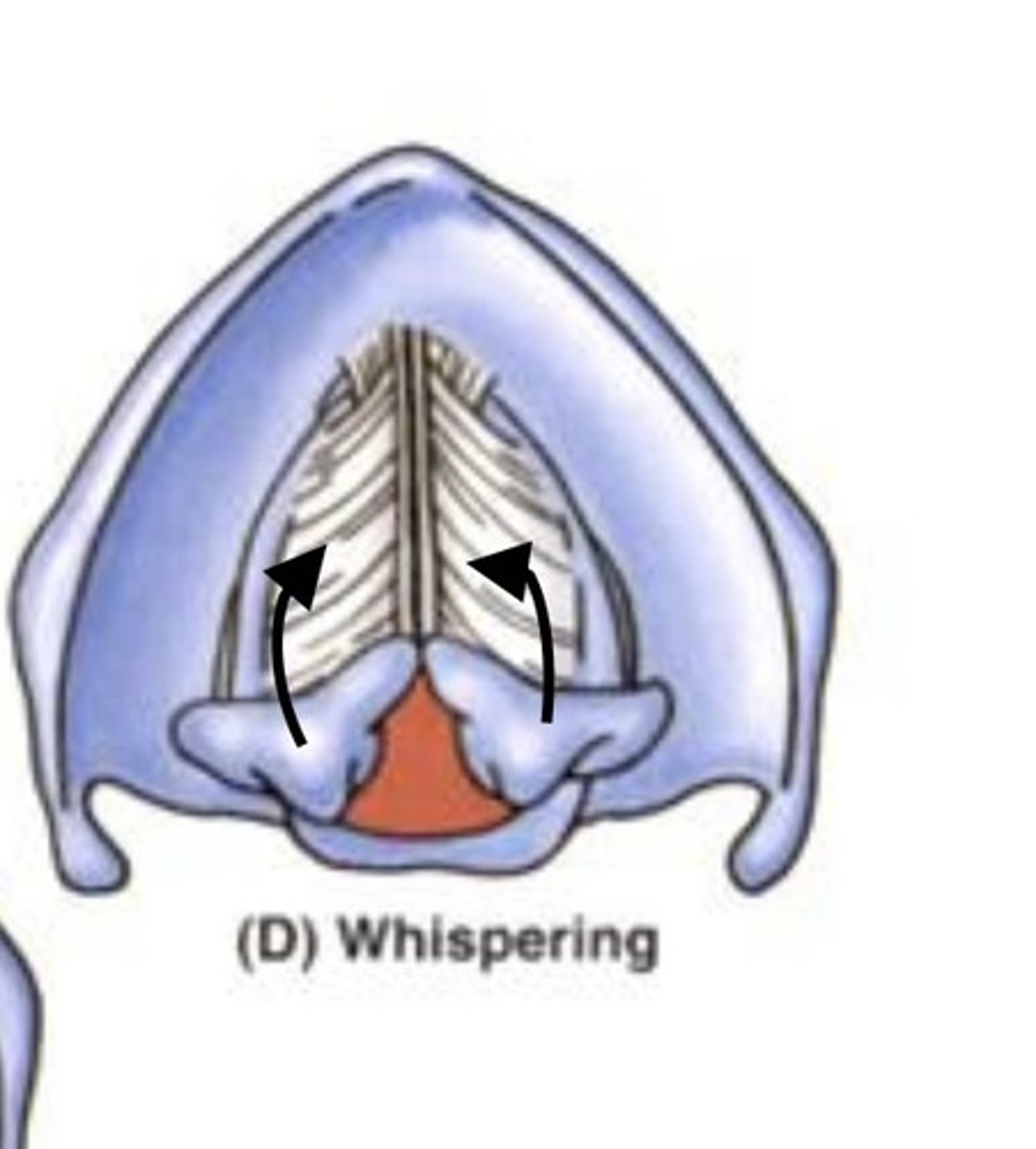
True or False: The tone of whispering is due to positioning the vocal cords and arytenoid cartilage as close together as possible.
False ==> Vocal cords are fully adducted when whispering but the posterior portion of the arytenoid cartilage is abducted (positioned away from each other) --> creates opening of rima glottidis behind the arytenoid cartilage --> air still able to pass thru--> produces sound w/o a tone = whisper
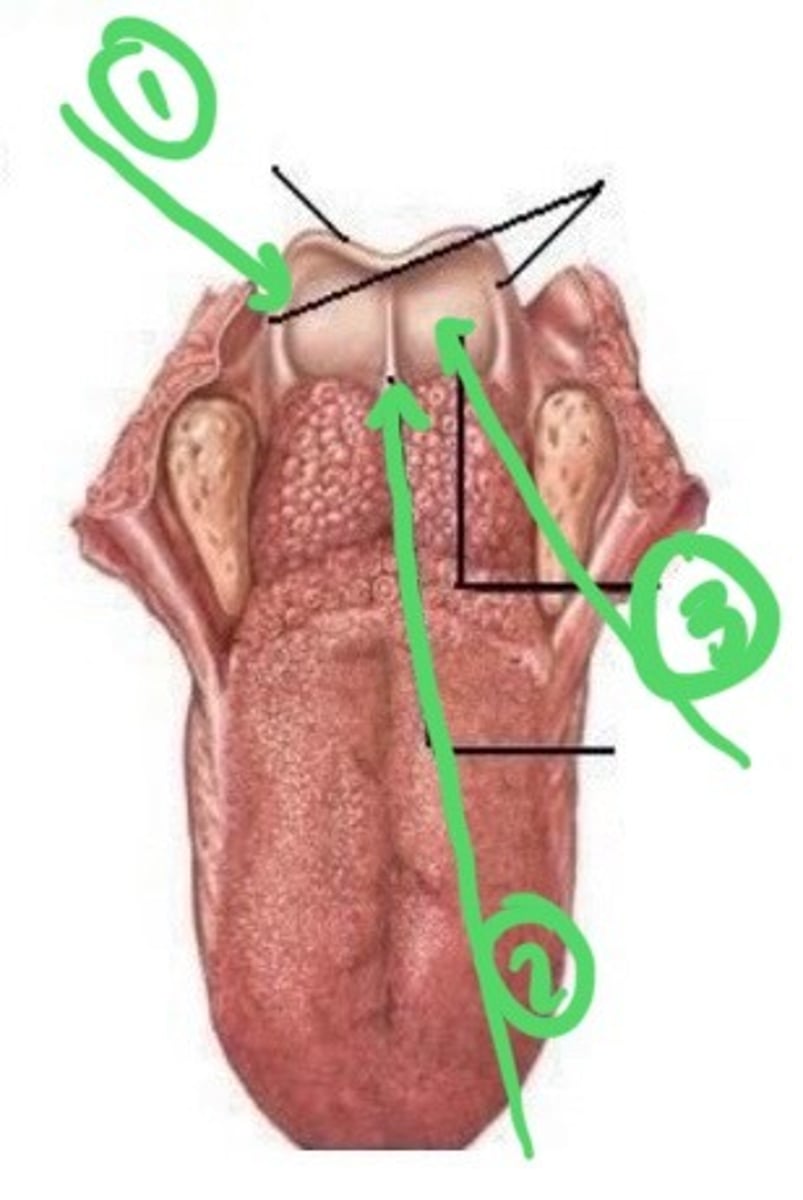
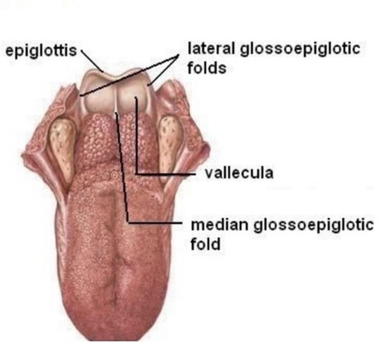
1.) lateral glossoepiglotic fold --> mucosa that attaches the side of the epiglottis to the back of the tongue
2.) median glossoepiglottic fold --> mucosa that attaches the median anterior aspect of the epiglottis to the back of the tongue
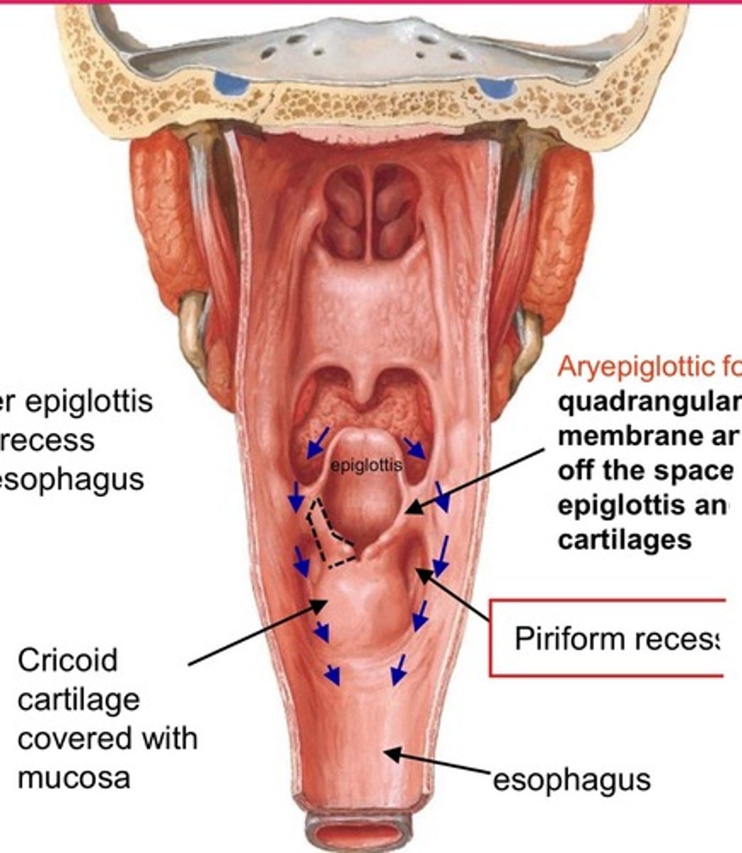
3.) vallecula --> the mucosa that attaches the epiglottis to the back of the tongue (be careful, the space around the epiglottis is also called vallecula)
Epiglottis
leaf shaped flap of elastic cartilage behind the tongue that blocks food from entering the larynx ==> food passes thru the vallecula (space bwtn tongue & epiglottis) --> passes by the sides of epiglottis--> enters piriform recess --> enters the esophagus
-attached to the front of the hyoid bone by hyoepiglottic ligament
-attached to the back of the th
yroid cartilage by thyroepiglottic ligament -lateral edges attached to aryepiglottic folds
-mucosa attached to the tongue anteriorly via lateral/median glossoepiglottic folds & valleculae
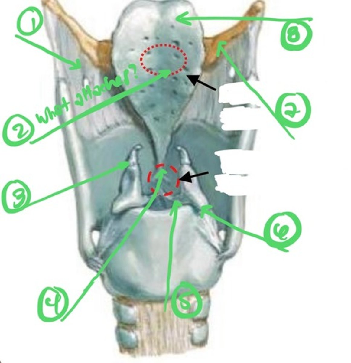
1.) thyrohyoid membrane
2.) hyoepiglottic ligament
3.) corniculate cartilage
4.) thyroepiglottic ligament
5.) vocal process of the arytenoid cartilage
6.) muscular process of arytenoid cartilage
7.) hyoid bone
8.) epiglottis
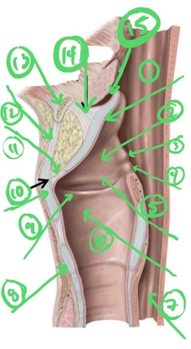
1.) epiglottis
2.) quadrangular membrane
3.) cuneiform tubercle
4.) corniculate tubercle --> from the corniculate cartilage that sits on top the arytenoid carilage
5.) vestibular ligament
6.) conus elasticus
7.) esophagus
8.) cricoid ligament
9.) vocal ligament
10.) thyroid ligament
11.) thyroepiglottic ligament
12.) thyrohyoid membrane
13.) hyoid bone
14.) hyoepiglottic ligament
15.) Vallecula
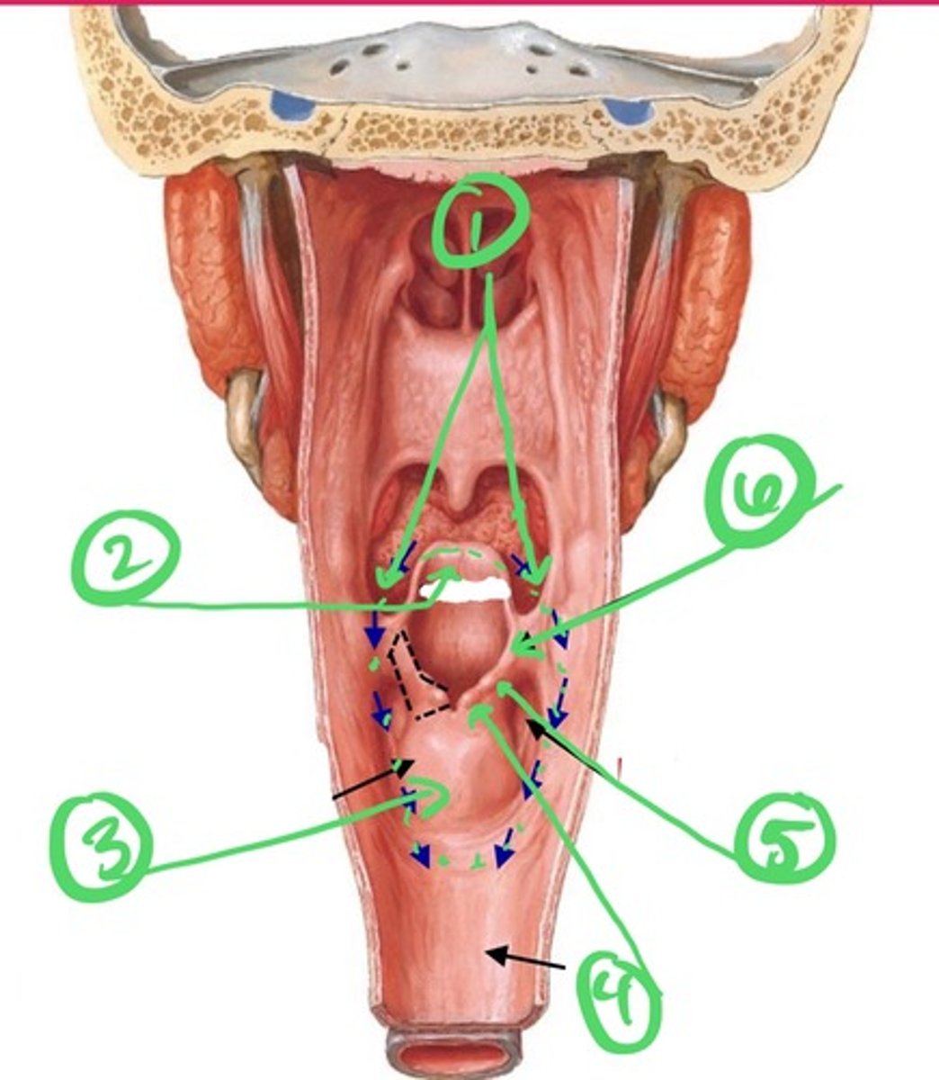
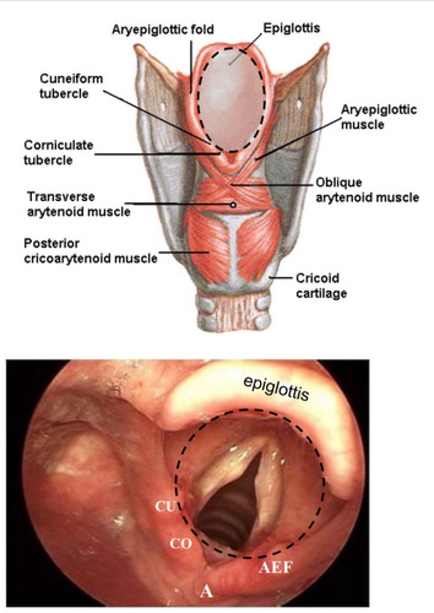
1.) vallecula
2.) epiglottis
3.) piriform recess
4.) corniculate tubercle
5.) cuneiform tubercle
6.) aryepiglottic fold
vallecula
space bwtn the posterior portion of the tongue & epiglottis (not the valleculae mucosa)==> food passes thru here & down into the piriform recess to the esophagus
*has tastebuds that are innervated by Vagus Nerve (CNX)
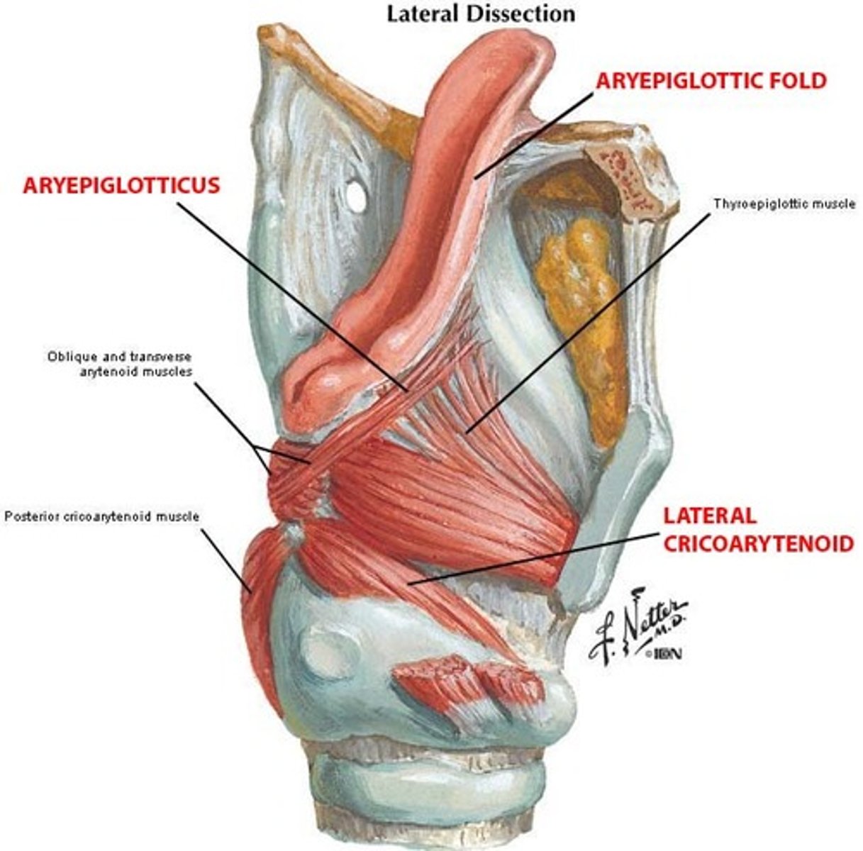
aryepiglottic folds
folds that overlie the quadrangular membrane of the larynx ==> closes off the space bwtn the epiglottis & arytenoid space during swallowing (closes larynx so food doesn't get into the trachea)
*supported by the corniculate & cuneiform cartilages
corniculate cartilage
small conical nodules of cartilage that sits on top of the arytenoid cartilage & supports the aryepiglottic folds (that close the larynx when swallowing)
cuneiform cartilage
small rod shaped cartilage that supports the aryepiglottic fold
*doesn't articulate w/ any other cartilage
True or False: The internal laryngeal nerve & superior laryngeal artery run through openings in the cricothyroid membrane to supply the larynx
False ==> internal laryngeal nerve & superior laryngeal artery run through the thyrohyoid membrane
Which membrane associated with the larynx is incised during cricothyroidotomy?
cricothyroid membrane
cricotracheal membrane
membrane that connects the cricoid cartilage to the 1st tracheal ring
Where is the incision made for a tracheotomy?
cricotracheal membrane
1.) epiglottis
2.) lateral thyroid hyoid ligament
3.) cricothyroid membrane
4.) cricotracheal membrane
5.) opening for the internal laryngeal nerve & superior laryngeal artery
6.) median thyrohyoid ligament
Quadrangular Membrane
membrane of the larynx that extends bwtn the epiglottis & the arytenoids cartilages
-upper edge = aryepiglottic folds
-lower edge = vestibular ligament ( apart of false vocal cords)
conus elasticus
vibrating membrane of the larynx that 's upper free edge forms the vocal ligament (a part of true vocal cords)
*vocal ligament attaches to thyroid cartilage anteriorly & vocal process of arytenoid cartilage posteriorly
*Also contributes to the increased pressure needed for the Valsalva Manuever (pushing a baby out or boo booing)--> closes the rima glottidis--> increase air pressure --> abdominal muscles contract -->>> push baby &/or boo boo out
1.) cuneiform tubercle
2.) epiglottis
3.) quadragular membrane
4.) arytenoid cartilage
5.) ventricle --> space bwtn the false & true vocal cords
6.) conus elasticus
7.) vocal ligament
8.) vestibular ligament
9.) corniculate tubercle
True or False: Closing the rima glottidis contributes to the pushing action of defecation & childbirth
True ==> When the Rima Glottidis is closed outflow of air is stopped & contributes to the increased pressure needed to push baby & boo boo out (along w/ the contraction of the abdominal muscles) = Valsalva Manuever
1.) thyroid cartilage
2.) laminae of cricoid cartilage
3.) conus elasticus
4.) colliculus
5.) corniculate cartilage
6.) muscular process of arytenoid cartilage
7.) vocal process of arytenoid cartilage
8.) arch of cricoid cartilage
9.) vocal ligament
10.) median cricothyroid membrane
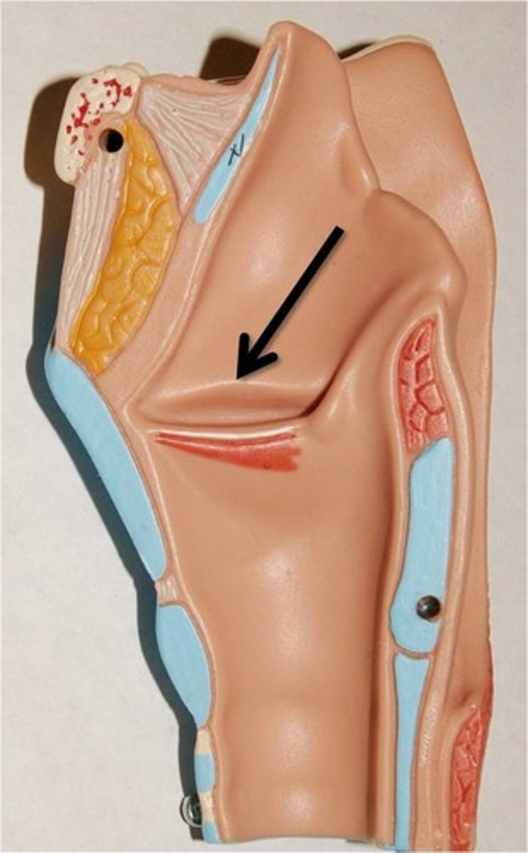
vestibule of larynx
inlet above the false vocal cords (space w/in the quadrangular membrane)
vestibular folds
mucosa of the false vocal cords made up by the lower edge of quadrangular membrane
-serve as protective function
-no part in voice production **
1.) ventricle
2.) true vocal cord
3.) cricoid cartilage
4.) inferior constrictor muscle of the pharynx
5.) vestibular fold (false vocal cord)
6.) vallecula fossa
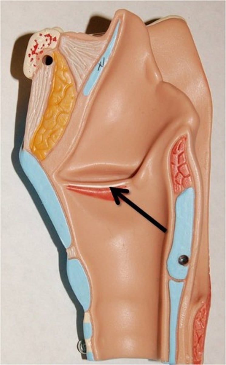
vocal cords
vocal ligaments + thyroarytenoid muscles + vocalis muscles + overlying mucosa
glottis
vocal cords (ligaments, muscles, mucosa) + rima glottidis space
1.) glottis
2.) corniculate cartilage
3.) aryepiglottic fold (cuneiform cartilage in this area)
4.) vocal fold
5.) epiglottis
6.) root of the tongue
7.) vestibular fold
8.) palatine tonsil
laryngeal inlet
2D area that faces backwards & upwards (slanted downward) to open into the laryngeal part of the pharynx
-anterior boundary = upper margin of the epiglottis
-posterior/lower boundary =arytenoid cartilage
-lateral boundary = aryepiglottic folds
rima vestibuli
space between the false vocal folds
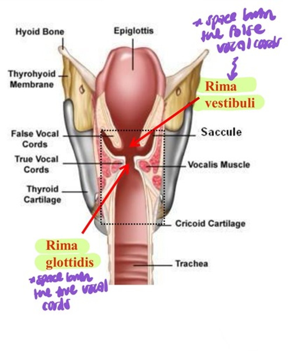
True or False: The rima vestibuli is the narrowest space in the laryngeal cavity
False ==> the rima glottidis is the narrowest space in the laryngeal cavity

extrinsic muscles of the larynx
muscles that elevate or depress the larynx
-suprahyoid muscles = elevators (the digastrics, stylohyoid, & myohyoid)
-sternothyroid muscle = depressor
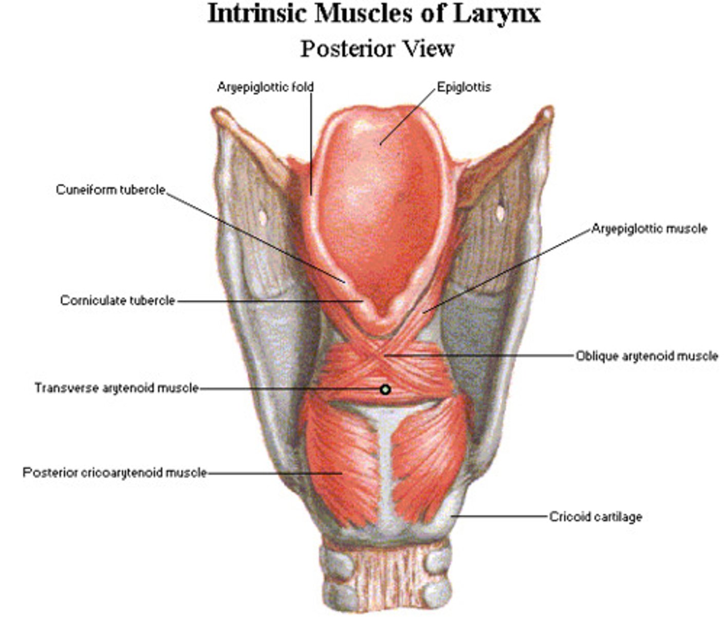
intrinsic muscles of the larynx
muscles that control the size of the laryngeal inlet or control the movements/tension of the vocal cords
-size of l. inlet --> oblique/transverse arytenoid muscles
- vocal cords m. --> cricothyroid, thyroarytenoid, vocalis, latera//posterior cricoarytenoid muscles
*innervated by the recurrent laryngeal nerve
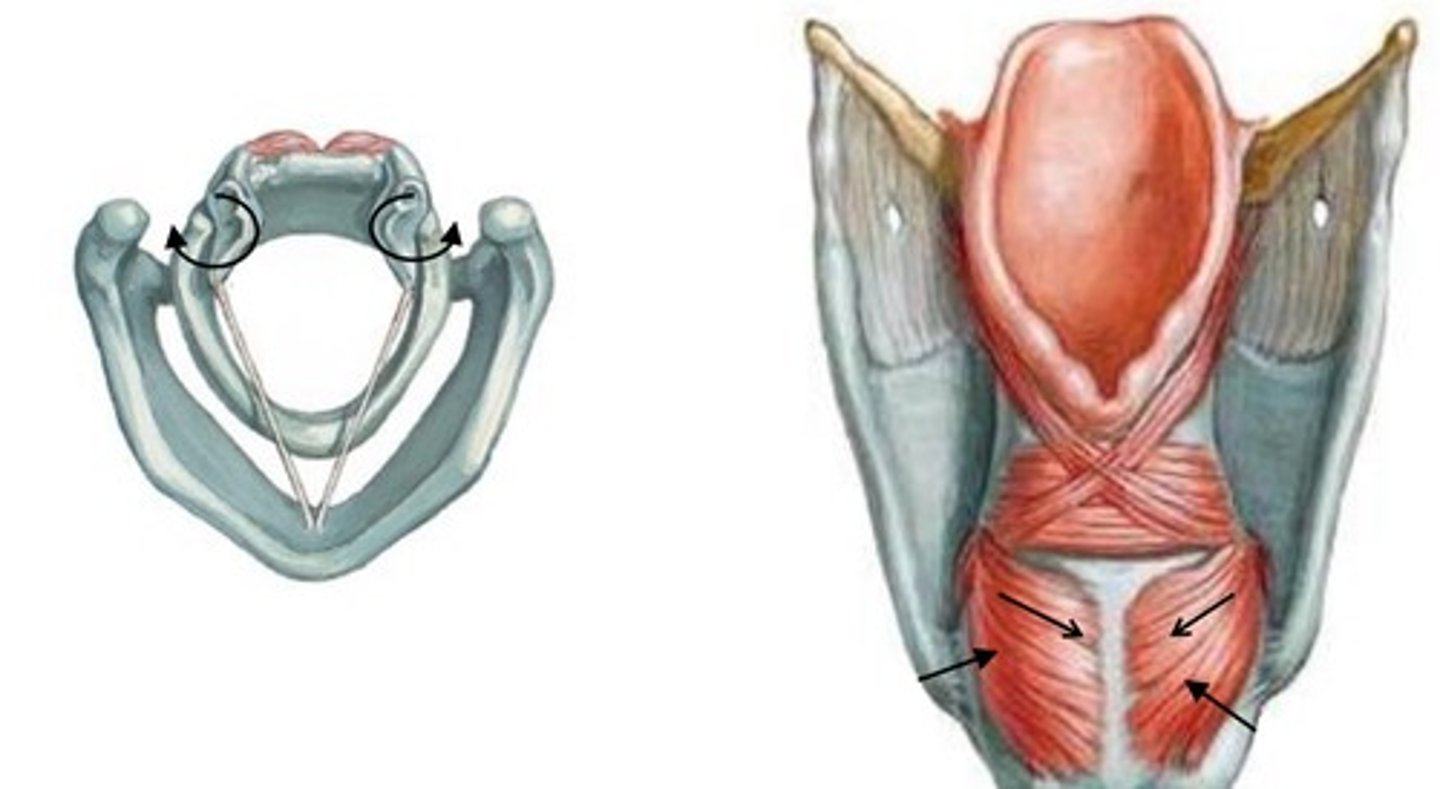
oblique arytenoid muscle
intrinsic laryngeal muscle that obliquely connects the posterior & lateral surfaces of both arytenoid cartilages ==> contracts to narrow the laryngeal inlet & adduct the vocal cords
*innervated by the recurrent laryngeal nerve

transverse arytenoid muscle
intrinsic laryngeal muscle that transversely connects the posterior & lateral surfaces of both arytenoid cartilages ==> contracts to narrow the laryngeal inlet & adducts the vocal cords
*innervated by the recurrent laryngeal nerve
aryepiglottic muscle
the upper fibers of the oblique arytenoid muscle that extends from the arytenoid cartilages to the lateral edge of the epiglottis==> acts as a sphincter of the laryngeal inlet (draws the aryepiglottic folds inward to narrow the inlet like a drawstring bag)
*innervated by the recurrent laryngeal nerve

cricothyroid muscle
intrinsic laryngeal muscle that increases the distance bwtn the angle of the thyroid cartilage & vocal processes of the arytenoid cartilage to increase the length & tension of the vocal cords ==> raises the pitch of the voice (paralysis of this muscle = low pitched voice)
-origin =cricoid cartilage
-insertion = deep surface of thyroid cartilage @ cricothyroid joint
*innervated by the external branch of superior laryngeal nerve (branch of Vagus Nerve CNX)
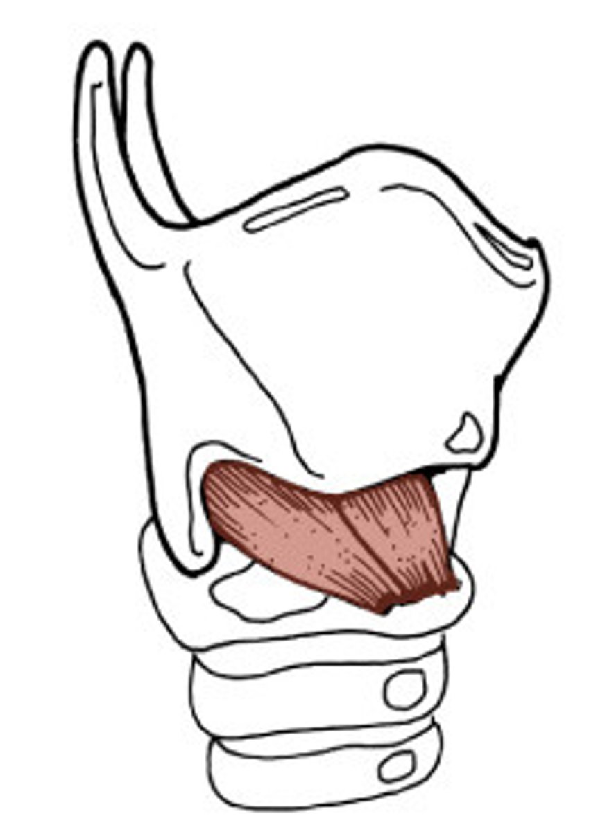
thyroarytenoid muscle
intrinsic laryngeal muscle that pulls the arytenoid cartilage forward towards the thyroid cartilage to shorten & relax the vocal cords ==> lowers the pitch of the voice
*upper part = thyroepiglottic muscle that widens the laryngeal inlet
*innervated by the recurrent laryngeal nerve
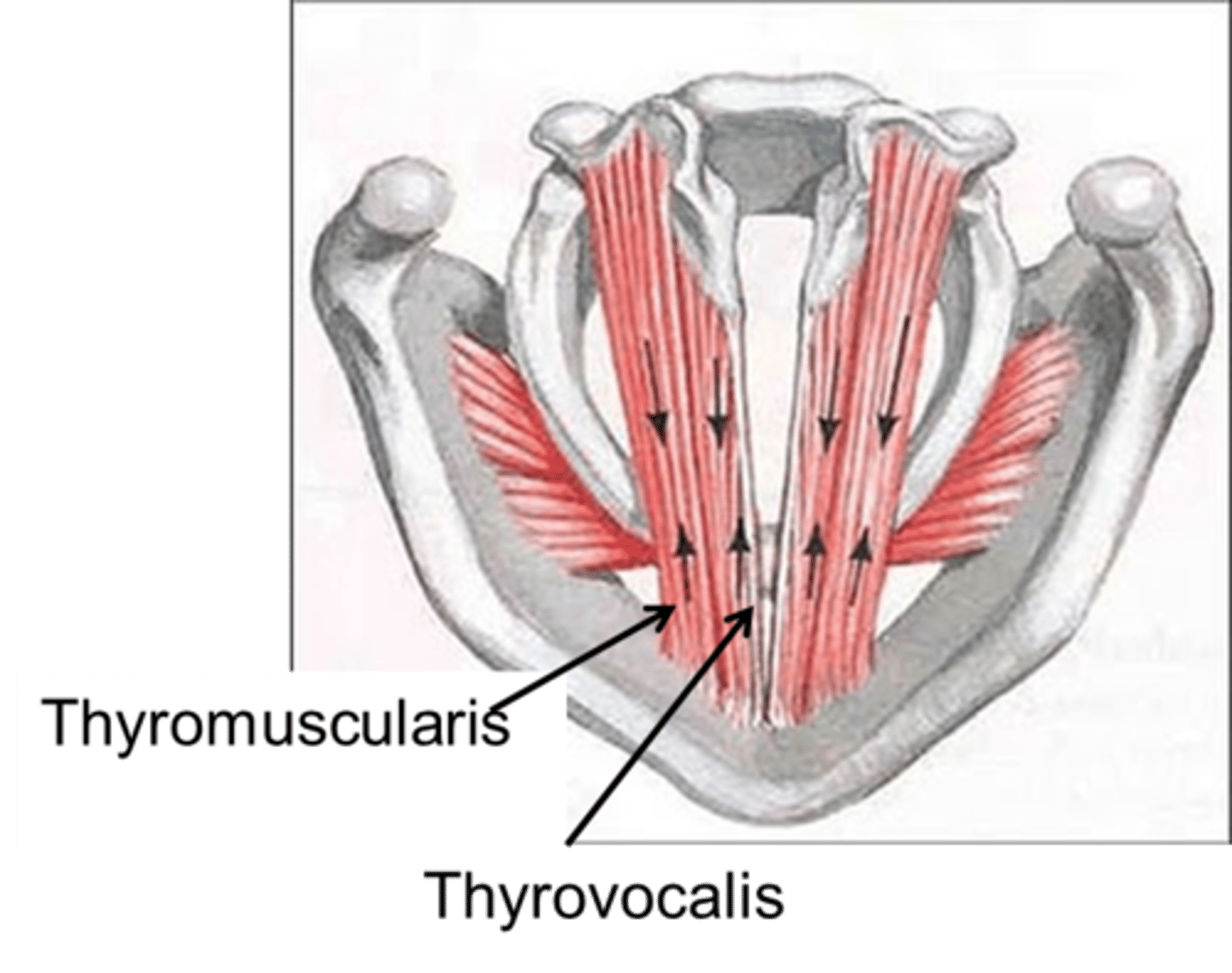
vocalis muscle
intrinsic laryngeal muscle that is in direct contact w/ the vocal ligament ==> responsible for fine control of the vocal cords
*have the ability to selectively tense or relax different parts of the vocalis muslce
*won't really be able to distinguish it in the dissection, but vocalis is more medial than thyroarytenoid muscle
*innervated by the recurrent laryngeal nerve

thyroepiglottic muscle
upper part of the thyroarytenoid muscle that pulls laterally on the aryepiglottic fold & epiglottis to widen the laryngeal inlet
*does the opposite of aryepiglottic muscle
*innervated by the recurrent laryngeal nerve
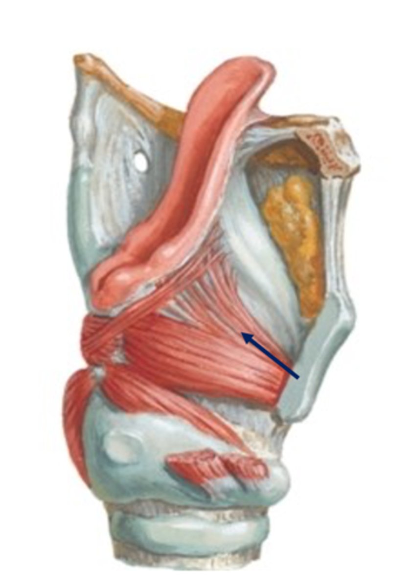
lateral cricoarytenoid muscle
intrinsic laryngeal muscle that medially rotates the arytenoid cartilage in a way that leaves open space posterior to the arytenoid cartilage ==> allows air to to escape w/o passing over the vocal cords & creates whisper = whisperer's muscle
*innervated by the recurrent laryngeal nerve
posterior cricoarytenoid muscle
intrinsic muscle laryngeal muscle that abducts the vocal cords (pivots the arytenoids so that the vocal processes move laterally)
-origin = laminae of cricoid cartilage
-insertion = posterior aspect of muscular process of arytenoid cartilage
*bilateral paralysis of posterior cricoarytenoid muscle adducts the vocal cords ->>> suffocation (cuz they need to be able to abduct to let air through)
*innervated by the recurrent laryngeal nerve
True or False: Branches of the Glossopharyngeal Nerve (CN IX) provides motor & sensory innervation to the larynx
False ==> Branches of the Vagus Nerve (CNX) innervates the larynx
-recurrent laryngeal branches --> dip under the aorta (left side) or subclavian artery (right side) to innervate the intrinsic laryngeal muscles
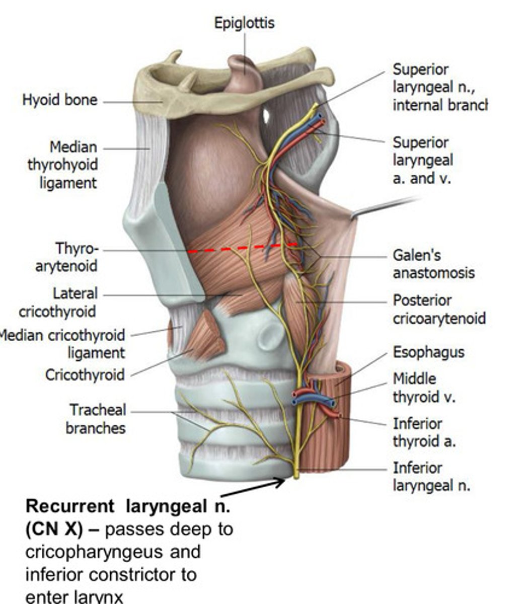
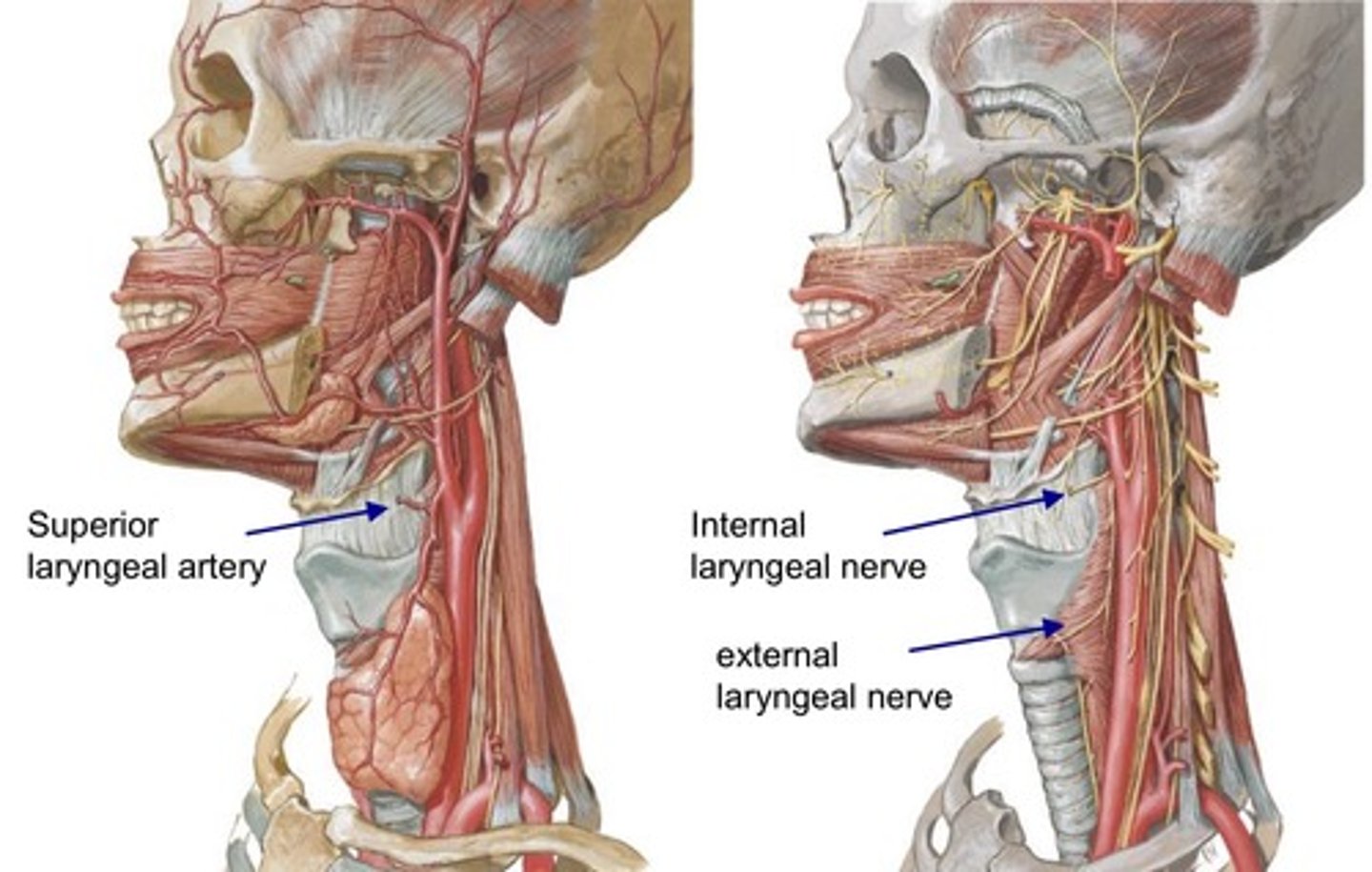
-superior laryngeal branch --> internal laryngeal branch + external laryngeal branch
what provides sensory innervation to the mucosa of the larynx that's above the vocal cords?
internal laryngeal nerve (branch of superior laryngeal branch of Vagus Nerve CN X)
what provides sensory innervation to the mucosa of the larynx below the vocal cords?
recurrent laryngeal nerve (branch of Vagus Nerve CNX)
True or False: All the intrinsic muscles are innervated by the recurrent laryngeal nerve.
False ==> Cricothyroid muscle is innervated by the external laryngeal nerve (the rest of the intrinsic muscles are innervated by the recurrent laryngeal nerve tho)
True or False: The recurrent laryngeal nerve passes deep to the cricopharyngeus & inferior constrictor muscles to enter the larynx
True
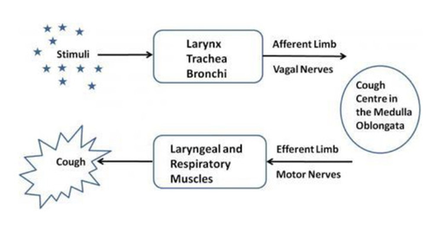
Cough Reflex
foreign object enters the larynx--> sensed by internal laryngeal nerve (afferent limb) --> tells the cerebral cortex & medulla --> send efferent limb signal to vagus nerve, phrenic nerve, & intercostal nerves--> air rushes into the lungs --> transverse arytenoid muscle & lateral cricoarytenoid muscles contract to close the glottis --> contraction of expiration muscles & abdominal muscles force the glottis back open = explosive release of air from larynx = cough
Superior Laryngeal Nerve Block
numbing of the internal laryngeal nerve to block the cough reflex for procedures in larynx (ex: laryngoscopy & bronchoscopy)
*inject at the lateral aspect of the thyrohyoid membrane
spasm of the laryngeal muscles . . .
happens when a foreign object id lodged into the vestibule to close the rima vestibuli & rima glottidis ==> may completely seal off the larynx & cause the person to choke (person is speechless too)
*Tx = Heimlech Maneuver to compress the abdomin--> make the diaphragm elevate --> compress the lungs --> force air out-> forces larynx to open & dislodges the object
Valsalva Manuever
forceful expiratory effort against a closed airway to increase abdominal pressure & push things out (coughing, sneezing, defecation, & childbirth)
*vestibular & vocal folds abduct widely--> lungs inflate (deep inspiration) --> then vestibular & vocal folds voluntarily adducted --> abdominal wall muscles contract --> intra-abdominal & subglottic pressure increases --> stuff gets pushes out (ex: boo boo or a baby)
Superior Laryngeal Nerve Injury
-loss of sensation to upper part of larynx (internal laryngeal branch) & piriform recess = no cough reflex
-paralysis of cricothyroid muscle (external laryngeal branch) = monotone voice b/c the vocal cords can't be tensed
Recurrent Laryngeal Nerve Injury
-bilateral injury --> most intrinsic laryngeal muscles won't work (no abduction, adduction, or relaxation of the vocal cords) = hoarseness & difficulty breathing during forced respiration
>stridor = high pitched sound during respiration
-inability to close the laryngeal inlet during swallowing ==> aspiration of food (food falling into the trachea)
A high pitched sound during respiration is indicative of . . .?
bilateral injury to the recurrent laryngeal nerve ==> most intrinsic laryngeal muscles won't work (no abduction, adduction, or relaxation of the vocal cords) = hoarseness & difficulty breathing during forced respiration
*stridor = the high pitched sound
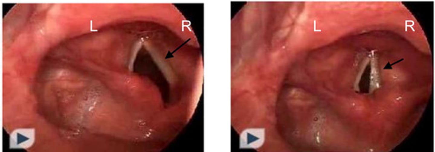
Unilateral Recurrent Laryngeal Nerve Paralysis
*apparent when the one vocal cord doesn't abduct during forced inspiration
Blood Supply of the Larynx
- superior laryngeal artery --> branch of superior thyroid artery (enters thyrohyoid membrane w/ internal laryngeal nerve) that supplies the upper half of larynx
- inferior laryngeal artery --> branch of inferior thyroid artery; supplies the lower half of the larynx
-superior/inferior laryngeal veins run the same way with the arteries
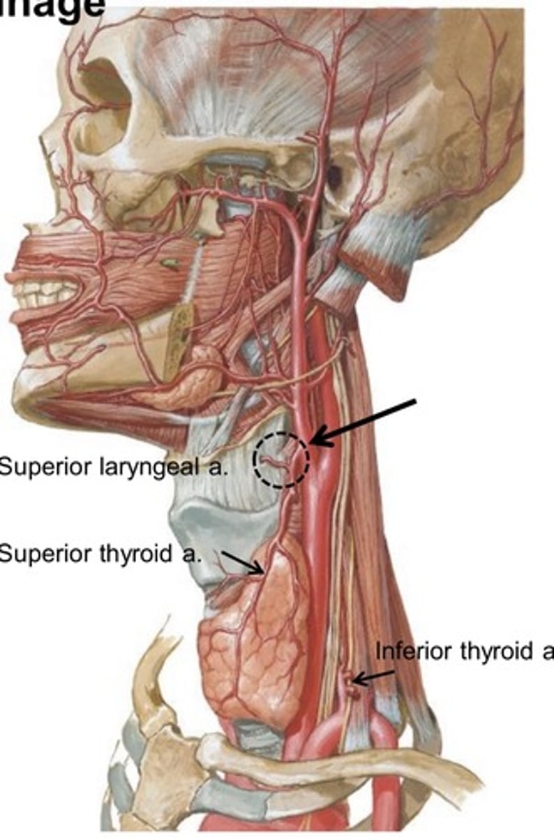
True or False: The external laryngeal nerve can be injured when the superior thyroid artery is ligated.
True ==> the external laryngeal nerve runs right beneath the superior thyroid artery.
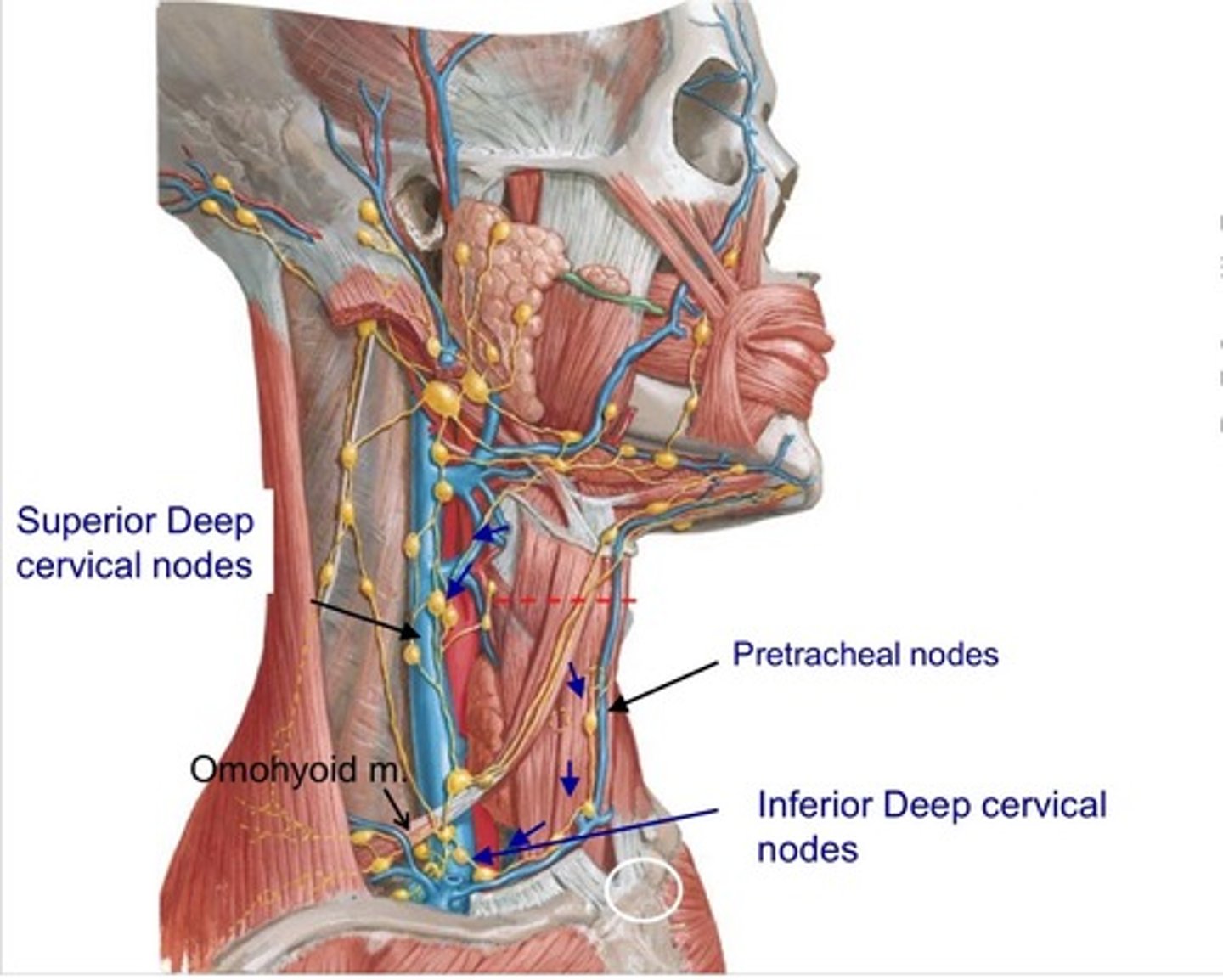
Lymphatic Drainage of the Larynx
-above the vocal cords ==> drain to superior deep cervical nodes (on IJV above the omohyoid muscle)
-below the vocal cords ==> drains to the pretracheal or paratracheal nodes --> inferior deep nodes (on the IJV below the omohyoid muscle)
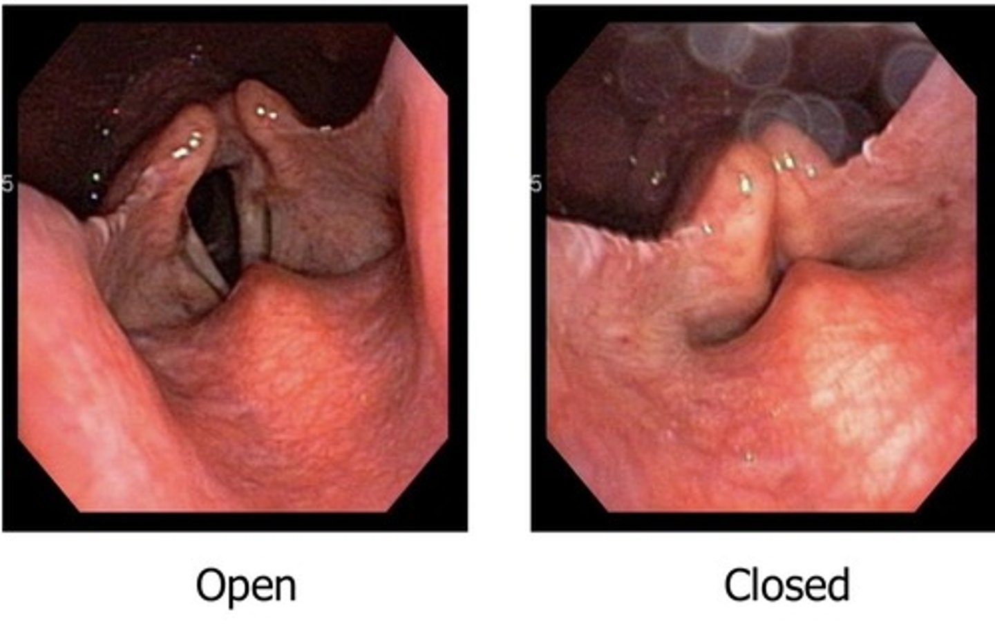
Glottis Open for Inspiration v. Closed for Swallowing
-closes up like drawstring bag due to aryepiglottic muscle
-open up due to thyroepiglottic muscle
Mechanics of Swallowing
bolus of food enters the oral cavity --> tongue & mylohyoid pushes it backwards into the oropharynx --> levator veli palatini elevates soft palate & tensor veli palatini tenses the soft palate + tongue elevated & retracted (via the styloglossus & palatoglossus) + epiglottis depressed--> laryngeal inlet closed by the aryepiglottic & oblique arytenoid muscles + glottis closed by the transverse arytenoid, oblique arytenoid, & the lateral cricoarytenoid muscles-->laryngeal pharynx elevated to catch the bolus of food via the stylopharyngeus, salpingopharyngeus, & palatopharyngeus muscles--> upper esophageal sphincter relaxed --> bolus of food enters the esophagus
*Innervation
-V3 of CNV --> mylohyoid, tensor veli palatini muscles
-CNXII --> styloglossus muscles
-CNIX --> stylopharyngeus muscle
-CNX --> palatoglossus, salpingopharyngeus, & palatopharyngeus muscles
>recurrent laryngeal branch of CNX --> transverse arytenoid, oblique arytenoid, lateral cricoarytenoid, aryepiglottic, oblique arytenoid muscles
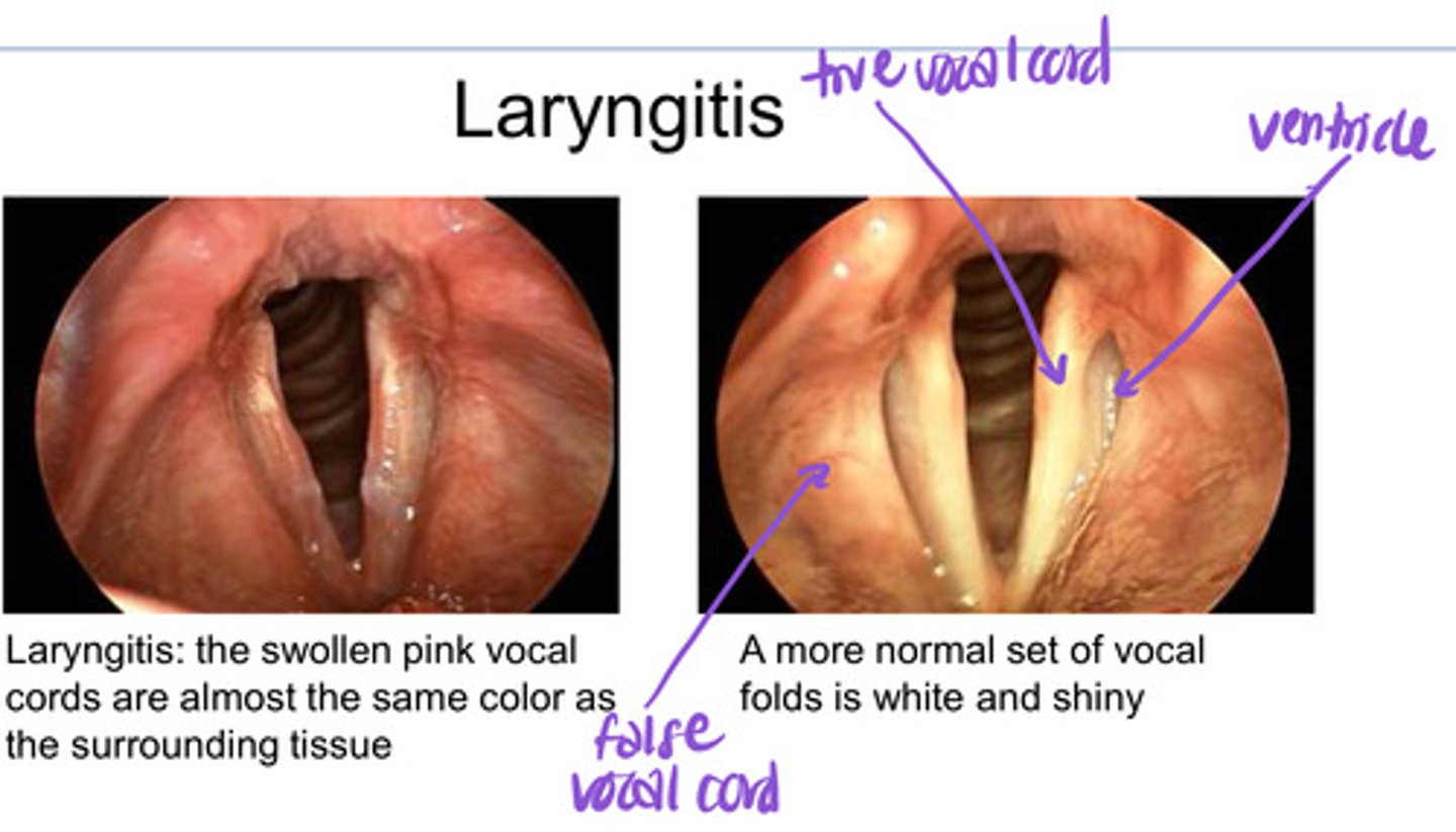
Laryngitis
swollen pink vocal cords due to viral/bacterial infection, excessive use of voice, smoking, excessive alcohol consumption, laryngeal trauma (vocal cords shouldn't be as pink as the surrounding tissue)
-symptoms ==> hoarseness, dry/burning throat, coughing, difficulty swallowing
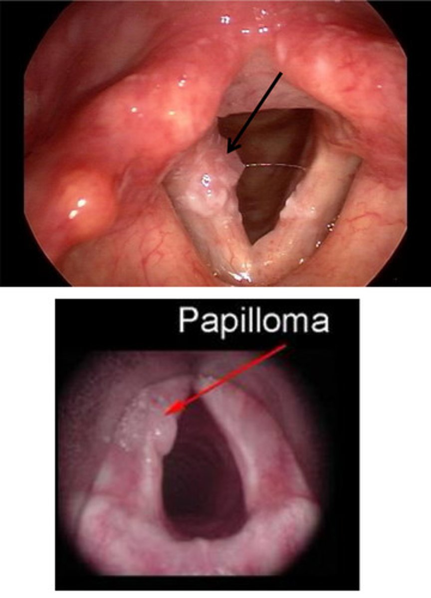
laryngeal papillomas
benign epithelial tumors on the vocal cords of the larynx that are caused by HPV infection (human papilloma virus) ==> causes hoarseness that's proportional to the size of the papilloma
*can eventually get big enough to block the airway
*Tx = laser surgery
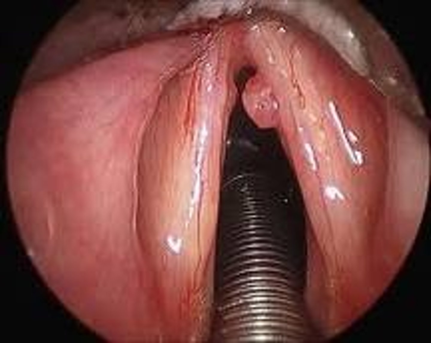
vocal cord polyps
small swellings in the mucous membranes of the vocal cords that develop from voice abuse, chronic allergic rxns in the larynx, & chronic inhalation of irritants (ex: industrial fumes & cigarette smoke)
*usually unilateral
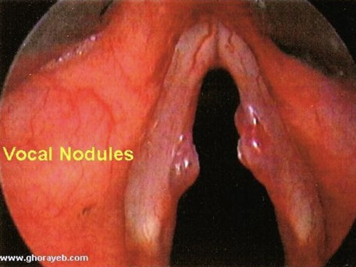
vocal cord nodule
small inflammatory or fibrous growth that develops on the vocal cords of people who constantly strain their voices ==> makes the voice breathy & hoarse
*usually appears in pairs & looks structurally similar to callus on a hand
Tracheotomy
an operative procedure that creates a surgical opening in the cervical trachea due to
-foreign body obstruction of airway
-bilateral vocal cord paralysis
-tumors of larynx & upper airway
-trauma to upper airway
-ventilation of comatose patient
*more dangerous than cricothyrotomy b/c must make the incision near major blood vessels (brachiocephalic trunk/veins)
Tracheostomy
creation of permanent or semi-permanent opening in the trachea ==> transverse incision made across the neck above the jugular notch --> surrounding tissues dissected to identify the thyroid --> isthmus of thyroid divided to expose the trachea --> 2nd tracheal ring divided--> tracheostomy tube placed into trachea
* palpable landmarks = thyroid cartilage notch, cricoid cartilage, trachea
Cricothyrotomy
incision made in cricothyroid membrane to create an artifical airway ==> used in emergencies when the patient can't be intubated or ventilated
*considered safer than tracheotomy due to fewer complications