biomechanics mmm combined
 Call Kai
 Learn
Learn Practice Test
 Spaced Repetition
 Match
Match Flashcards
 Knowt Play
1/219

There's no tags or description
Looks like no tags are added yet.
Name | Mastery | Learn | Test | Matching | Spaced | Call with Kai | Chat |
|---|
No analytics yet
Send a link to your students to track their progress
220 Terms

This term associated with the gait cycle is defined as the interval between two sequential initial floor contacts of the same foot. It is represented by the block arrow in the following diagram:
stride
Double stance refers to:
the time in gait during which both feet are contacting the ground
Double stance occurs twice in the normal gait cycle. When does double stance occur:
at the beginning and end of the stance phase, during initial contact and terminal stance
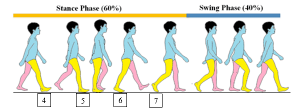
The following diagram depicts several phases of gait:We are considering only the actions of the right lower extremity. For each number listed above, choose the corresponding phase of the gait cycle.
4. This phase of gait begins the stance phase of the gait cycle.
initial contact
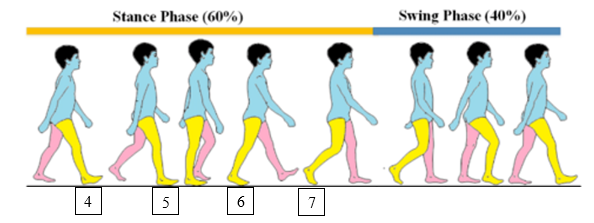
5. This phase of gait occurs next. It is the phase of the gait when the foot is lowered down to the ground and conforms to whatever surface the individual is walking on:
loading response

This phase of gait occurs after mid stance as the gastrocnemius begins to contract to propel the body forward:
loading response

This phase of gait occurs at the very end of stance phase. It is the last phase before the limb enters into the swing phase:
pre-swing
As the gait cycle moves from initial contact to loading response, which of the following muscle activities occurs to lower the foot to the ground:
eccentric contraction by the tibialis anterior and pre-tibial group
At what point in the gait cycle is the quadriceps most active:
just after initial contact or heel strike as the knee flexes to absorb shock and to prevent the knee from collapsing as the body weight is transferred on to the stance limb
Which of the following characteristics is typical of an antalgic gait:
decreased stance time on the painful side
The names of the movements occurring in the primary planes change when considering the movements of the foot and ankle. Movement in the frontal plane was typically called abduction and adduction. When considering foot movement, frontal plane movement is now called:
eversion and inversion
Movement in the horizontal plane was typically called internal and external rotation. When considering foot movement, horizontal plane movement is now called:
abduction and adduction
Tri-planar movement is movement of the foot and ankle that occurs simultaneously in all three primary planes. The terms pronation and supination are used to represent this movement. Open chain pronation consists of what three movements:
dorsiflexion, eversion, and abduction
Movement of the talocrural joint occurs primarily in the sagittal plane, although some movement occurs in the other two primary planes. This movement could be called supination and pronation because it is a tri-planar movement. However, because most of the movement occurs in the sagittal plane, talocrural movement is called:
dorsiflexion and plantarflexion
Which of the following statements best describes the arthrokinematics of the talocrural joint during dorsiflexion:
talus rolls anterior and glides posterior
Movement of the subtalar joint is truly tri-planar movement. Because of the tri-planar nature of the movement, movements of the subtalar joint are called:
pronation and supination
The movements of the subtalar joint change slightly in an open chain versus closed chain situation. During closed chain pronation, what movements occur and what joints are moving:
calcaneus everts, talus adducts and plantarflexes
Movement of the talus in the horizontal plane creates rotation of the tibia. During closed chain subtalar pronation in which direction does the talus and tibia move:
talus adducts, tibia internally rotates
Movement of the subtalar joint controls the amount of movement that can occur in the mid tarsal joints. When the subtalar joint pronates, what occurs at the midtarsal joints:
midtarsal joints unlock and move into their LPP
The pronation and supination of the subtalar joint along with the unlocking and locking of the midtarsal joints, allows the foot to accomplish its two main functions: 1) mobile adaptor - conforming to the walking surface and absorbing shock, and 2) rigid lever - pushing off the ground to propel the body forward. During the loading response phase of gait what is the subtalar joint doing and what foot function is created:
subtalar joint pronates, foot becomes a mobile adaptor

The picture below represents a patient attempting to perform a straight leg raise. This patient has some quadriceps weakness and is unable to maintain the knee in an extended position while performing the straight leg raise. Fully extending the knee through terminal knee extension places large demands on the quadriceps. This inability to maintain the knee in full extension during a straight leg raise is known as:
extensor or quadriceps lag
There are several 2 joint muscles that cross both the hip and the knee joint. Which of the following is NOT a two joint muscle that can move both the hip and the knee:
biceps femoris short head
This 2 joint muscle can flex the hip AND flex the knee:
sartorius
what are medial rotators of the knee:
semimembranosus, sartorius, popliteus
what are the 3 muscles of pes anserine
sartorius, gracilis, semitendinosus
what are functions of the menisci of the knee:
dissipate pressure of the femoral condyles on the tibial plateau
deepen the concavity of the tibial plateaus
improve the congruity between the femoral condyles and the tibial plateau
This part of the meniscus is considered to have the most abundant blood supply and because of that is sometimes referred to as the red-red zone. Tears in this area of the meniscus are often surgically repaired because of the potential for these tears to heal:
outer third

A valgus force at the knee (a force applied to the lateral aspect of the knee and directed medially, abducting the tibia on the femur as pictured below) will most stress which of the following ligaments:
medial or tibial collateral ligament

An anterior drawer test (attempting to pull the tibia anteriorly on the femur as pictured below) will most stress which of the following ligaments:
anterior cruciate ligament
The concave-convex rule dictates the pattern of rolling and gliding that occurs at a joint. Which of the following statements best describes the arthrokinematics that occur with OPEN chain knee extension (tibia moving on femur):
tibia rolls and glides anteriorly on the femur
Which of the following statements best describes the arthrokinematics that occur with CLOSED chain knee extension (femur moving on tibia)
femur rolls anteriorly and glides posteriorly
At the end of CLOSED chain knee extension, some rotation occurs to put the knee in the screw home position. Which bone rotates and in which direction to create the screw home position in CLOSED chain knee extension:
femur internally rotates
Unlocking the knee or moving it out of the screw home position is a necessary first step to allow knee flexion to occur. Which muscle is primarily responsible for the rotation that occurs to unlock the knee and to allow knee flexion to take place:
popliteus
The quadriceps angle or Q angle is an angle that measures the lateral pull of the quadriceps muscle. It is the angle between a line connecting the mid patella and the ASIS and a line connecting:
the mid patella to the tibial tuberosity
An increased Q angle does not correlate with patello-femoral pain. However, an increased dynamic Q angle does correlate with patello-femoral pain. During a single leg squat, what would you observe to indicate an increased dynamic Q angle:
the knee would move medially creating an increased angle of knee valgus
In which of the following muscles, would you expect the highest innervation ratio:
gluteus maximus
A muscle will produce the greatest force when it is held at what percentage of its resting length:
110% stretched slightly
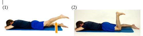
Your patient is lying prone and fully extends his hip (1). He then flexes his knee and tries again to extend his hip but cannot extend his hip as far (2). This lack of hip extension occurs because the hamstrings cannot shorten enough to move the hip through full range with the knee flexed. The hamstrings lack the functional excursion to move all the joints over which they pass through the full range of motion. The hamstrings are experiencing:
active insufficiency

Your patient is lying supine and flexes her knee and hip bringing her knee to her chest (1). She then extends her knee and again tries to flex her hip as far as possible but cannot flex her hip as far (2). In this case, the hamstrings are experiencing:
passive insufficiency
What terms would you use to describe the fiber arrangement of the rectus femoris.
Would it be 1) parallel or oblique and 2) what type of fiber arrangement:
1) oblique, 2) bipennate
What muscle attaches to the medial border of the tibia:
no muscle attaches to the medial border of the tibia
What nerve typically passes around the neck of the fibula:
common peroneal (fibular)
The distal tibia and fibula form the ankle mortise. These structures articulate with which tarsal bone:
talus
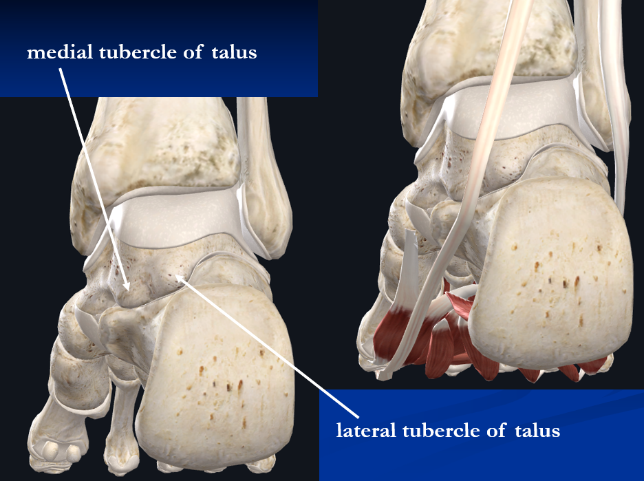
The medial and lateral tubercles of the talus are pictured below. Together they form a small channel for the passage of one of the tendons of the foot. What is the name of the tendon that passes between these tubercles:
flexor hallucis longus
The subtalar joint involves the articulation between the talus and the:
calcaneus
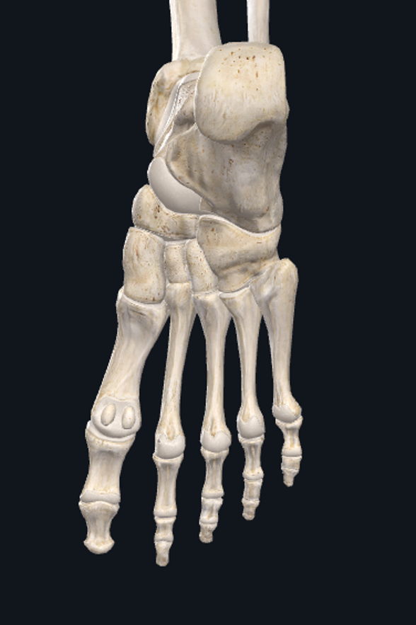
The medial process of the calcaneus pictured below is the attachment of what ligamentous structure:
plantar fascia
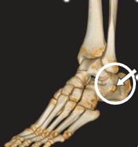
What is the bony landmark of the foot circled in the following picture:
peroneal tubercle
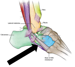
One tendon passes superior to the landmark circled above in Question #7, and one tendon passes inferior to this landmark. Which tendon, pointed to by the arrow in the following picture, passes inferior to this landmark:
peroneus longus
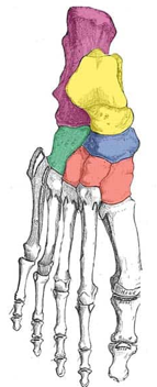
The most prominent tuberosity on the medial aspect of the foot is pictured below. This tuberosity is part of the bone that functions as the keystone of the medial longitudinal arch. What is the name of this tuberosity:
navicular tuberosity
The foot consists of both longitudinal and transverse arches. The keystone of the transverse arch that runs across the midfoot is this bone. This bone is also the high point of the dorsum of the foot:
middle cuneiform
Two of these muscles have distal attachments around the base of the 5th metatarsal. Which one of these muscles does NOT have an attachment to the 5th metatarsal:
peroneus longus
Which of the following statements about the tibialis anterior is NOT correct:
the proximal attachment of the tibialis anterior is the medial tibia and interosseous membrane
Which of the following muscles could still plantarflex the ankle if the Achilles tendon were completely torn:
tibialis posterior
Which of the following muscles is both a talocrural dorsiflexor and a subtalar everter:
extensor digitorum longus
Which of the following muscles is both a talocrural plantarflexer and a subtalar inverter:
flexor hallucis longus
There is only one foot intrinsic muscle located on the dorsum of the foot. This muscle is the:
extensor digitorum brevis
Which of the following is a 2 joint muscle:
gastrocnemius
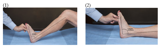
Your patient is lying on a table with her knee flexed. She maximally dorsiflexes her ankle and is able to dorsiflex her ankle 20 degrees (#1 in picture below). She then extends her knee and again maximally dorsiflexes her ankle. This time she is only able to dorsiflex her ankle 5 degrees (#2 in picture below). What is causing this change in available ROM:
the gastrocnemius becomes passively insufficient when the knee is extended and does not allow as much dorsiflexion
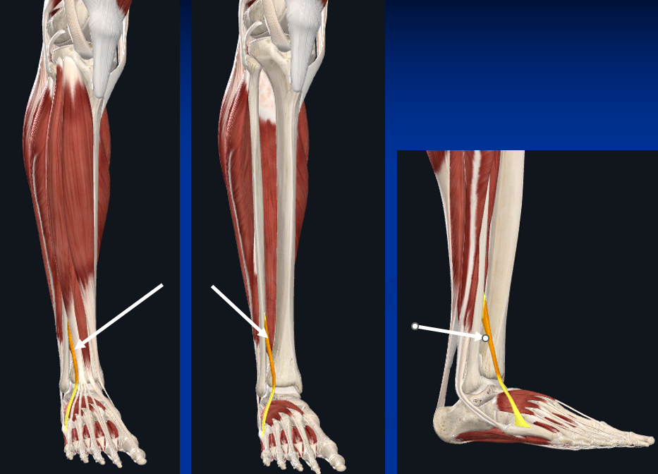
There are 3 different "peroneal" muscles. To which peroneal muscle are the arrows in the following picture pointing:
peroneus tertius
What artery lies between the extensor hallucis longus tendon and the extensor digitorum longus tendons:
dorsalis pedis
The shoulder complex consists of 5 joints. Which of the following joints of the shoulder complex is NOT a true synovial joint:
scapulothoracic joint

What is the name of the bony landmark circled in the picture below:
lesser tubercle
What muscle attaches to the landmark pictured in Question #2 to the shaded area within the circle:
subscapularis
The rotator cuff muscles create one of the deepest layers of muscles in the shoulder joint, assist with moving the humerus on the glenoid, and help to stabilize the humeral head in the glenoid fossa. The rotator cuff muscles include the supraspinatus, infraspinatus, teres minor, and the subscapularis. However, only one of these rotator cuff muscles is an internal rotator. Which of the following rotator cuff muscles is an internal rotator of the glenohumeral joint:
subscapularis
Only one of the rotator cuff muscles is an abductor. Which of the following muscles is an abductor of the glenohumeral joint:
supraspinatus
What is the name of the tendon that passes through the intertubercular groove between the greater and lesser tubercles:
biceps long head tendon
In the anatomic position the glenoid fossa of the scapula does not face directly laterally. Considering the plane of the scapula and the orientation of the glenoid, the glenoid fossa actually faces:
lateral, anterior, and superior
Three muscles attach to the anterior humerus over the crest of the greater tubercle, the crest of the lesser tubercle, and the bone in between the two crests. Which of the following muscles does NOT have an attachment to the anterior humerus near these crests:
teres minor
The three other muscles listed as incorrect answers in Question #8 have very similar actions. All three of these muscles can perform which of the following actions:
internal rotation and adduction

The levator scapulae is a muscle that can elevate the scapula, downwardly rotate the scapula, and retract the scapula. Choose the letter below that best marks the attachment of the levator scapulae to the scapula:
A
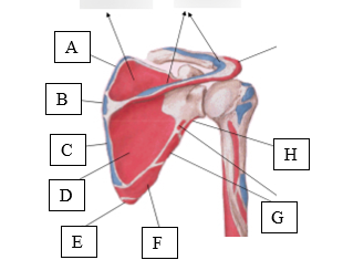
Choose the letter from the following picture that best marks the attachment of the teres major:
F
There are several muscles that attach to the coracoid process. Which of the following muscles does NOT attach to the coracoid process:
pectoralis major
This ligament blends with and reinforces the anterior glenohumeral joint. It pulls tight during external rotation and may be torn when the shoulder dislocates. This ligament is known as the:
glenohumeral or Z ligament
The upper trapezius and levator scapulae work together to create what motion of the scapula:
elevation
During elevation of the scapula, the clavicle must move to accommodate the movements of the scapula. Which of the following statements best describes the arthrokinematics that occur at the sternoclavicular joint during scapula elevation:
proximal end of the clavicle rolls superiorly and glides inferiorly
All of the following can cause or allow depression of the scapula EXCEPT:
all of these can cause depression of the scapula
pectoralis minor
lower trapezius
gravity and eccentric lowering of the upper trapezius
The upward rotation of the scapula occurs simultaneously with elevation of the humerus to allow the humerus to be raised 180 degrees. This simultaneous movement is called scapulohumeral rhythm. Approximately what is the ratio of humeral movement to scapula movement:
2:1
The upward rotation of the scapula is important not only in providing additional movement of the humerus overhead but also in stabilizing the scapula to create a firm base for the glenohumeral muscles. If your patient is abducting his shoulder against resistance and the scapula upward rotators are weak, the deltoid will overpower the scapular muscles and cause the scapula to downward rotate and “wing.” What muscle is the primary scapular stabilizer during upward rotation to prevent this winging:
serratus anterior
Which of the following muscles is NOT a glenohumeral internal rotator:
teres minor
What occurs arthrokinematically at the glenohumeral joint during abduction:
humeral head rolls superiorly and glides inferiorly
Which of the following joints is NOT located inside the capsule that anatomically defines the “elbow” joint:
distal radio-ulnar joint
Which of the following joints of the elbow and forearm complex is NOT a true synovial joint:
mid radio-ulnar joint
The humero-radial joint is formed by which two bony structures:
the capitulum and the radial head
Mary Smith is referred to physical therapy for treatment of right elbow pain. You assess and measure the carrying angle of her elbows. The carrying angle of her right elbow is 27 degrees, and her left elbow 18 degrees. Based on this finding, you could make which of the following statements:
she has excessive cubital valgus in her right elbow, but her left elbow is within normal limits
This nerve passes through the spiral groove of the humerus and may be injured by humeral shaft fractures.
radial
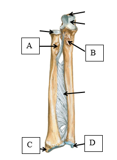
The biceps brachii and the brachialis brachii are both elbow flexors with distal attachments to the forearm. Use the above figure to identify their attachments. Which of the following statements is correct with regard to their attachments:
the biceps attaches at letter A and the brachialis attaches at letter B
Because of its attachment to the radius this muscle can function not only as an elbow flexor but also a forearm supinator:
biceps brachii
This muscle is the "work horse" of the elbow flexors and functions in elbow flexion with the forearm in any position and at any speed of contraction. It does not have a role in supination and pronation:
brachialis brachii
Energy efficiency is always a concern for the body as it decides which muscles to recruit. Recruiting the biceps to flex the elbow requires what shoulder muscle to provide adequate stabilization at the shoulder and to prevent the shoulder flexion that the biceps may create:
posterior deltoid
Two joint muscles may be recruited because they can more efficiently complete a complex task that involves movement at both joints that the muscle crosses. Which muscle is often recruited for a "pulling" motion because as it shortens at the elbow, it lengthens at the shoulder and maintains a better length-tension ratio:
biceps brachii
As the name suggests, the triceps has 3 heads. Which head functions as a two joint muscle, being able to extend the elbow and extend the shoulder:
long head
This is a common bursitis that occurs about the elbow. It occurs when students lean on their elbow in class or when individuals confined to wheelchairs lean on their elbow on the arm rest of the chair. It involves irritation of the bursa between the point of the elbow and the skin. This bursitis is known as:
olecranon bursitis
The ulnar collateral ligament or medial collateral ligament of the elbow is stressed by 2 mechanisms: 1) a throwing motion with the shoulder moving to an extreme position of external rotation and 2) which of the following forces at the elbow joint:
valgus force – trying to pull the forearm into abduction
Tearing of the ulnar collateral ligament or fractures around the elbow joint may lead to irritation of a nerve that passes through the cubital tunnel under this ligament. Irritation of this nerve may lead to numbness in the medial side of the hand and weakness of the hypothenar eminence muscles. This nerve is the:
ulnar nerve
This ligament encircles the radial head and holds the radial head against the ulna. If the radial head pulls out of this ligament, it creates a condition known as a “pulled elbow” or “nurse maid’s elbow”. This ligament is the:
annular ligament
Moving into extension from slight flexion, how does the head of the radius move on the capitulum at the humero-radial joint:
radial head rolls and glides dorsally
Moving into extension from slight flexion, how does the trochlear notch move on the trochlea at the humero-ulnar joint:
trochlear notch rolls and glides dorsally
The loose packed position of the humero-radial joint is which of the following:
full extension and full supination
You are trying to determine the end feel of elbow extension. You passively extend your patient’s elbow and then apply overpressure to push farther into the limitation of motion. In a normal elbow with elbow extension, you should feel what kind of end feel:
hard
During supination from the anatomical position, how does the head of the radius move on the radial notch of the ulna at the proximal radio-ulnar joint:
radial head rolls dorsolateral and glides volarmedial