asthma and other respiratory disorders
 Call Kai
 Learn
Learn Practice Test
 Spaced Repetition
 Match
Match Flashcards
 Knowt Play
1/75

There's no tags or description
Looks like no tags are added yet.
Name | Mastery | Learn | Test | Matching | Spaced | Call with Kai | Chat |
|---|
No analytics yet
Send a link to your students to track their progress
76 Terms
croup
epiglottitis
bacterial tracheitis
laryngomalacia
vocal cord paralysis
foreign body aspiration
Type of Noise | Suggests Obstruction |
|---|---|
Inspiratory stridor | Supraglottic or glottic (upper airway) |
Biphasic stridor | Fixed lesion at the glottis or subglottis |
Expiratory stridor/wheeze | Intrathoracic trachea or bronchi (lower airway) |
Sound | Think | Location | Examples |
|---|---|---|---|
Inspiratory stridor | Upper airway obstruction | Larynx/trachea | Croup, epiglottitis, foreign body |
Expiratory wheezing | Lower airway obstruction | Bronchi/bronchioles | Asthma, bronchiolitis (RSV) |
Biphasic stridor | Fixed/central airway obstruction | Subglottis/trachea | Vascular ring, subglottic stenosis |
differential diagnosis for stridor
- Asthma
-Bronchiolitis (RSV)
- Viral-induced wheeze
- Allergic reaction/anaphylaxis
- Foreign body aspiration
- Tracheomalacia
differential diagnosis for wheezing
Choanal stenosis/atresia
-congenital problem in the neonatal period, relatively rare
-obstruction off nasal passages----> Cyanosis that WORSENS with feeding and IMPROVES with crying
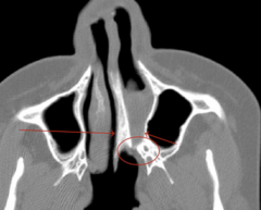
Inability to easily pass a small catheter through the nostrils should raise the suspicion of choanal atresia.
The diagnosis is confirmed by CT scan and by inspecting the area directly with a flexible nasopharyngoscope.
An oral airway may be useful in the short term, but the definitive treatment is surgery
how do you diagnose choanal atresia?
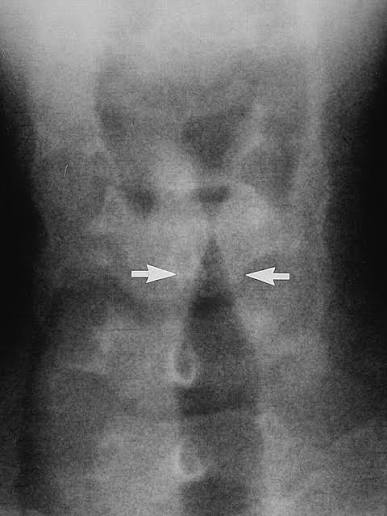
Croup (laryngotracheobronchitis)
Parainfluenza virus (most common),
6mo’s-3yrs
Inspiratory stridor
Barking or brassy harsh cough
Hoarseness,
worst at night, Low grade fever
CROUP: (laryngotracheobronchitis)
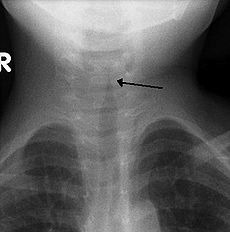
steeple sign due to subglottic narrowing on CXR
diagnostic finding of ?
Mild: dexamethasone PO.
Moderate-severe: nebulized racemic epinephrine + dexamethasone.
Oxygen if hypoxic.
treatment of croup
HiB »
GAS, staph a.
—
note: 2–7 yr (less common after Hib vaccine)
most common agents of epiglottitis
epiglottitis
Prefer sitting head held forward, mouth open, jaw thrust forward (sniffing position)
Muffled voice, sore throat, fever, drooling/secretions, dyspnea or stridor
epiglottis:
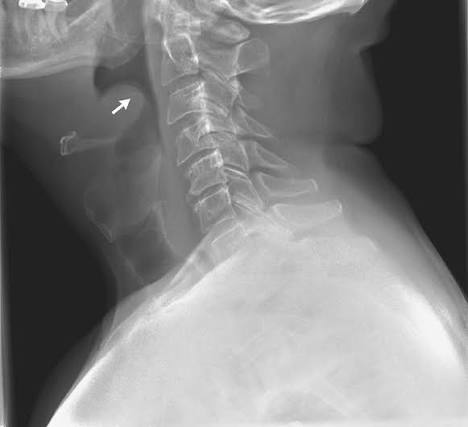
thickened and bulging epiglottis (thumb print sign)
CXR finding of?
Secure airway first (ET tube),
then IV ceftriaxone or cefotaxime ± vancomycin
can be extubated 48-72 hours
management of epiglottitis
Laryngomalacia
Floppy/immature supraglottic structures collapse during inspiration
dx: Flexible laryngoscopy
Infant presents within the first few weeks of life with:
inspiratory stridor
WORSE with feeding, crying, or lying SUPINE
and improves when PRONE/upright.
resolve on own usually by 20 months (12-24mo’s)
severe cases → supraglottoplasty
management of laryngomalacia
bacterial tracheitis
-staph aureus
preschool/school age children
Superinfection of the trachea that may follow viral croup and most commonly caused by S. aureus
High fever, toxic appearance, stridor, thick purulent secretions, poor response to racemic epinephrine
Irregular tracheal narrowing, subglottic edema in imagining |
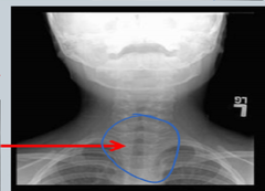
Toxic appearing
Spasmodic croup usually at night
inspiratory stridor
thick purulent secretions
signs and symptoms of bacterial tracheitis
subglottic stenosis
Narrowing of the space below the glottis and above the first tracheal ring;
involves narrowing of the cricoid, the only complete cartilage ring in the airway
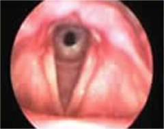
often occurs after long periods of intubation and ventilation for respiratory problems
cause of acquired subglottic stenosis
Recurring croup
Respiratory distress
Noisy breathing
Inability to breathe without tracheostomy
signs and symptoms of subglottic stenosis
damage to recurrent laryngeal nerve (CN X) or the vagus nerve itself.
Category | Associated Conditions | Board Pearls |
|---|---|---|
Birth trauma | Difficult vaginal delivery, forceps delivery, shoulder dystocia | Most common cause of unilateral VCP in neonates. |
Congenital CNS abnormalities | Arnold-Chiari II malformation, hydrocephalus, meningomyelocele | ⭐ Chiari II is the most tested congenital association, especially with bilateral VCP. |
Cardiac surgery | PDA ligation, aortic arch repair, congenital heart surgery | Injury to the left recurrent laryngeal nerve is common because it loops under the aortic arch. |
Neck/thoracic surgery | Thyroid surgery, tracheoesophageal fistula repair, ECMO cannulation | Surgical nerve injury is a common acquired cause. |
Neurologic disorders | Brainstem tumors, cerebral palsy, hypoxic-ischemic injury, cranial neuropathies | Consider when VCP occurs with other neurologic deficits. |
Trauma | Intubation trauma, neck trauma | May cause temporary or permanent paralysis. |
Mass lesions | Mediastinal tumors, vascular rings, enlarged lymph nodes | Compression of the recurrent laryngeal nerve. |
Idiopathic | No identifiable cause | Accounts for a significant proportion of pediatric cases. |
conditions associated with vocal cord paralysis
low dose ICS
Medication | Typical Low Daily Dose* | Notes |
|---|---|---|
Fluticasone propionate (Flovent HFA) | 88 mcg/day (44 mcg, 1 puff BID) to 176 mcg/day | Very commonly used in children |
Budesonide (Pulmicort Flexhaler) | 180–360 mcg/day | Dry powder inhaler (older children) |
Budesonide nebulizer (Pulmicort Respules) | 0.5 mg/day | Preferred ICS for infants and young children who need nebulizer therapy |
Beclomethasone (QVAR RediHaler) | 80–160 mcg/day | Breath-actuated inhaler |
Mometasone (Asmanex) | 110 mcg once daily (age-dependent) | Once-daily option in many patients |
Ciclesonide (Alvesco) | 80 mcg/day | Lower oropharyngeal deposition due to prodrug activation |
Step | Treatment |
|---|---|
Step 1 (Intermittent) | Albuterol (SABA) as needed |
Step 2 (Mild persistent) | ⭐ Low-dose ICS daily + albuterol PRN |
Step 3 | Low-dose ICS/LABA or medium-dose ICS |
Step 4 | Medium-dose ICS/LABA |
Step 5–6 | High-dose ICS/LABA ± biologic therapy ± oral corticosteroids (specialist management) |
treatment of step 2 persistent asthma
Low dose ICS + LABA
or
medium dose ICS
Regimen | Examples | When Used | Board Pearl |
|---|---|---|---|
Low-dose ICS + LABA | Fluticasone/salmeterol (Advair), Budesonide/formoterol (Symbicort), Mometasone/formoterol (Dulera) | Preferred Step 3 option for many children ≥5–6 years and adolescents | ⭐ Preferred over simply increasing the ICS dose in many current guidelines. |
Medium-dose ICS | Increase fluticasone, budesonide, beclomethasone, etc., to the medium-dose range | Alternative if LABA cannot be used or in younger children | Common alternative, especially in younger children. |
Age | Preferred Step 3 |
|---|---|
0–4 years | ⭐ Increase to medium-dose ICS (LABAs are generally not first-line in this age group) |
5–11 years | Low-dose ICS + LABA or medium-dose ICS |
≥12 years | ⭐ Low-dose ICS + LABA is generally preferred; SMART therapy may be appropriate for some patients using an ICS-formoterol inhaler |
Medication | ICS | LABA |
|---|---|---|
Advair | Fluticasone | Salmeterol |
Symbicort | Budesonide | Formoterol |
Dulera | Mometasone | Formoterol |
Breo Ellipta | Fluticasone furoate | Vilanterol (typically for older adolescents/adults, not commonly used in younger children) |
treatment of step 3 persistent asthma
medium dose ICS + LABA
treatment of step 4 persistent asthma
high dose ICS + LABA
treatment of stepp 5 persistent asthma
high dose IC + LABA + oral corticosteroid
Therapy | Examples | Purpose |
|---|---|---|
High-dose ICS | High-dose fluticasone, budesonide, beclomethasone | Maximize anti-inflammatory effect |
LABA | Salmeterol or formoterol (always combined with an ICS) | Long-acting bronchodilation |
Oral corticosteroid | Prednisone or prednisolone | Reserved for severe, refractory asthma after specialist evaluation |
Biologic therapy | Omalizumab, Mepolizumab, Benralizumab, Dupilumab (depending on age and asthma phenotype) | Preferred add-on for many eligible patients to reduce exacerbations and minimize steroid exposure |
Step | Treatment |
|---|---|
Step 1 | Albuterol as needed |
Step 2 | Low-dose ICS |
Step 3 | Low-dose ICS + LABA or medium-dose ICS |
Step 4 | Medium-dose ICS + LABA |
Step 5 | High-dose ICS + LABA ± biologic (specialist referral) |
Step 6 | High-dose ICS + LABA + consider maintenance oral corticosteroids if still uncontrolled |
treatment of step 6 persistent asthma
intermittent asthma
Symptoms less than 2 times/week and <2 nighttime symptoms/month
Tx with SABA
mild persistent asthma
Symptoms: >2x/wk but <1x/d
Night Sxs: >2x/mo
FEV: >80% prediction
Tx: *Low-dose ICS* > Cromolyn, leukotriene (S/E: ↑ mucous production)
moderate persistent asthma
FEV1/PEF 60-80%, daily symptoms. SABA, daily moderate dose ICS, or low dose ICS and LABA
severe persistent asthma
FEV1/PEF <60%, symptoms most of day. SABA, high dose ICS, LABA, oral steroid daily
-nightly wakenings
Ongoing self-management, detecting early decline before symptoms
indication to measure peak expiratory flow
peak expiratory flow meter
Device measuring maximum airflow during exhalation.
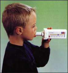
spirometry
-gold standard for diagnosis of asthma and assessing severity
normal ~0.75-0.80 in adults,
≥0.85 in children
what is a normal FEV1/FVC ratio
↑ FEV₁ ≥ 12% and ≥ 200 mL = significant reversibility
post bronchodilator response FEV1 value supporting asthma diagnosis
FVC
total air exhaled after full inspiration
Asthma episode not controlled in 30-60 min with standard treatment
Hypoxemia, hypercarbia, secondary respiratory failure
define status astmaticus
O2
SABA
anticholinergic
systemic corticosteroidsd
management of status asthmaticus
Tracheomalacia | |
|---|---|
Classic presentation | Infant with noisy breathing + barking/brassy cough |
Breath sound | Expiratory stridor/wheeze |
Worse with | Crying, coughing, feeding, respiratory infections |
Why expiration? | Intrathoracic pressure rises → weak trachea collapses |
Associated with | Tracheoesophageal fistula/esophageal atresia, vascular compression, prolonged intubation |
Diagnosis | Dynamic bronchoscopy = gold standard |
Treatment | Most cases → observation/supportive care |
Severe disease | CPAP may splint airway open; occasionally surgery |
Prognosis | Congenital cases often improve as cartilage strengthens |
congenital Floppy tracheal cartilage → airway collapse during expiration
newborn screening (IRT)
IRT = Immunoreactive Trypsinogen
Sweat chloride test (>60 mmol/L),
CFTR genetic testing,
diagnostic approach to cystic fibrosis
👶 Meconium ileus
🫁 Recurrent infections → bronchiectasis → respiratory failure
🥞 Pancreatic insufficiency → steatorrhea + ↓ A,D,E,K
🍬 CF-related diabetes
🟢 Biliary cirrhosis
👃 Nasal polyps/chronic sinusitis
♂ Absent vas deferens → infertility
🧂 Salt loss → hypochloremic metabolic alkalosis
sequelae of cystic fibrosis
Expiratory stridor, barking cough, worse with crying/URI, recurrent respiratory infections
signs and symptoms of tracheomalacia
Mild: observation (improves by 18-24 mo);
Severe: CPAP, surgical aortopexy/tracheal stenting
management of tracheomalacia
bronchopulmonary dysplasia
Prevention:
• Antenatal steroids • Surfactant therapy • Gentle ventilation strategies
Treatment:
• Oxygen supplementation • Diuretics (furosemide) if pulmonary edema • Nutrition optimization • Bronchodilators if reactive airway symptoms
Chronic lung disease in preterm infants (<32 wks) with prolonged oxygen/mechanical ventilation → alveolar simplification & fibrosis
Oxygen dependence >28 day
Chest X-ray: hazy opacities, hyperinflation
clinical dx
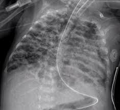
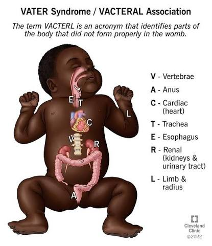
tracheoesophageal fistula
associated with VACTERL
Failed separation of trachea & esophagus (most often esophageal atresia + distal TEF)
tracheoesophageal fistula
Polyhydramnios prenatally;
newborn with choking, coughing, cyanosis, drooling during feeds; inability to pass NG tube
AR defect in dynein arms → impaired ciliary motility → defective mucociliary clearance
etiologgy of primary ciliary dyskinesia
Neonatal respiratory distress, chronic sinusitis/otitis, recurrent pneumonia, situs inversus (Kartagener: PCD + situs inversus + chronic sinusitis), infertility
signs of primary ciliary dyskinesia
Primary Ciliary Dyskinesia
-Inherited abnormality of ultrastructure of the cilia
-Caused by dynein arm defect
-Pt's present w/ recurrent infections, bronchiectasis, Kartagener syndrome
Dx: Electron microscopy of respiratory cilia
Tx: Preventative abx, surveillance cultures, sinus surgeries
pectus excavatum
Posterior depression of sternum & costal cartilages
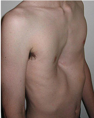
pectus carinatum
Anterior protrusion of sternum & costal cartilages
-often asymptomatic

Retropharyngeal abscess
Deep neck-space infection; |
tx with Abx and surgical drainage
Fever + drooling + dysphagia + neck stiffness/limited ROM + muffled voice |
caused by: Staph, Strep, anaerobes; typically <5 yr
contrast CT neck → retropharyngeal fluid collection/rim enhancement.
note: Lateral neck X-ray → widened prevertebral space
1st line imaging for suspected Retropharyngeal abscess
peritonsillar abscess
Infection between tonsillar capsule and pharyngeal muscles;
usually after tonsillitis; GAS + anaerobes
tx with abx and surgical drainage
*can spread to retro structures
severe unilateral throat pain
+ hot-potato voice
+ TRISMUS
+ drooling
+uvula dev’s AWAY
foriegn body aspiration
Sudden choking/coughing.
Upper airway → stridor.
Bronchial → unilateral wheeze/dec’d breath sounds
RIGHT BRONCHIAL MAINSTEM
most common location of foriegn body aspiration
CXR first ± inspiratory/expiratory or decubitus views
→ unilateral air trapping/hyperinflation or atelectasis.
first imaging for foriegn body aspiration
rigid bronchoscopy = diagnosis + treatment
gold standard for foriegn body aspiration
decreases due to hypervent.
ABG PaCO2 in an acute asthma attack
PaCO2 will be normal in fatigue
and will be increased in resp failure
how do you know if asthma is getting worst even though patient doesnt seem to be stuggling (silent chest)
Vascular ring
= congenital aortic arch anomaly that surrounds/compresses the trachea and/or esophagus.
dx w/ CT // tx: surgical correction
biphasic stridor that worsens with feeding or crying
improves with neck extension
worst in prone
Feature | B. PERTUSSIS (WHOOPING COUGH) |
|---|---|
Organism | Gram-negative coccobacillus, aerobic |
Transmission | Respiratory droplets |
Major virulence factor | Pertussis toxin → ↑ cAMP |
Mechanism | ADP-ribosylates Gi → inhibits Gi → ↑ adenylate cyclase → ↑ cAMP |
Classic CBC | ⭐ Lymphocytosis |
Classic symptom | Paroxysmal coughing → inspiratory “whoop” → post-tussive vomiting |
Infants | May have apnea/cyanosis instead of classic whoop |
Diagnosis | Nasopharyngeal PCR |
Treatment | Macrolide (usually azithromycin) |
Prevention | DTaP/Tdap |
Close contacts | Macrolide prophylaxis, regardless of vaccination status |
Child with weeks of severe coughing fits + inspiratory whoop + vomiting after coughing + lymphocytosis =
Bordetella pertussis
Catarrhal → Paroxysmal → Convalescent
Catarrhal: runny nose, low fever, mild cough → MOST contagious
Paroxysmal: coughing fits + whoop + post-tussive vomiting
Convalescent: cough gradually improves but may persist for weeks
Macrolides (azithromycin)
B. pertussis
infant with severe cooughing fits followed by vomitting and spells of apnea is tx with?:
Group B Strep (Streptococcus agalactiae)
E. coli
What are the most common causes of community-acquired pneumonia in neonates?
virus
What is the most common cause of community-acquired pneumonia in infants <1 year?
Viruses
Most common cause of pneumonia overall in this age group
Respiratory syncytial virus, influenza A and B, human metapneumovirus, adenoviruses, parainfluenza viruses, enterovirus, coronaviruses
Bacteria
Streptococcus pneumoniae: most common cause of typical bacterial pneumonia in all patients beyond the first few weeks after birth
Staphylococcus aureus: may occur as a secondary infection following influenza* pneumonia
Streptococcus pyogenes: may occur as a secondary infection following varicella infection
Atypical pathogens: Mycoplasma pneumoniae, Chlamydia pneumoniae
What is the most common cause of pneumonia overall in children 1–4 years?
Bacteria
S. pneumoniae: most common cause of typical bacterial pneumonia in this age group
M. pneumoniae: most common cause of bacterial pneumonia overall in this age group
C. pneumoniae
What are the common causes of community-acquired pneumonia in children ≥ 5 years of age?
pneumo
note:
Typical bacterial:
🔥 Higher fever + focal crackles + lobar consolidation
Viral/atypical:
🌬 More diffuse findings + bilateral/interstitial infiltrates
Most important physical sign in kids: Tachypnea is a particularly useful clue for pneumonia.
Finding | What you'll see |
|---|---|
Symptoms | ⭐ Fever + cough + tachypnea |
Breathing | Tachypnea, dyspnea, retractions, nasal flaring |
Infant clues | Poor feeding, irritability, lethargy; may have grunting |
Auscultation | ⭐ Crackles (rales), decreased breath sounds; possible bronchial breath sounds |
Percussion | Dullness over consolidated lung |
Oxygen | May have hypoxemia/cyanosis if severe |
CXR – bacterial | ⭐ Focal/lobar consolidation ± air bronchograms |
CXR – viral/atypical | Diffuse/interstitial infiltrates, often bilateral |
Fever + cough + tachypnea + crackles → think
early onset neonate pneumo (48 hours to 6 days of age)
Transmission
Intrauterine aspiration of amniotic fluid
Transplacental transmission
Aspiration of infected amniotic fluid during or after birth
Risk Factors
Prematurity and low birth weight (typically <2500 g [5 lb 8 oz])
Intraamniotic infection
Premature or prolonged rupture of membranes
Maternal Group B Streptococcus colonization
Maternal intrapartum fever
Most Common Bacterial Pathogens
Group B Streptococcus
Escherichia coli
Initial Empiric Therapy: Ampicillin + gentamicin
broad gram + and - coverage
early onset neonate pneumo (48 hours to 6 days of age)
is treated with?
late onsent neonate pneumo (>7 days of age)
Transmission
Maternal vertical transmission in the community
OR horizontal transmission if still hospitalized (ie, ventilator-associated)
Risk Factors
Prematurity and low birth weight (typically <2500 g [5 lb 8 oz])
Invasive mechanical ventilation
Anatomic anomalies (eg, choanal atresia, tracheoesophageal fistula)
Prolonged hospitalization
Underlying severe pulmonary disease
Most Common Bacterial Pathogens
Group B Streptococcus
Escherichia coli
Staphylococcus aureus
Klebsiella pneumoniae
Coagulase-negative staphylococci
Initial Empiric Therapy
Ampicillin OR vancomycin
PLUS
Aminoglycoside (typically gentamicin) OR
Extended-spectrum cephalosporin
late onsent neonate pneumo (>7 days of age)
is treated with?
typical pneumo strep pneumo
RX
Amoxicillin**
Levaquin/levofloxacin OR Clindamycin (if penicillin allergy)
Linezolid (if high rates of resistance)
Augmentin/amoxicillin-clavulanate (if aged 6 months to 5 years, use others if ≥ 5 years)
5yo male: high fever, productive cough with green sputum, crackles, and egophony (EEE —> AAA = +lung console.)
Child looks relatively well → “walking pneumonia”
Mycoplasma, which usually causes a drier, more gradual “walking pneumonia” presentation.
> 5-year-old, remember:
S. pneumoniae = most common typical bacterial pneumonia
Mycoplasma = most common bacterial pneumonia overall in older children/school-age kids
rx:
For atypical pneumonia due to Mycoplasma pneumoniae in a child:
⭐ Azithromycin = first-line (macrolide 50S) // Alternative: clarithromycin
Doxycycline can be used in older children when appropriate.
mycoplasma has NO cell wall so pencillins WONT work !!
10-year-old comes in with:
Gradual onset over several days
Low-grade fever
Dry, nonproductive cough
Headache, malaise, sore throat
Lung exam may have diffuse crackles
CXR → diffuse/interstitial infiltrates rather than focal lobar consolidation
staph a » pseudomonas
Pneumo in a ped with CF is likely?
Mycoplasma Species Pneumonia
Epidemiology
Respiratory droplet transmission
Close quarters (military, college dormitories, day care)
Summer and early fall
Clinical Features
Insidious onset of headache, malaise, low-grade fever, persistent nonproductive cough
Macular/vesicular rash
± upper respiratory tract symptoms, pharyngitis
Diagnostics
Hemolytic anemia, normal leukocyte count
High titers of serum IgM
Patchy or diffuse interstitial infiltrates on chest x-ray
Treatment
First-line: macrolides (eg, azithromycin)
Alternatives: tetracyclines (eg, doxycycline) or fluoroquinolones (eg, levofloxacin)
Bronchiolitis
Pathophysiology
Viruses infect the terminal bronchiolar epithelial cells, causing direct damage and inflammation in the small bronchi and bronchioles
Edema, excessive mucus, and sloughed epithelial cells lead to obstruction of small airways and atelectasis
Causes
Respiratory syncytial virus (most common)
Parainfluenza virus, Influenza virus, Adenovirus, Coronavirus
Clinical Manifestations
Generally presents with fever (usually ≤ 38.3°C [100.9°F]), cough, and respiratory distress (eg, increased respiratory rate, retractions, expire. wheezing, crackles)
Often preceded by a 1–3 day history of upper respiratory tract symptoms (eg, nasal congestion and/or discharge)