4. appendicitis
 Call Kai
 Learn
Learn Practice Test
 Spaced Repetition
 Match
Match Flashcards
 Knowt Play
1/39

There's no tags or description
Looks like no tags are added yet.
Name | Mastery | Learn | Test | Matching | Spaced | Call with Kai | Chat |
|---|
No analytics yet
Send a link to your students to track their progress
40 Terms

appendix
Most common position: Retrocecal (~65%)
blind ended tube off the cecum
Origin: arises from the posteromedial cecum, ~2–3 cm below the ileocecal valve
RLQ
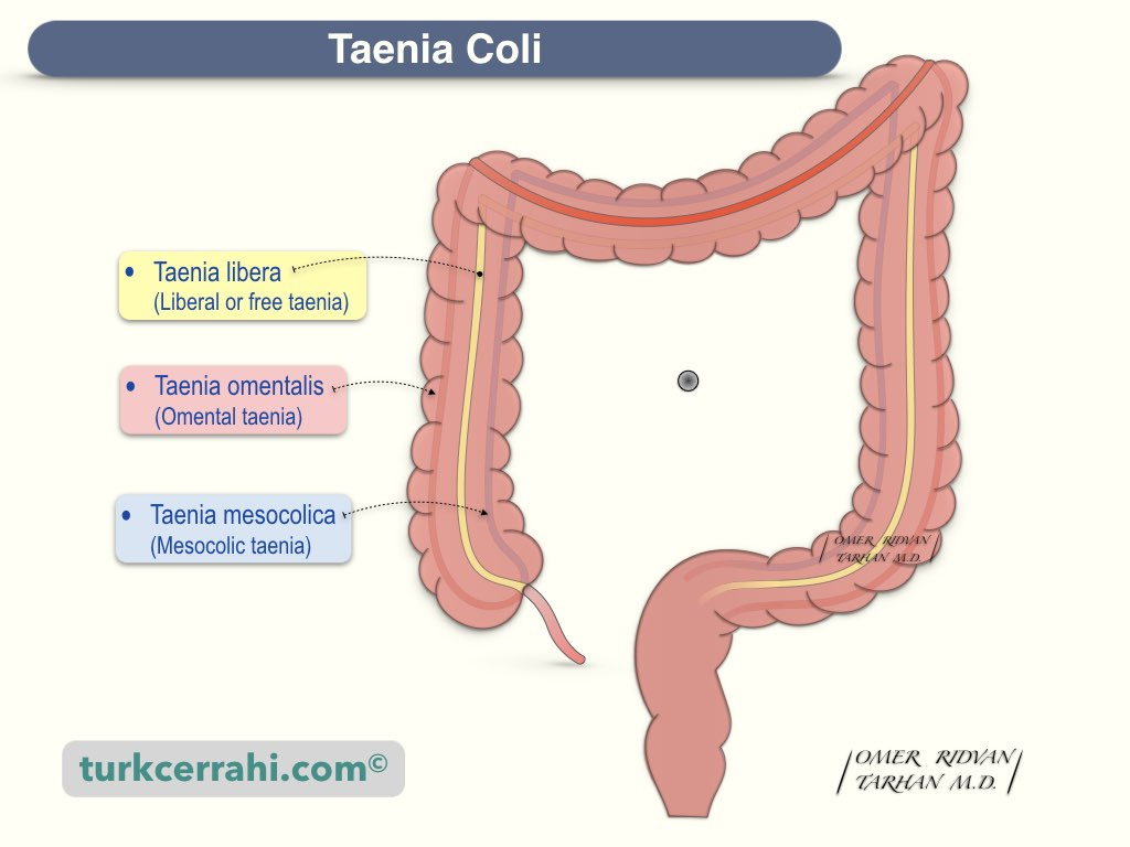
where the teniae coli converge
where to locate the base of the appendix

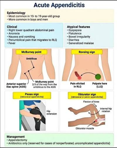
1/3 from ASIS → umbilicus
location of McBurney point
retrocecal
-results in less peritoneal irritation
most common position of the appendi
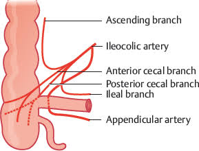
appendicular artery
(branch of ileocolic → from the SMA)
blood supply to the appendix
T10 // VAGUS
Sympathetic: T10 spinal level via the lesser splanchnic nerves → superior mesenteric plexus
Parasympathetic: Vagus nerve (CN X)
High-yield: Visceral pain from early appendicitis refers to the periumbilical region (T10).
→ Once the parietal peritoneum becomes irritated, pain localizes to the RLQ/McBurney point via somatic nerves.
visceral inervation of appendix
(sympathetic and parasympathic)
fecalith,
lymphoid hyperplasia,
tumor
causes of luminal obstruction of the appendix (3)
Luminal obstruction (fecalith, lymphoid hyperplasia, tumor)
↑ intraluminal pressure
Venous congestion → ischemia
Bacterial overgrowth
Perforation if untreated
pathophysiology of appendicitis

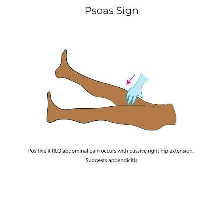
LLQ palpation → RLQ pain
retrocecal appendix
(most common position at 65%)
a positive psoas sign in appendicitis indicates what position of appendix?

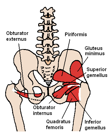
pelvic appendix
a positive obturator sign indicates what position of the appendix


pain in the lower abdomen or inside of thigh when the hip is flexed and internally rotated; a sign of appendicitis
Rapid progression → early perforation
More diffuse pain, vomiting prominent
appendicitis complication associated with children
minimal symptoms
Higher risk of perforation
have lower threshold to dx
presentation of appendicitis more commonly seen in elderly
Appendicitis
Crohn's disease
Meckel diverticulum
Gynecologic causes
differential diagnosis of RLQ pain
Patient | Best/first imaging |
|---|---|
Nonpregnant adult | CT abdomen/pelvis with IV contrast ⭐ |
Child | Ultrasound first |
Pregnant | Ultrasound first → MRI if nondiagnostic |
Unstable/peritonitis | Surgery consult — don't delay for imaging |
best imaging & gold standard for appendicits
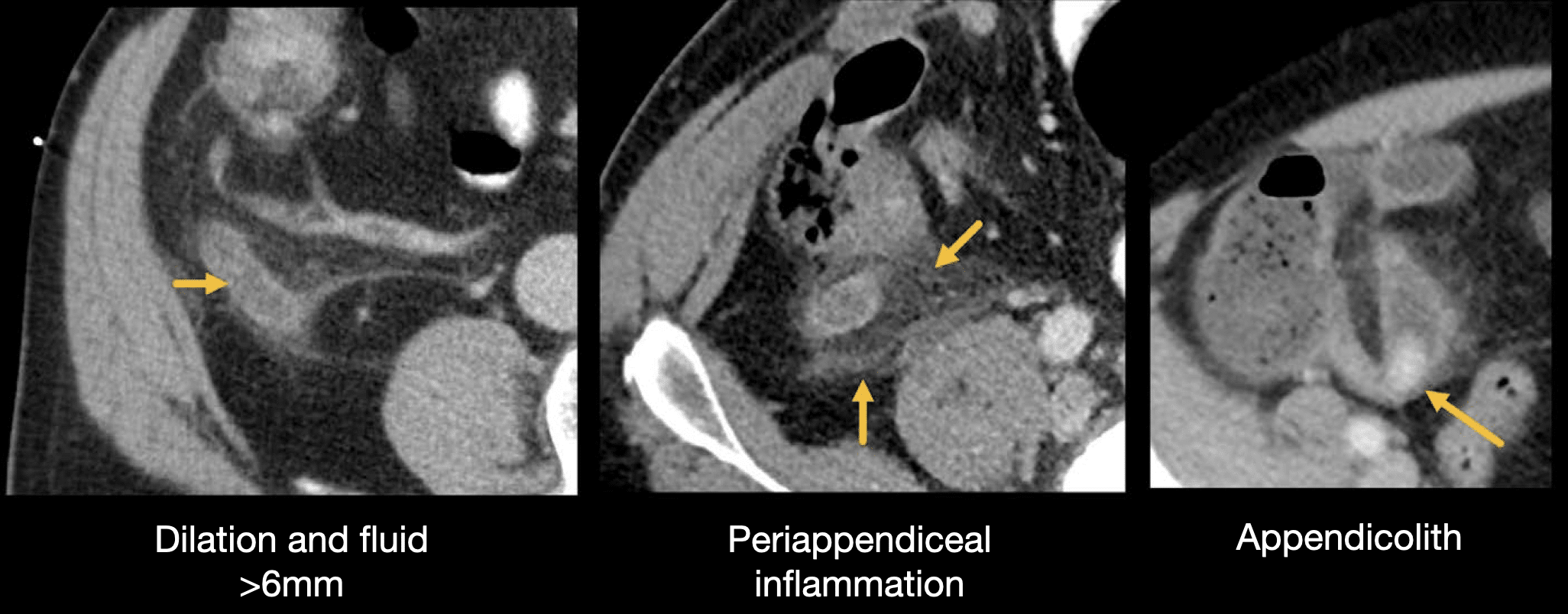
Enlarged appendix (>6 mm)
Wall thickening
Periappendiceal fat stranding
imaging findings associated with appendicitis (3)
fat stranding
On CT, normal fat looks dark/black. When inflamed, the fat develops streaky, hazy, whiter lines → fat stranding.
For appendicitis:
Enlarged appendix (>6 mm) + periappendiceal fat stranding = classic CT finding.
Think: “The inflammation spills into the surrounding fat.”
Other classic examples:
Diverticulitis → pericolic fat stranding
Pancreatitis → peripancreatic fat stranding
Cholecystitis → pericholecystic inflammatory changes
Increased attenuation in fat from edema, inflammation, or hemorrhage
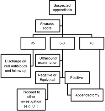
Alvarado score (MANTRELS)
for suspected acute appendicitis.
This scoring system combines findings to assist evaluation in patients with RLQ pain:
MANTRELS | Finding | Points |
|---|---|---|
M | Migration of pain to RLQ | 1 |
A | Anorexia | 1 |
N | Nausea/vomiting | 1 |
T | Tenderness in RLQ | 2 |
R | Rebound tenderness | 1 |
E | Elevated temperature | 1 |
L | Leukocytosis | 2 |
S | Shift to left (neutrophilia) | 1 |
Total | 10 |
≤4 → appendicitis unlikely; high value for ruling out
5–6 → possible → further evaluation/imaging
≥7 → high probability of appendicitis
High-yield: The two 2-point findings are RLQ tenderness + leukocytosis → everything else is 1 point.
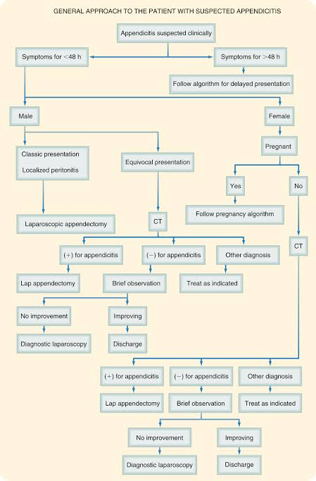
Laparoscopic appendectomy
standard treatment of uncomplicated appendicitis
NPO
IV fluids
Antibiotics (broad-spectrum)
Situation | Common regimen |
|---|---|
Uncomplicated appendicitis | Ceftriaxone + metronidazole |
Alternative | Cefoxitin alone |
Perforated/complicated | Piperacillin-tazobactam OR ceftriaxone + metronidazole |
pre-op care of appendicitis
Sudden relief of pain → then worsening diffuse pain
Signs of peritonitis
Fever, tachycardia
signs of a perforated appendix
perforated appendix
sudden relief of pain----> then worsening diffuse pain
palpable mass in RLQ
signs of a appendiceal abscess
emergency surgery
management of perforation with peritonitis
Contained abscess → ABX + drain → cool it down
vs.
Free perforation/generalized peritonitis → surgery now.
management of appendix abscess
recurrent appendicitis
-multiple episodes of RLQ pain -> resolved after appendectomy
-sx proven to be result of inflamed appendix by histology
chronic appendicitis
An inflammatory process with increasing pain
(other symptoms may be present or absent)
Minimal pain
No fever
No leukocytosis
Delayed presentation → high perforation rate
describe the atypical presentation of appendicitis in the elderly
appendicitis in the elderly
Vague abdominal pain
Altered mental status
Late signs of peritonitis
neuroendocrine (carcinoid)
Usually found incidentally after appendectomy
Most commonly at the tip of the appendix
Usually small and low-grade
<2 cm → appendectomy alone usually sufficient
>2 cm → generally consider right hemicolectomy based on pathologic risk factors
Feature | Board pearl |
|---|---|
Location | Tip of appendix ⭐ |
Presentation | Usually incidental after appendectomy |
Histology | “Salt-and-pepper” chromatin ⭐ |
Markers | Chromogranin A+, synaptophysin+ |
Hormone | Can produce serotonin (5-HT) |
Carcinoid syndrome | Rare with appendiceal NET; usually requires significant metastatic disease — must mets to liver, otherwise liver will filter and prevent symptoms |
Carcinoid syndrome symptoms | Flushing, diarrhea, bronchospasm, right-sided valvular disease |
Urine test | ↑ 5-HIAA |
Small tumor | Appendectomy generally sufficient |
Large/high-risk tumor | May require right hemicolectomy |
most common type of appendix cancer
Type | High-yield feature |
|---|---|
Neuroendocrine tumor (carcinoid) ⭐ | Most common appendiceal tumor; usually at tip, incidental |
Low-grade appendiceal mucinous neoplasm (LAMN) | Produces mucin; rupture → pseudomyxoma peritonei (jelly belly) |
Adenocarcinoma | Malignant epithelial tumor; resembles colorectal adenocarcinoma |
Mucinous adenocarcinoma | Mucin-producing invasive adenocarcinoma; can cause pseudomyxoma peritonei |
Goblet cell adenocarcinoma | Has both goblet/mucinous + neuroendocrine-like features; more aggressive than classic NET |
Signet-ring cell adenocarcinoma | Rare, very aggressive, poor prognosis |
name the types of appendiceal cancer
(know highlighted)
abdominal distension
because…
Pseudomyxoma peritonei = accumulation of mucin (“jelly”) throughout the peritoneal cavity, usually from a ruptured appendiceal mucinous neoplasm (especially LAMN).
High-yield
Appendiceal mucinous tumor → ruptures → mucin-producing cells seed peritoneum → progressive mucinous ascites
Typical presentation:
Increasing abdominal girth/distention ⭐
Abdominal discomfort
Mucinous/gelatinous ascites
Can eventually cause bowel obstruction
Classic CT: Scalloping of the liver/spleen surfaces from mucinous deposits.
Treatment: Cytoreductive surgery + HIPEC (heated intraperitoneal chemotherapy) in appropriately selected patients.
most common physial exam finding of mucinous appendix tumor
<2cm: appendectomy
>2 cm: right hemicolectomy
treatment of carcinoid appendix tumor
(tumor markers: chromogranin A and 5-HIAA)
right hemicolectomy
tx of adenocarcinoma of the appendix
(tumor markers: CEA, ca-19-9, ±ca 125)
Cytoreduction ± HIPEC
treatment of mucinous tumor of the appendix
(tumor marker: CEA, CA 19-9, CA-125.)
CEA → usually used to monitor
CA 19-9 → GI/mucinous tumor marker
CA-125 → can reflect peritoneal involvement, including pseudomyxoma peritonei
CEA, CA 19-9, CA-125.
highlights:
Can you treat with antibiotics alone?
Yes—in selected patients.
Imaging-confirmed uncomplicated appendicitis can sometimes be treated nonoperatively with antibiotics after discussing risks and benefits.
Benefits: avoids immediate operation/anesthesia.
Downside: significant risk of treatment failure or recurrent appendicitis, meaning appendectomy may eventually be required.
Appendicolith = higher risk of failure/complications with antibiotic-only management → surgery is generally favored.
Board answer:
Uncomplicated appendicitis → NPO + IV fluids + analgesia + preoperative antibiotics → laparoscopic appendectomy.
Key distinction:
Uncomplicated → appendectomy
Free perforation/peritonitis → urgent source control
Contained abscess/phlegmon → antibiotics ± percutaneous drainage, with operative timing individualized.
dont drained if not contained