Oncogenesis Tumor nomenclature
 Call Kai
 Learn
Learn Practice Test
 Spaced Repetition
 Match
Match Flashcards
 Knowt Play
1/33

There's no tags or description
Looks like no tags are added yet.
Name | Mastery | Learn | Test | Matching | Spaced | Call with Kai | Chat |
|---|
No analytics yet
Send a link to your students to track their progress
34 Terms
Hamartoma
extra “normal tissue” at “normal” site
Choristoma
“normal tissue” at “abnormal” site
Vascular metastasis’s main sites:
Spleen, right auricle, skin
Ademona
Benign glandular tumor
Adenocarcinoma
Malignant Glander tumor
Squamous papilloma
Benign squamous tumor
Squamous cell carcinoma
Malignant Squamous epithelial tumor.
Osteoma
Benign Bone tumor
Osteosarcoma
Malignant bone tumor
Benign epithelial tumors:
Adenoma, papilloma
Malignant Epithelial tumors
Carcinoma, squamous cell carcinoma, adenocarcinoma
Papilloma
exophytic (outward) growth, squamous cell origin (skin)
Polyp
exophytic (outward) growth, mucosal surfaces (intestines, ear/nose)
Adenoma
not necessarily outward growth, Glandular epithelial origin (mammary gland, thyroid gland, and also intestines)
What is similar between polyp and adenoma?
all are benign epithelial neoplasms
surgically curative
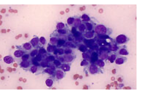
Epithelial tumor cytology
Cohesive clusters, defined borders, cytoplasmic vascuoles
Cell shape: cuboidal, columnar, or polygonal
Anisocytosis/anisokaryosis
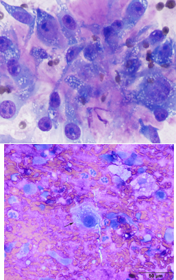
Cytology of mesenchymal cells
Spindle and oval-shaped cells with elongated nuclei
Individualized
Often have associated matrix (in the background or with the neoplastic cells)
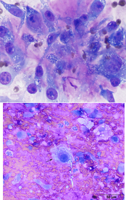
What is this a cytology of?
Mesenchymal cells
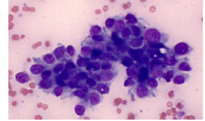
What is this a cytology of?
Epithelial tumor cells
What are the 5 stages of lymphoma:
1) A single lymph node is involved
2) Regional lymphadenopathy
3) Generalized lymphadenopathy
4)Hepatosplenomegaly
5) Involvement of bone marrow, central nervous system, or extranodal sites
What is it called what lymphoma is stage 5?
Leukemia
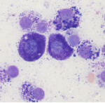
What is this?
Mast cell tumor
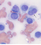
What is this?
Histiocytoma
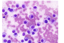
What is this?
Plasma cell tumor
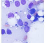
What is this?
Lymphoma
Mast cell tumor
Purple cytoplasmic granules
Eccentric nuclei
Well-differentiated tumors → usually more granules
Histocytoma
Epidermal Langerhans cell origin
Round to oval “big cells” with abundant light blue cytoplasm
Minimal cellular atypia
Can spontaneously regress
Plasma cell tumor
Round to oval cells
Round to oval eccentric nuclei
Bi/multinucleation
More atypia than histocytoma
Lymphoma
Round to oval cells
highest N:C ratio (almost no cytoplasm)
Round nuclei
Needs histopathology (biopsy) for discerning B vs T cell lymphoma
Cytology of round cell tumors contains:
Round, discrete cells, high nucleus:cytoplasm ratio (N:C ratio)
Neuroendocrine tumors
Secretory epithelial cells (produce hormones/neurotransmitters)
often boring looking but aggressive biological behavior
Histopathology (biopsy) is more definitive
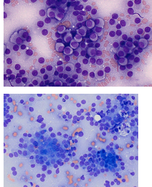
Neuroendocrine tumors:
Round to polygonal cells in packets/small sheets
Round to oval nuclei
Indistinct cell borders
“naked nuclei”
AGASACA cytology:
has neuroendocrine appearance
What is this?
Neuroendocrine tumor