Ch 6 Assignment 1
 Call Kai
 Learn
Learn Practice Test
 Spaced Repetition
 Match
Match Flashcards
 Knowt Play
1/33

There's no tags or description
Looks like no tags are added yet.
Name | Mastery | Learn | Test | Matching | Spaced | Call with Kai | Chat |
|---|
No analytics yet
Send a link to your students to track their progress
34 Terms
Which metatarsal bone of the foot has a prominent tuberosity frequently
Fractured?
Fifth
Which term describes the top or anterior surface of the foot?
Dorsum
The medial malleolus is part of the
Tibia
To decrease the angle between the anterior surface of the foot and anterior surface of the lower leg is described as
Dorsiflexion
To reduce scatter radiation during tabletop procedures, the Bucky tray should not be positioned directly under the lower limb being radiographed
True
When using computed radiography, lead masking should not be placed on the regions of the imaging plate not within the collimation field.
False
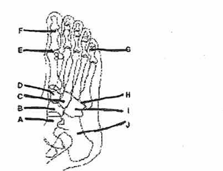
Which of these labeled structures or bones identifies the"cuneiform? (Place a check by the best choice
D
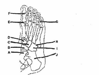
Which projection/position of the foot is represented by the drawing of the foot in Fig. 6-1?
AP oblique, 55° medial rotation
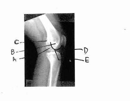
Which one of the labeled structures is the medial condyle?
A
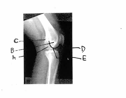
Which of the labeled structures is the adductor tubercle?
C
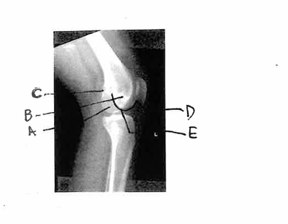
Which of the labeled structures is the lateral condyle?
B
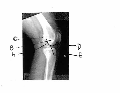
This radiograph represents which of the following positions?
Lateral knee under-rotated
Extending the ankle joint or pointing the foot and toes downward is called
Plantar flexion
How much CR angulation (if any) should be used for an AP projection of the
toes?
10° to 15° toward calcaneus
Which of the following routines should be performed for a study of the second
toe?
AP, AP oblique with medial rotation, lateromedial projection
A lateral knee radiograph that is over-rotated toward the image receptor can be recognized by which of the following
The fibular head will appear less superimposed by the tibia than a true lateral
To properly visualize the joint spaces with the AP projection of the foot, the CR must be
Perpendicular to the metatarsals
What CR angulation is required for the AP oblique projection of the foot
CR is perpendicular to the image receptor
How much CR angulation to the long axis of the foot is required for the
plantodorsal (axial) projection of the calcaneus
40°
How much rotation from an AP of the ankle will typically produce a mortise
view
15° to 20°
Which projection of the ankle best demonstrates the distal tibiofibular joint
AP oblique with 45° rotation
To ensure that both joints are included on an AP projection of the tibia and fibula on an adult, the technologist should
Turn image receptor diagonally
What CR angulation is required for an AP projection of the knee on a patient with an ASIS-to-tabletop measurement of 18 cm
3° to 5° caudad
What CR angle should be used for a lateral projection of the knee on a normal size pelvis
No CR angle is required
5° to 7° cephalad
Follow-up radiographs for a fractured tibia and fibula may include only the joint closest to the site of injury
True
How much flexion of the knee is best for the lateral projection of the knee
20° to 30°
A radiograph of an AP oblique projection of the foot, if positioned correctly,
should demonstrate
Third through fifth metatarsals free of superimposition
A radiograph of an AP knee reveals rotation with almost total superimposition of the fibular head and the proximal tibia. What must the technologist do to correct this positioning error on the repeat exposure
Rotate the knee medially
A patient has a lower leg fracture reduced and cast in ER. The following factors were used initially: 65 kVp, 10 mAs, 40-in SID, detail screens. A large plaster cast is applied. Which of the following exposure factors should be used on the postreduction study
65 kVp, 20 mAs, 40-in SID, detail-speed screens
A patient comes to the radiology department for a knee study with special interest in the region of the proximal tibiofibular joint and the lateral condyle of the tibia.
Which of the following projections should the technologist obtain
AP, lateral, and medial oblique knee
A patent enters the ER with a possible transverse fracture of the patella. Which of the following routines would safely provide the best images of the knee
AP and horizontal beam lateral, no flexion
A 3° to 5° caudad CR angle should be used for an AP knee projection for patients with thick thighs
False
The correct CR placement for an AP projection of the knee is mid-patella
False
A correctly positioned lateral ankle will demonstrate the lateral malleolus
superimposed over the posterior half of the tibia
True