Lecture 17 Functional Retina Part 2
1/59
There's no tags or description
Looks like no tags are added yet.
Name | Mastery | Learn | Test | Matching | Spaced |
|---|
No study sessions yet.
60 Terms
What are the two types of photoreceptors?
Rods and cones
What is the outer segment of photoreceptors used for?
Light absorption
What does the inner segment of photoreceptors house?
The biomachinery
What type of receptors are photoreceptors?
Specialized sensory receptors
What do photoreceptors contain that is crucial for their function?
Photosensitive pigment
What happens to light quanta in photoreceptors?
It is converted into electrical activity
What is the resting membrane potential of photoreceptors?
~ -50mV
-Photoreceptors are slightly depolarized (typical neuron resting potential is -70mV
What happens to photoreceptors when exposed to light?
They hyperpolarize
-Potential goes from -50mV to -70mV
What is phototransduction?
Phototransduction is the process by which light is translated into electrical signals.
-This process alters electrical currents in order to change the membrane potential across the photoreceptor
What happens to the membrane potential of a photoreceptor in the dark?
In the dark, the membrane potential of a photoreceptor is around -40mV due to the balance of outward K+ and inward Na+ currents.
What neurotransmitter is released by depolarized photoreceptors?
Depolarized photoreceptors release the neurotransmitter glutamate.
What effect does glutamate have on bipolar cells?
Glutamate prevents bipolar cells from firing.
Phototransduction
When light strikes the photoreceptor, it starts a process that eventually causes the Na+ channels to close. Without the counterbalancing effect of the inward Na+ current, the outward K+ current causes the membrane potential to hyperpolarize to -70mV. In turn, glutamate is no longer released to the bipolar cell, and the membrane potential of the bipolar cell changes, which allows it to release glutamate.
What are the two parts of rhodopsin?
Light-absorbing retinal and the membrane-bound protein opsin.
What happens to retinal when it absorbs light?
It changes shape from the 11-cis to the all-trans conformation and dissociates from opsin.
What is the result of retinal dissociating from opsin?
Rhodopsin becomes activated, represented as rhodopsin*.
What does activated rhodopsin* do?
It activates hundreds of molecules of the protein transducin by exchanging GDP for GTP.
What does each activated transducin* molecule stimulate?
A cGMP phosphodiesterase (PDE) molecule.
What is the effect of activated cGMP PDE?
It hydrolyzes cGMP to 5'-GMP.
How many molecules of cGMP can each PDE hydrolyze per second?
Over 1000 molecules.
What happens to cGMP levels during phototransduction?
The number of cGMP molecules decreases.
What is the consequence of decreased cGMP levels?
The cGMP-gated channels close, decreasing the inward current of Na+.
What is the effect of decreased inward Na+ ions on the cell's membrane potential?
It hyperpolarizes the cell from -40mV to -70mV.
What happens to the phototransduction process after activation?
It rapidly shuts down and restores the cell to the dark state.
What occurs during the restoration of the dark state in phototransduction?
Activated molecules return to their resting states, 5'-GMP is converted back to cGMP, and Na+ channels reopen.
Graded potential
-Not 'all or nothing' like some neurons
-Greater intensity stimulus causes greater hyperpolarization
Dark current
-Na+ ions flow through ion channels into rod outer segment
-Produces the slight -50mV depolarization
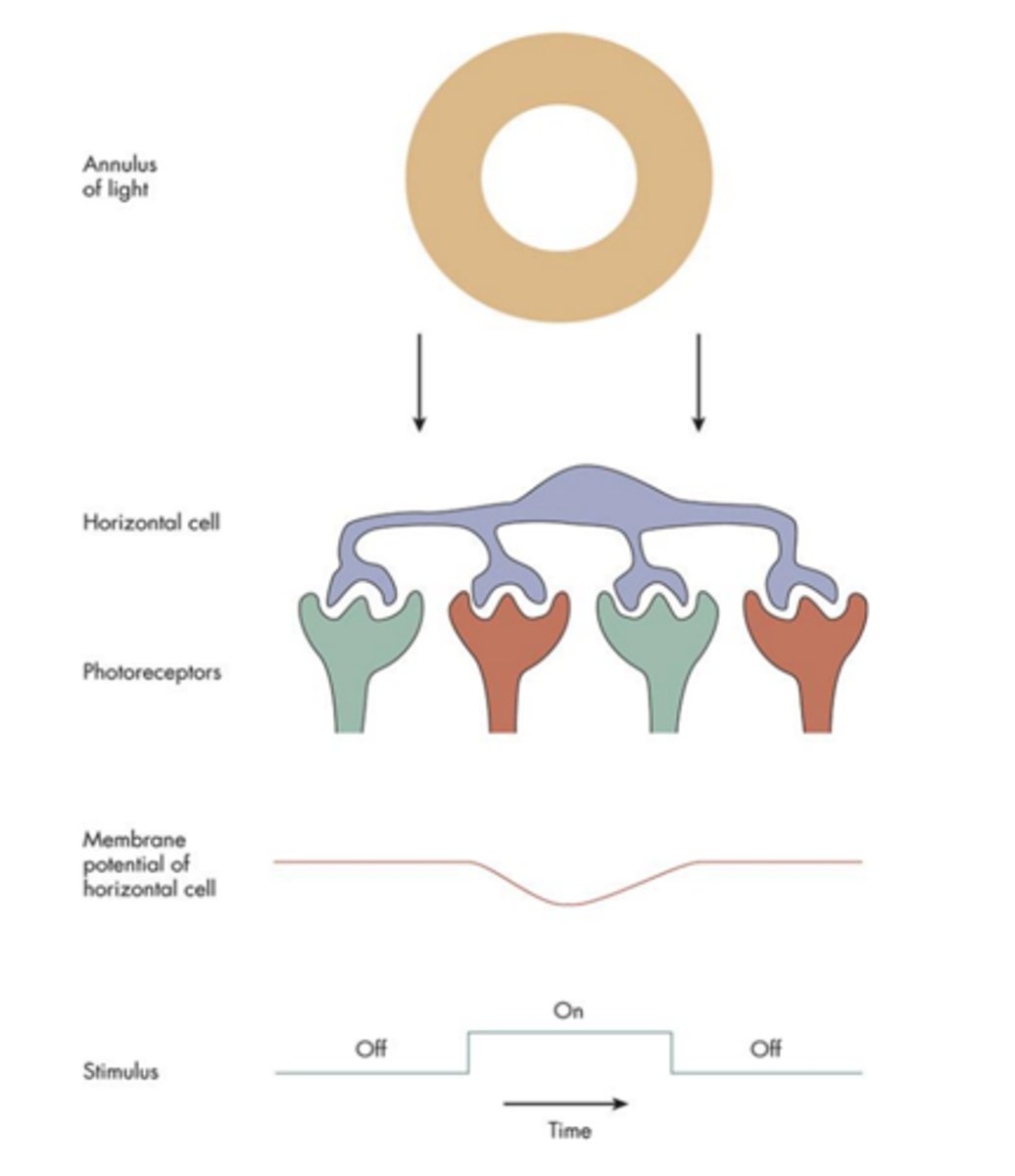
Horizontal cells
-Widely dispersed dendritic tree synapses with many photoreceptors
-Substantial spatial summation
-H1- input from M- and L- cones
-H2 - input from S-cones
-Graded potential
-Sign-conserving synapses

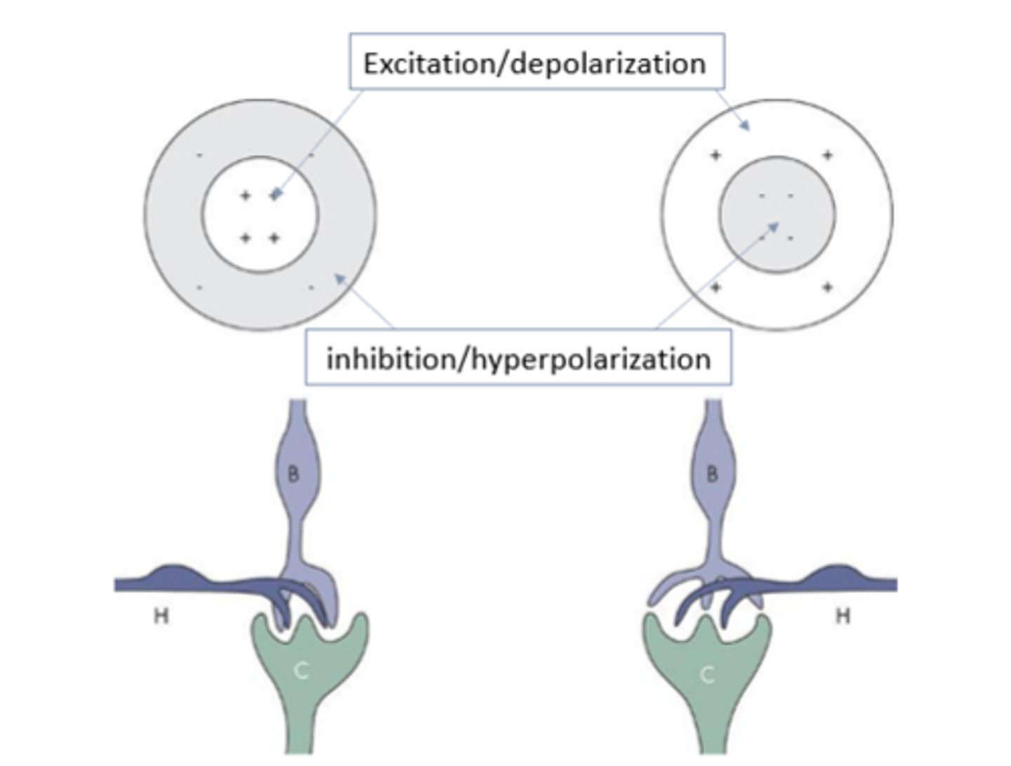
On-center bipolar cells
-Invaginating synapse
-Sign-inverting
-Inner sublayer (IPL)
-Glutamate is inhibitory

Off-center bipolar cells
-Flat synapse
-Sign -conserving
-Outer sublayer (IPL)
-Glutamate is excitatory

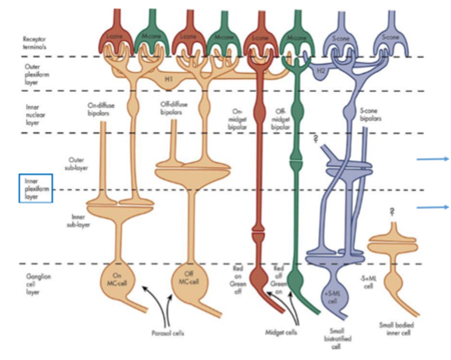
Both on- and off-center bipolar cells synapse with
ganglion cells in the inner plexiform layer (IPL), but in different sublayers.
-Off-center cells synapse in the outer sublayer of the inner plexiform layer
-On-center cells synapse in the inner sublayer.

Midget bipolar cell
-Midget - small spread
-Receives input from L & M cones.
-4 types: Red-ON, Red-OFF, Green-ON, and Green-OFF
-Fovea: one-to-one with cone
->15 degree: one to many cones
Diffuse bipolar cell
-Diffuse - large spread (even in fovea)
-Receives input from L & M cones, and S-cones. Less selective of stimulus wavelength than midget. Responds to wider spectral range.
-6 types: 3-ON and 3-OFF types.
-Each cone (L & M) inputs to two midget bipolar cells and six diffuse bipolar cells
Blue cones
have their own bipolar cells
-More closely resembles rod bipolar cells
-Receive multiple inputs from only ON type cells.
-Both blue and rod bipolar terminate closer to ganglion cells than any other bipolar cells.
Bipolar cells
-Graded potential
-Display spatial antagonism-Midget bipolar cells have smaller soma, smaller dendritic tree, and smaller receptive field.
-Diffuse bipolar cells center formed by 5 to 10 M- and L-cones. Center spectral sensitivity is similar to the surround.
Amacrine cells
-Center-surround (some)
-Action potentials
-Brief transient response at stimulus onset and offset
-Coding movement: amacrine cells, might regulate the transfer of signals from the bipolar cells to ganglion cells, supplying the information needed for motion detection.
What type of bipolar cells are associated with rods?
Rods have their own bipolar cells. All of them are ON bipolar cells.
What type of cells do the output from rod bipolar cells go to?
The output goes to small amacrine cells (known as All type) whose output is onto the terminal end of cone bipolar cells through gap junction.
What is the flow of information from rods to ganglion cells?
Rods -> Rod bipolar cell -> All amacrine cell -> Cone bipolar cell -> Ganglion cell.
Why do rods need amacrine cells?
Rods need amacrine cells to transport signals out of the retina.
Midget Ganglion cell
-Midget - small spread
-Receives input from midget bipolar cells
-Only one midget bipolar cells contacts one midget ganglion cell (small).
-Two types: ON & OFF types. 2 OFF types and 5 ON types pathways.
-ON type midget bipolar contacts only ON midget ganglion cell. And, similarly for OFF types.
-Have wavelength info embedded in their signal, but doesn't coney (bistratified does)
Parasol ganglion cell
-Extensive unstratified branching
-Receives input from diffuse bipolar cells.
-Two types: ON & OFF types. 3 OFF types and 4 ON types pathways.
Midget and Parasol ganglion cells
-Found in all retinal locations, fovea to periphery.
-Both account for 80% of all ganglion cells.
-Remaining 20% of the cells are bistratified (<10%) and shrubs (>10%) ganglion cell types.
-Small bistratified cells
--Receive input from S-cone bipolar cells
--8% of ganglion cells
--On-center formed exclusively by S-cones
--Convey specific wavelength information
How do dendritic fields of ganglion cells vary on the retina?
Dendritic fields of ganglion cells at any particular location on the retina differ in size.
What is the relationship between receptive field center size and sensitivity function sharpness?
Receptive field center size is related to sharpness of the peak in the sensitivity function.
What characterizes small receptive field centers?
Small receptive field centers have a narrow, sharp peak sensitivity function (less blur).
What characterizes large receptive field centers?
Large receptive field centers have a broader and flatter peak sensitivity function (more blur).
Ganglion cell projections
the pathways through which retinal ganglion cells (GCs) send visual information from the eyes to different brain regions.
-LGN
-Superior Colliculus
-Pulvinar
Parallel pathways: vertical
different wiring patterns between photoreceptors, bipolar cells, and ganglion cells:
-One-to-one wiring
-Divergent wiring
-Convergent Wiring
LGN (Retinogeniculate)
It receives inputs from different types of ganglion cells (GCs):
Parvo (P-cells) → Midget GCs (associated with fine detail and color vision)
Magno (M-cells) → Parasol GCs (associated with motion detection and contrast sensitivity)
Konio (K-cells) → Bistratified GCs (associated with blue-yellow color processing)
Superior colliculus
This midbrain structure helps control eye movements and reflexive visual tracking.
Pulvinar
A thalamic region involved in:
-Visual attention
-Motion processing
-Visually guided movement (e.g., coordinating vision with body movements)
One-to-one wiring
The photoreceptor makes synaptic contact with a bipolar cell's dendritic end and the axon terminal end of the bipolar contacts ganglion cell.
-Good for resolving fine details.
Divergent wiring
Cones normally contact several bipolar cells, and thus more than one ganglion cell
Convergent wiring
Characteristic of peripheral retina. A many to one.
-A number of photoreceptors contact a smaller number of bipolar cells and in turn contact a single ganglion cell.
-In this type of wiring, the cell report on the aggregate activities of many photoreceptors
-This is good for detecting and reporting the presence of small amounts of light, not good for resolving find details.
What are the two types of lateral pathways in the retina?
Horizontal cells (OPL) and amacrine cells (IPL).
What type of input do bipolar cells receive in vertical pathways?
Direct photoreceptor input and input from surrounding photoreceptors mediated by horizontal cells.
How does the sign of lateral receptor input affect the lateral pathway's effect?
The effect depends on the sign or polarity of the lateral receptor input.
What is the effect of positive lateral receptor input?
The effect is positive if the lateral receptor input is of the same sign, which coarsens resolution.
What is the effect of negative lateral receptor input?
It signals illumination contrast between photoreceptors and their neighbors, helping spatial resolution.