Respiratory System - Notes
 Call Kai
 Learn
Learn Practice Test
 Spaced Repetition
 Match
Match Flashcards
 Knowt Play
1/112

There's no tags or description
Looks like no tags are added yet.
Name | Mastery | Learn | Test | Matching | Spaced | Call with Kai | Chat |
|---|
No analytics yet
Send a link to your students to track their progress
113 Terms
Respiratory System
Provides the means for gas exchange
Consists of respiratory passageways in head, neck, and trunk, and the lungs
Respiration
Gas exchange: O2 and CO2
Occurs between atmosphere and body cells
Cells need O2 for aerobic ATP production and need to dispose of CO2 that process produces
Functions of Respiratory System
Air passageway
Site for exchange of oxygen and carbon dioxide
Detection of odors
Sound production
Function: Air passageway
Air moves between atmosphere and alveoli as we breathe
Function: Exchanging Gas
Oxygen diffuses from alveoli into blood
Carbon dioxide diffuses from blood into alveoli
Function: Detection of Odors
Olfactory receptors in superior nasal cavity
Function: Sound Production
Air moves across vocal cords of the larynx (voice box)
Vocal cords vibrate, producing sound
Structural Organization
Upper respiratory tract: Nose, nasal cavity, pharynx, larynx
Lower respiratory tract: Trachea, bronchi, bronchioles, alveolar ducts, alveoli

Function Organization
Structures of the conduction zone transport air
Nose to terminal bronchioles
Structures of the respiratory zone participate in gas exchange
Respiratory bronchioles, alveolar ducts, alveoli

Mucosa
Mucous membrane: respiratory lining
Epithelium resting on a basement membrane
Underlying lamina propria made of areolar connective tissue
Respiratory epithelium Structure
Thinner from the nasal cavity to the alveoli
Starts out as pseudostratified ciliated columnar
Changes to simple ciliated columnar
Changes to simple cuboidal
Changes to simple squamous
Exceptions: stratified squamous found in high abrasion areas – oropharynx, laryngopharynx, vocal cords, superior portion of larynx
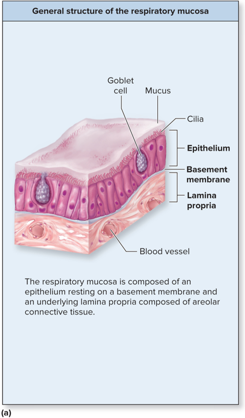
Progressively Thinner Epithelium

Respiratory Mucosa: Mucous Secretions
Produced from secretions of goblet cells of epithelial lining
Mucous and serous glands of lamina propria
Contains mucin protein
Contains defenses against microbes
Lysozymes (antibacterial enzyme)
Defensins (antibacterial proteins)
Immunoglobulin A (antibody)
Called sputum when coughed up with saliva and trapped substances
Mucin
Increases mucus viscosity and serves to trap dust, dirt, pollen, etc.
Clinical View: Cystic Fibrosis
Defective chloride channels prevent chloride ion from being pumped from epithelial cells lining respiratory tract into the lumen of respiratory tract
Without chloride on cell surface, water fails to hydrate the cells’ surface
Mucus becomes thick and cilia not able to mobilize it, so the mucus blocks the respiratory tract
Pulmonary infections are common
Ducts of pancreas and salivary glands can also be blocked
Backup of digestive enzyme can destroy pancreas
Which respiratory structure is associated with the exchange of respiratory gases?
Thin barrier between alveoli and pulmonary capillaries
What four structures compose the upper respiratory tract? What three structures compose the respiratory zone?
The nasal cavity (nose), pharynx (throat), larynx (voice box), and mouth
Nose
First conducting structure for inhaled air
Formed by bone, hyaline cartilage, dense irregular connective tissue, and skin
Bridge of nose formed by paired nasal bones
One pair of lateral cartilages and two pairs of alar cartilages
Flared part of nostrils (nares) made of dense irregular connective tissue

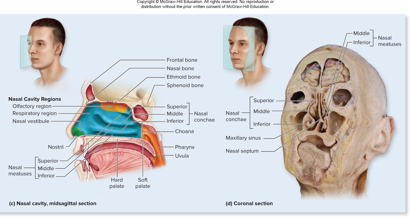
Nasal Cavity
From nostrils to choanae
An oblong-shaped internal shape
Floor formed by palate
Roof made of nasal, frontal, ethmoid, and sphenoid bones plus some cartilage
Choanae
Posterior Nasal Apertures
Paired openings that lead to pharynx
Nasal Septum
Divides left and right sides
Anterior part is septal nasal cartilage
Posterior part is bony perpendicular plate of ethmoid plate and vomer bone
Nasal Conchae
Three paired, bony projections on lateral walls of nasal cavity
Superior, middle, and inferior nasal conchae
Also called turbinate bones
Produce turbulence in inhaled air
Partition the nasal cavity into separate passages
Each passage called a nasal meatus
Each meatus immediately inferior to its corresponding concha
Nasal Cavity Parts: Nasal Vestibule
Just inside nostrils
Lined by skin and particle-trapping hairs called vibrissae

Nasal Cavity Parts: Olfactory Region
Superior part of nasal cavity containing olfactory epithelium
Airborne molecules stimulate receptors for odor detection

Nasal Cavity Parts: Respiratory Region
Lined by pseudostratified ciliated columnar epithelium
Has an extensive vascular network
Nosebleeds (epistaxis)
Common due to large numbers of superficial vessels
Nasolacrimal Ducts
Drain lacrimal secretions from eye surfaces to nasal cavity
Nasal Cavity Conditions the Air
Air is warmed by extensive blood vessels
Mucus traps dust, microbes, and foreign material
Cilia sweep mucous toward the pharynx to be swallowed
Moist environment humidifies
Air turbulence created by conchae enhances all three processes
Clinical View: Rhinorrhea (runny hose)
Increased production of mucus (allergies, virus)
Increased secretions from lacrimal glands draining into the nasal cavity (crying)
Exposure to cold air (water condensation)
Paranasal Sinuses
Spaces within skull bones
Named for specific bone in which they are housed
All connected by ducts to nasal cavity
Lined by pseudostratified ciliated columnar epithelium
Mucus swept into pharynx and swallowed

Paranasal Sinuses: Superior to Inferior
Frontal sinuses
Ethmoidal sinuses
Sphenoid sinuses posterior to ethmoidal sinuses
Maxillary sinuses
Clinical View: Sinus Infections and Sinus Headaches
Respiratory infection or allergy can cause inflammation of the ducts that drain from the paranasal sinuses.
Drainage of mucus decreases and accumulates in the sinuses.
Germs can grow in the accumulated mucous, causing a sinus infection.
Inflamed and blocked sinuses and pressure changes can cause sinus headaches.
Pharynx (throat)
Funnel-shaped passageway posterior to nasal cavity, oral cavity, and larynx
Lateral walls composed of skeletal muscles
Partitioned into
Nasopharynx
Oropharynx
Laryngopharynx

Nasopharynx
Most superior part of pharynx, posterior to nasal cavity, superior to soft palate
Lined by pseudostratified ciliated columnar epithelium
An air passage-not for food
Connects to middle ear via auditory (eustachian) tube
Contains tonsils-infection-fighting lymphatic tissue

Auditory tube
Opening tubes allows equalization of pressure on each side of tympanic membrane
Tubal Tonsils
Located near auditory tube opening
Pharyngeal tonsil on posterior nasopharynx wall
Called adenoids when enlarged
Oropharynx
Middle pharyngeal region
Posterior to oral cavity
Extends from soft palate to hyoid bone
Passageway for both food and air
Lined by nonkeratinized stratified squamous epithelium
Contains tonsils
Palatine tonsils on the lateral walls
Lingual tonsils at base of tongue

Laryngopharynx
Inferior, narrow region of pharynx, posterior to the larynx
From level of hyoid down to esophagus
Passageway for both food and air
Lined by nonkeratinized stratified squamous epithelium

Larynx (voice box)
Cylindrical airway between laryngopharynx and trachea
Function of Larynx
Produces sound
Air passageway
Prevents ingested materials from entering respiratory tract
Epiglottis covers superior opening during swallowing
Assists in increasing pressure in abdominal cavity
Participates in sneeze and cough reflexes
Valsalva maneuver
Vocal folds close off rima glottidis (opening between the folds) and contraction of abdominal muscles
Increased pressure facilitates urination, defecation, childbirth
Larynx: Sneezing and Coughing
Help remove irritants from nasal cavity or lower respiratory tract
Abdominal muscles contract increasing thoracic pressure
Vocal cords are forcibly opened by pressure from below
Explosive blast of exhaled air is a cough or sneeze
Larynx anatomy: Laryngeal Inlet
Also known as the laryngeal aperture, connects pharynx and larynx
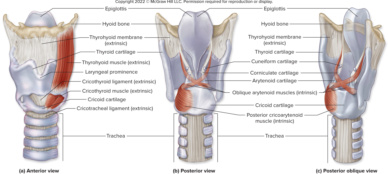
Formed and supported by nine pieces of cartilage
Cartilages held in place by ligaments and muscles
Single thyroid, cricoid, and epiglottis cartilages
Paired arytenoid, corniculate, and cuneiform cartilages
Larynx anatomy: Thyroid Cartilage
Large, shield-shaped
Forms lateral and anterior walls of larynx
Attached to lateral surface of cricoid cartilage
Anterior protrusion = laryngeal prominence (Adam’s apple)
Larger in males; enlarge during puberty
Larynx anatomy: Cricoid Cartilage
Ring-shaped
Just inferior to thyroid cartilage
Larynx anatomy: Epiglottis
Spoon-shaped
Anchored to inner aspect of thyroid cartilage
Projects postersuperiorly into the pharynx
Closes over laryngeal inlet during swallowing
Larynx anatomy: Arytenoid, corniculate, and cuneiform
All laryngeal cartilages are made of hyaline cartilage, except the epiglottis, which is made of elastic cartilage
Laryngeal ligaments are extrinsic or intrinsic
Extrinsic Ligaments
Attach external surface of larynx to other structures (for example: hyoid bone)
Intrinsic Ligaments
Are located within the larynx
Include the vocal ligaments and the vestibular ligaments
Larynx
Larynx anatomy: Vocal Ligaments
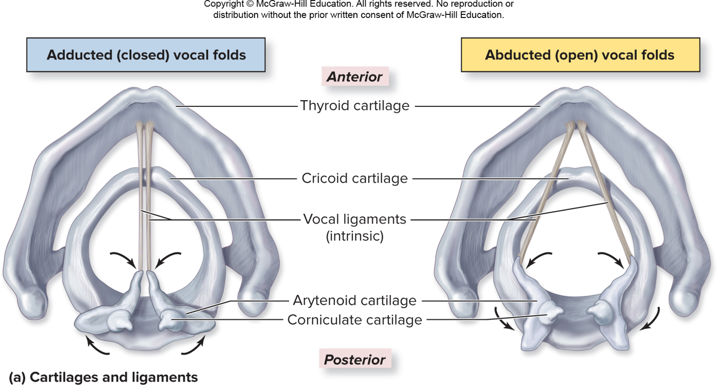
Extend between thyroid and arytenoid cartilages
Composed primarily of avascular elastic connective tissue
Covered with mucosa to form the vocal folds (true vocal cords)
Produce sound when air passes between them
Opening between ligaments = rima glottidis
Rima glottidis + vocal folds = glottis
Larynx anatomy: Vestibular Ligaments
Extend from thyroid cartilage to arytenoid and corniculate cartilages (superior to vocal folds)
Covered with mucosa to form the vestibular folds (false vocal cords)
Play no role in sound production; protect vocal cords
Opening between vestibular folds = rima vestibuli

Larynx anatomy: Extrinsic muscles
Skeletal muscles that stabilize larynx and help it move during swallowing
Originate on hyoid bone or sternum; insert on thyroid cartilage
Larynx anatomy: Intrinsic Muscles
Skeletal muscles located within larynx
Attach to arytenoid and corniculate cartilages
Contraction results in change in dimension of rima glottidis
Narrowing with adduction; widening with abduction
Involved in voice production and swallowing
Sound Production
Vocal cord vibration
Intrinsic laryngeal muscles narrow opening of rima glottidis
Air is forced past vocal cords during expiration
Sound Production: Range
Voice determined by length, thickness of vocal cords
Males have longer and thicker folds, and so deeper voices
Folds increase in length with growth, deepening range
Sound Production: Pitch (frequency)
Determined by tension on vocal cords
Increased tension = folds vibrate more = higher pitch
Regulated by intrinsic laryngeal muscles
Sound Production: Loudness
Depends on force of air passing across vocal cords
More air = loud sound
Clinical View: Laryngitis
Inflammation of the larynx
Symptoms of hoarse voice, sore throat, sometimes fever
Caused by bacterial or viral infection, or overuse (yelling)
Serve cases can extend to the epiglottis
May lead to sudden airway obstruction, especially in children
Lower Respiratory Tract
Includes conducting pathways from trachea to terminal bronchioles
Includes structures involved in gas exchange: respiratory bronchioles, alveolar ducts, and alveoli
Trachea (windpipe)
Open tube connecting larynx to main bronchi
Anterior to esophagus, posterior to part of sternum
About 13 cm long, 2.5 cm in diameter

Gross anatomy of the trachea: Tracheal Cartilages
Support anterior and lateral walls
C-shaped rings of hyaline cartilages
Ensheathed in perichondrium and dense fibrous membrane
Rings are connected to each other by anular ligaments
Gross anatomy of the trachea: Trachealis Muscle
On trachea’s posterior surface
Connects open ends of C-shaped cartilages
Allow accommodation for esophagus when bulge of food passes
Trachealis contracts during coughing
Gross anatomy of the trachea: Carina
Internal ridge at inferior end of trachea (where it splits) containing many sensory receptors
Initiates cough reflex when irritants are present
Clinical View: Tracheotomy
Incision into trachea to facilitate breathing when:
Airway is blocked
Ventilation compromised by disease or injury
Clinical View: Cricothyrotomy
Incision between cricoid cartilage and thyroid cartilage
Tube placed in opening to facilitate air exchange
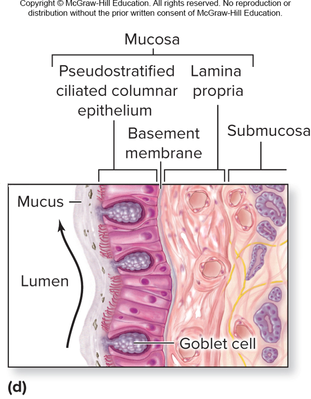
Histology of the Tracheal Wall: Mucosa
Innermost layer
Pseudostratified ciliated columnar epithelium and lamina propria
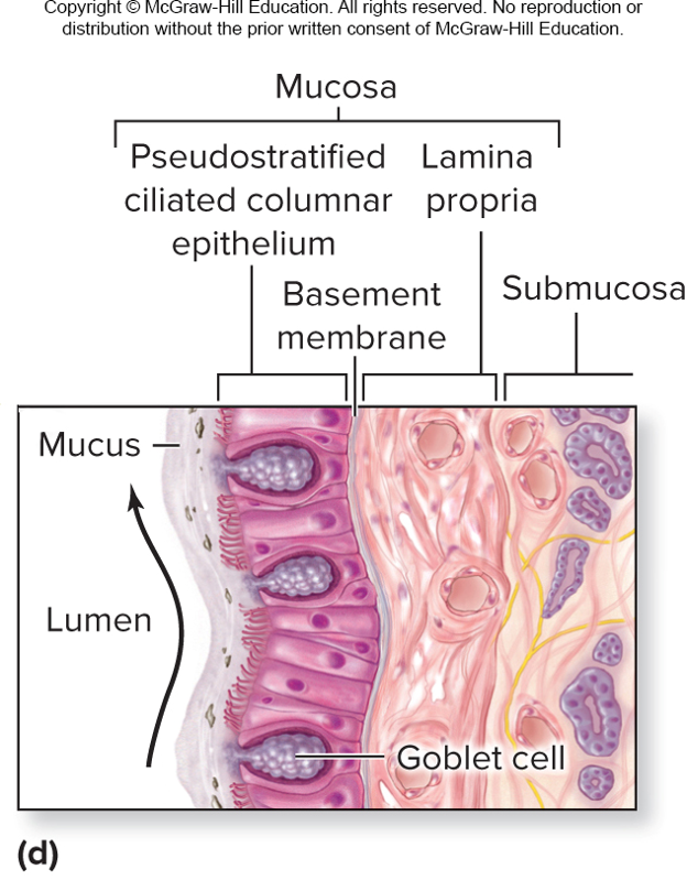
Histology of the Tracheal Wall: Submucosa
Middle layer
Areolar connective tissue with blood vessels, nerves, serous, and mucous glands, lymphatic tissue

Histology of the Tracheal Wall: Tracheal Cartilage
Adventitia: Elastic connective tissue
Bronchial Tree
System of highly branched air passages
Originates at main bronchi, branches to more narrow tubes
Ends in small bronchiole passageways

Gross anatomy of the bronchial tree
Trachea splits into right and left main bronchi (primary bronchi) at level of sternal angle
Each main bronchus branches into lobar bronchi (secondary bronchi)
Further divide into segmental bronchi (tertiary bronchi)
Tree continues to divide into smaller passageways
Leads to tubes of <1mm, the bronchioles
Leads to terminal bronchioles (last part of conducting zone)
Clinical View: Bronchitis
Inflammation of the bronchi caused by bacterial or viral infection or inhaled irritants
Clinical View: Acute Bronchitis
Occurs during or after an infection
Coughing, sneezing, pain with inhalation, fever
Most cases resolving in 10 to 14 days
Clinical View: Chronic Bronchitis
Occurs after long-term irritant exposure
Large amounts of mucus, and cough lasting 3 months
Permanent changes to bronchi occur
Increases likelihood of future bacterial infections
Histology of Bronchial Tree
Main bronchi are supported by incompleted rings of hyaline cartilage (keeps them open)
Wall support (cartilage) lessens as bronchi divide
Bronchioles have no cartilage (thicker layer of smooth muscles)
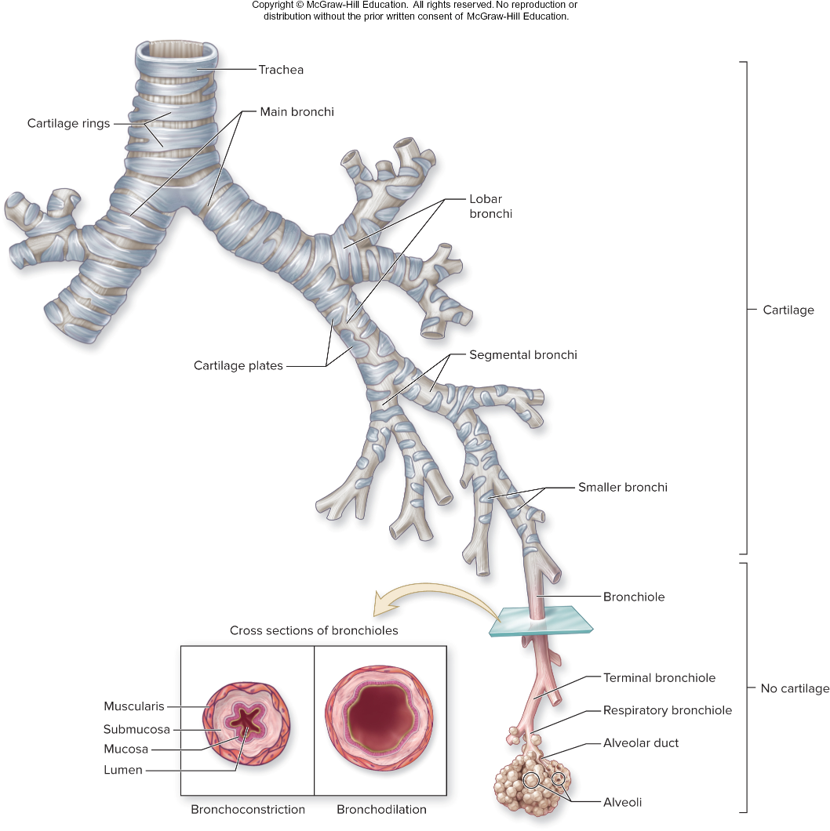
Bronchoconstriction
Muscle contraction narrows bronchiole diameter
Less air through bronchial tree (less entry of potentially harmful substances)
Bronchodilation
Muscle relaxation increases bronchiole diameter
More air through the bronchial tree
Clinical View: Asthma
Episodes of bronchoconstriction, wheezing, coughing, shortness of breath, and excess mucus
Asthmatic with sensitivity to airborne agent
Localized immune reaction occurs in bronchi and bronchioles
Walls of the bronchi becoming permanently thickened
Primary treatments
Inhaled steroids
Bronchodilators
Exercise-induced asthma (bronchoconstriction) leads to excessive constriction of airways during exertion
Respiratory Zones
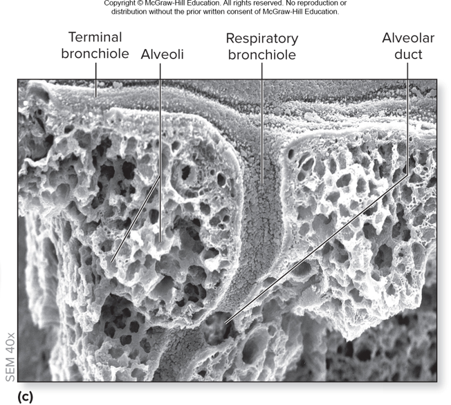
Respiratory bronchioles subdivide to alveolar ducts
Alveolar ducts lead to alveolar sacs, clusters of alveoli
Alveoli = saccular out pocketing

Epithelium
Respiratory bronchioles lined with simple cuboidal epithelium
Alveoli and alveolar ducts lined by simple squamous
Thinness facilitates gas exchange
Alveoli
Each lung contains 300 to 400 million
Alveolar pores: openings providing collateral ventilation
Surrounded by pulmonary capillaries
Divided by interalveolar septum
Contain elastic fibers
Alveolar type I cells
Squamous alveolar cells
Most common of the two cell types making up alveolar wall
95% of alveolar surface area
Form the alveolar epithelium for the respiratory membrane
Alveolar type II cells
Septal cells
Secrete oil pulmonary surfactant
Coats inside of alveolus and opposes collapse during expiration
Alveolar macrophage
Dust cells
Leukocytes that engulf microorganisms
Either fixed in alveolar wall or free to migrate
Respiratory Membrane Anatomy
Thin barrier (0.5 microns) separating air in alveoli and blood in pulmonary capillaries
Consists of:
Alveolar epithelium and Capillary epithelium and its basement membrane
Basement membranes are fused

Respiratory Membrane Function
Oxygen diffuses from alveolus into capillaries
Erythrocytes become oxygenated
Carbon dioxide diffuses from blood to alveolus
Expired to external environment
Clinical View: Pneumonia
Infection of the lung, resulting in alveoli filling with fluid exudate, or pus
Usually caused by bacterial or viral infection
Cough, fever, difficulty breathing, weakness, chills, increased heart rate, and chest pain while inhaling
Pneumonia with milder symptoms = walking pneumonia
Older or immunocompromised individuals have a longer illness than younger adults
Gross Anatomy of the Lung
Lungs are in the thorax
House bronchial tree and all respiratory portions of respiratory system
Each lung has a conical shape
Wide concave base atop diaphragm
Apex (cupula) points superiorly just behind clavicle

Lung Surfaces
Costal surface adjacent to ribs
Mediastinal surface adjacent to mediastinum
Diaphragmatic surface adjacent to diaphragm
Hilum
Indented region on lung’s mediastinal side
Bronchi, pulmonary vessels, autonomic nerves, lymph vessels pass through here
These structures collectively termed the root of the lung
Right Lung
Larger and wider than left lung
Has three lobes divided by two fissures
Horizontal fissure separates superior (upper) lobe from middle lobe
Oblique fissure separates middle lobe from inferior (lower) lobe

Left Lung
Smaller than right due to heart’s position
Has two lobes divided by one fissure
Oblique fissure separates superior and inferior lobes
Lingula: projection from superior lobe that is homologous to right lung’s middle lobe

Three surface indentations accommodate heart and aorta
Cardiac impression on medial surface
Cardiac notch on anterior surface
Groovelike impression for aorta on medial surface
Bronchopulmonary Segments
10 segments in right lung; 8-10 in left lung
Autonomous units encapsulated with connective tissues
Each supplied with its own segmental bronchus
Supplied with its own pulmonary artery and vein and lymph vessels
Can be removed individually in cases of disease
Each segment organized into lobules
Each supplied by terminal bronchiole, arteriole, venule, and lymph vessel

Clinical View: Smoking
Respiratory infections
Cellular or genetic changes to the lungs
Emphysema
Cancer of the lungs, esophagus, stomach, and pancreas
Stomach ulcers
Atherosclerosis
Lower birth weight babies in pregnant women
Poor delivery of oxygen and nutrients to all systemic tissues
Bronchitis, asthma, ear infections from secondhand smoke
Clinical View: Lung Cancer
Highly aggressive and frequently fatal malignancy originating in respiratory epithelium
Smoking causes about 85% of all lung cancer
Symptoms include chronic cough, coughing up blood, excess pulmonary mucus, and increased pulmonary
Blood Supply
Two types of circulation in the lungs
Pulmonary circulation
Bronchial circulation
Pulmonary Circulation
Replenishes O2 and eliminates CO2
Pulmonary arteries carry deoxygenated blood to pulmonary capillaries
Blood is reoxygenated
Blood enters pulmonary venules and veins,

Bronch8ial Circulation
Transports oxygenated blood to tissues of lungs
Bronchial arteries (3 to 4) branch off descending aorta
Bronchial veins collect venous blood
Some drains into the pulmonary veins