COG PSYCH 2
 Call Kai
 Learn
Learn Practice Test
 Spaced Repetition
 Match
Match Flashcards
 Knowt Play
1/25

There's no tags or description
Looks like no tags are added yet.
Name | Mastery | Learn | Test | Matching | Spaced | Call with Kai | Chat |
|---|
No analytics yet
Send a link to your students to track their progress
26 Terms
Patients can recognize loved ones, but think that they’re actually imposters.
Under the belief that the real loved one was kidnapped (or worse)
May even see slight differences between the real loved one and the “imposter”
Eventually leads to paranoia due to believing that ‘someone is lying in plain sight’
Damage from the brain that can come from diverse backgrounds.
What is Capgras syndrome?
Cognitive Appraisal
Looking at someone and recognizing them from their facial properties.
Emotional Appraisal
Requires hypothalamus and thalamus
Trying to tell you how to feel about a certain face
* In Capgras syndrome, this part is damaged/disrupted
“I’m looking at my [ loved one ], but I feel nothing?”
What are the two systems of normal facial recognition?
The Amygdala is linked to emotional processing, which is also linked to:
Feelings of familiarity
Memory for emotional events
Emotional decision making
A close neighbour to the hippocampus
Even simple processings in the brain involve multiple regions
Data from these different regions must be assembled or connected
What can we learn from Capgras Syndrome?
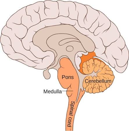
Top of the Spinal Cord
Helps with key life functions
Heart rate, breathing, etc
Cerebellum - the largest region of the hindbrain
Also includes the pons and medulla
What is the Hindbrain?
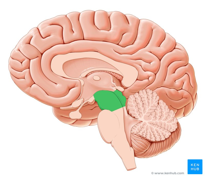
Serves as a hub to boost information from the brain
Coordinating precise eye movement
Rapid small movements to keep sustained tracked movement to an object
Relaying auditory information from ears to forebrain
Regulates pain experiences
What is the Midbrain?
Surrounds the midbrain and most of the hindbrain
Includes the cortex, lobes, and subcortical structures
Cortex - outer surface of the forebrain covered in convolutions, which takes up 80% of the brain
Divided into two cerebral hemispheres
What is the Forebrain?
Frontal lobes
Parietal lobes
Temporal lobes
Occipital lobes
Fissures divide the cerebral cortex into each hemisphere into what different lobes?
Thalamus - sensory relay station
Different neurons go to the thalamus and the thalamus helps direct them to the specific brain region
Hypothalamus - controls behaviours that serve specific biological needs (i.e. eating, body temp, stress, etc.)
Manages endocrine system
Limbic System, two parts:
Amygdala - emotional processing
The olfactory system is close to this, meaning certain smells could give certain emotions.
Hippocampus - learning and memory
Relating to the amygdala, it can help recognize and learn if certain things are threatful
What are the Subortical Structures?
The brain is divided into (roughly) symmetrical left and right hemispheres
Contralateral cortical organization
Connected by commissures (thick bundles of fibers that carry information)
Largest commissure is the corpus callosum
What is Lateralization?
Both hemispheres are damaged
Severed corpus callosum
Was a last-resort treatment for severe epilepsy
Severely limits communication between the hemispheres
Evidence for some hemispheric specializations of functions
What are Split-brain Patients?
Using structural neuroimaging techniques
Records the ‘structures’ of the brain
Blood distribution in general
Computerized Axial Tomography (CT) scans
Magnetic Resonance Imaging (MRI) scans
Uses magnets that ‘shake’ to show the amount of blood in each portion of the brain
Using functional neuroimaging techniques
Records ‘function’ of the brain
“Where in the brain is activity taking place?”
Where is the blood going when performing a certain task?
Position Emission Tomography (PET) scans
What is structural and functional neuroimaging techniques?
Communication between neurons is chemical
Neurons communicate with one another via neurotransmitters
Communication within a neuron is electrical
The input end of a neuron receives neurotransmitters, the output end releases neurotransmitters
An electrical impulse conveys the signal from the input end to the output end
How do neurons communicate between and within?
Recording of the electrical communication within neurons
It is used to study:
Broad rhythms (i.e. sleep stages)
Event-related Potentials (ERPs)
What is an EEG? (Electroencephalogram)
Brain function can also be studied through techniques that manipulate the functions
Chemical effects on neurotransmitters
EX) Depression medications. Allows serotonin reuptake.
EX) Taking pain medications (i.e. acetaminophen). Inhibiting certain pain receptors.
Electrical stimulation
Gene manipulation
Ethical considerations
Institutional review boards
How can we manipulate certain brain function?
EEG:
S: temporally locating neural activity (when?)
W: spatially locating neural activity (where?)
fMRI:
S: spatially locating neural activity (where?)
W: temporally locating neural activity (when?)
MRI scans:
Detecting brain structures but NOT activity
What are the strengths and weaknesses of an EEG, fMRI, and an MRI?
By combining techniques.
In the previous method, how can we limit the weaknesses?
Most neuroimaging techniques used to study brain activity and structures provide only correlational data, meaning no causality can be determined
That said, what are the sources of causal data?
Brain lesions
Transcranial Magnetic Stimulation (TMS)
Produces magnetic pulses that activate neurons
But also creates temporary lesions, to ‘take specific parts out of the game’
Deep TMS
Conventional TMS
Creates functional lesions for a period of time
Transcranial Direct Current Stimulation (TDCS)
What is the problem with neuroimaging techniques? How is it solved?
Largest portion of the human brain
A thin layer of tissue covering the cerebrum (i.e. forebrain)
Regions of the cortex
What is the cerebral cortex?
Primary sensory projection areas - arrival points in the motor cortex for signals from the sensory
Primary motor projection areas - departure points in the motor cortex for signals that control muscle movement
Contralateral Control
More cortical coverage reflects greater motor precision
What are the motor areas of the cerebral cortex?
Somatosensory Area - skin sensations
Primary Auditory Cortex - auditory sensations
Primary Visual Cortex - visual sensations
Contralateral organization
Cortical space assigned based on acuity
What are the sensory areas of the cerebral cortex?
Contains many specialized subregions, damage to which can result in:
Apraxia - brain struggles to plan and sequence physical movements
Agnosia - loss of recognition of familiar objects, smells, people, shapes, sounds, etc., despite having intact senses
Unilateral Neglect Syndrome
Aphasia
What can happen to the many specialized subregions of the cerebral cortex?
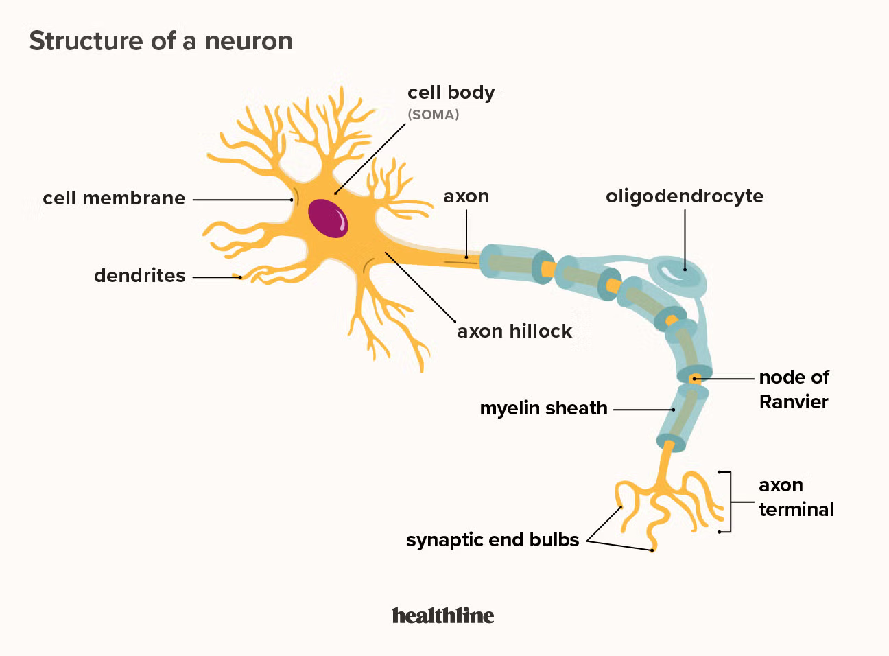
Dendrites - detects incoming signals from other neurons
Cell Body - contains the nucleus and cellular machinery
Axon - transmits signals to other neurons
What are the basic parts of a neuron?
Guides development of the nervous system
Repairs damage in the nervous system
Controls nutrient flow to neurons
Electrical insulation
What are glias?
When a neuron receives a message:
A neuron is stimulated and is either ready to fire or it doesn’t (all-or-nothing)
Neurotransmitters change the postsynaptic membrane
If there is sufficient ionic flow to surpass the cell’s threshold, an action potential is produced
More than 100 neurotransmitters have been identified so far
What are synapses?
Allows a neuron to receive and integrate information from many other neurons
Neurons can compare incoming signals and adjust responses to input as per other incoming signals
Strengths of synaptic connections are adjustable:
They are altered by experience
Considered crucial for learning
What is synaptic transmission?
How is information represented (or coded) by neurons?
Specific neurons can, in some cases, represent specific stimuli
“Pattern coding” or distributed representation
More complex movements (i.e. which parts of the brain are activating to activate specific muscle regions and what not?)
What is Coding?