Week 1 Anatomy lab quiz
 Call Kai
 Learn
Learn Practice Test
 Spaced Repetition
 Match
Match Flashcards
 Knowt Play
1/19

There's no tags or description
Looks like no tags are added yet.
Name | Mastery | Learn | Test | Matching | Spaced | Call with Kai | Chat |
|---|
No analytics yet
Send a link to your students to track their progress
20 Terms
Where does nervous system occur?
Brain
- CNS
- Perception and processing of sensory stimuli (somatic/autonomic)
- Execution of voluntary motor responses (somatic-voluntary)
- Regulation of homeostatic mechanisms (autonomic- involuntary or unconscious)Nerves
- PNS
- Fibers of sensory and motor neurons (somatic/autonomic)Digestive tract
- ENS: enteric nervous systen
- located in digestive tract
- responsible for autonomic functions and can operate independently of the brain and spinal cordSpinal cord
- CNS
- initiation of reflexes from ventral horn (somatic) and lateral horn (autonomic) gray matter
- Pathways for sensory and motor functions between periphery (outer edges) and brain (somatic/autonomic)Ganglia
- PNS
- Reception of sensory stimuli by dorsal root and cranial ganglia (somatic/autonomic)
- Relay of visceral motor responses by autonomic ganglia (autonomic)
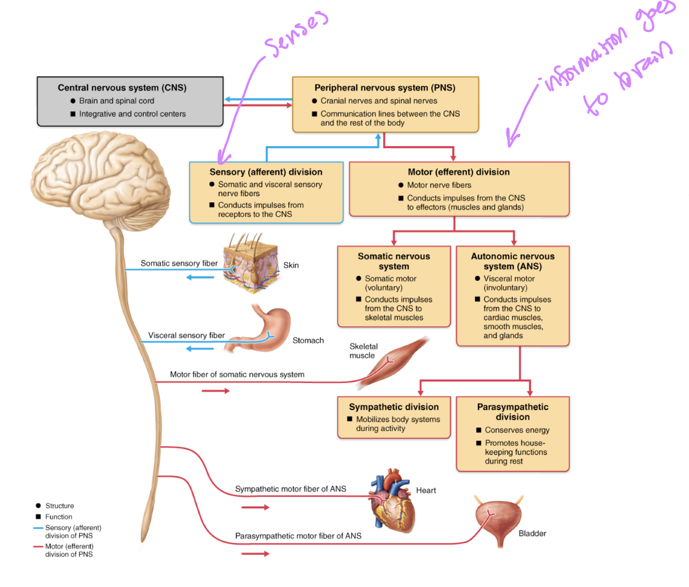
What compromises the nervous system?
top level of division:
- Central Nervous System (CNS)
- Structure: consists of the brain and spinal cord
- Function: acts as the integrative and control center of the body
- Peripheral Nervous System (PNS)
- Structure: consists of cranial nerves (12) and spinal nerves (32)
- Function: acts as the communication lines linking all parts of the body to the CNSFunctional divisions of the PNS
- Sensory (Afferent) Division
- Structure: comprises somatic (superficial) AND visceral (deep) sensory nerve fibers
- Function: Conducts electrical impulses from sensory receptors located throughout the body to the CNS
- information gathered by sensory receptors (like your skin and stomach) travel inward TOWARD the central processing units (spinal cord and brain) so the body can perceive and process what is happening in or around it
- Sub-pathways that originate here:
- somatic sensory fibers: carry incoming sensory information from external receptors in the skin
- visceral sensory fibers: carry incoming sensory information from internal organs (stomach)
- Motor (Efferent) Division
- Structure: compromises motor nerve fibers
- Function: conducts outgoing impulses away from the CNS to effectors, which are muscles and glands
- This division further branches into two distinct systems based on the type of effector controlled:Subdivisions of the motor division:
A. Somatic Nervous system
- control type: somatic motor (voluntary control)
- function: conducts motor impulses from the CNS to the skeletal muscles
B. Autonomic nervous system (ANS)
- control type: visceral motor (involuntary control)
- function: conducts motor impulses from the CNS to cardiac muscle, smooth muscles, and glands
- ANS splits further:
- sympathetic division: mobilizes body systems during activity (flight or fight), sends sympathetic motor fibers of the ANS to visceral effectors like the heart (increases activity) and the bladder
- parasympathetic division: conserves energy and promotes “housekeeping” functions during rest (rest and digest), sends parasympathetic motor fibers of the ANS to regulate organs like the heart (decreases activity) and bladder
Nervous System Anatomy
Plexus: bundle of intersecting nerves that exit from the spinal cord
CNS:
- nuclei: collection of somas
- tracts: bundle of axonsPNS:
- Ganglia: collection of somas
- Nerves: bundle of axons
Structure of a nueron
Soma (cell body): contains all normal cell structures, Nissl bodies, neurofibrils, and microtubules
Dendrites: receive incoming signal to neuron
- ReceiversAxons: transmit outgoing signal away from neuron
- transmitters
- axon hillock: structure that generates the signal that travels down the axon (not an actual physical structure)→ action potential propagates and goes to axon terminals and release neurotransmitters→ can potentially go to another neuron or a target tissue (muscle)
- Neurotransmitters: a chemical→ converted into an electrical signal because it is converted by the axon hillock and created into an action potential
- Axon terminals: structure that releases neurotransmitter allowing communication with the other neurons/cellsMyelin Sheath: surrounds the axon and provides insulation that increases conduction speed, insulates neuron to increase conduction velocity
- spaces between the myelin sheath are nodes of Ranvier, and these house sodium-potassium channels
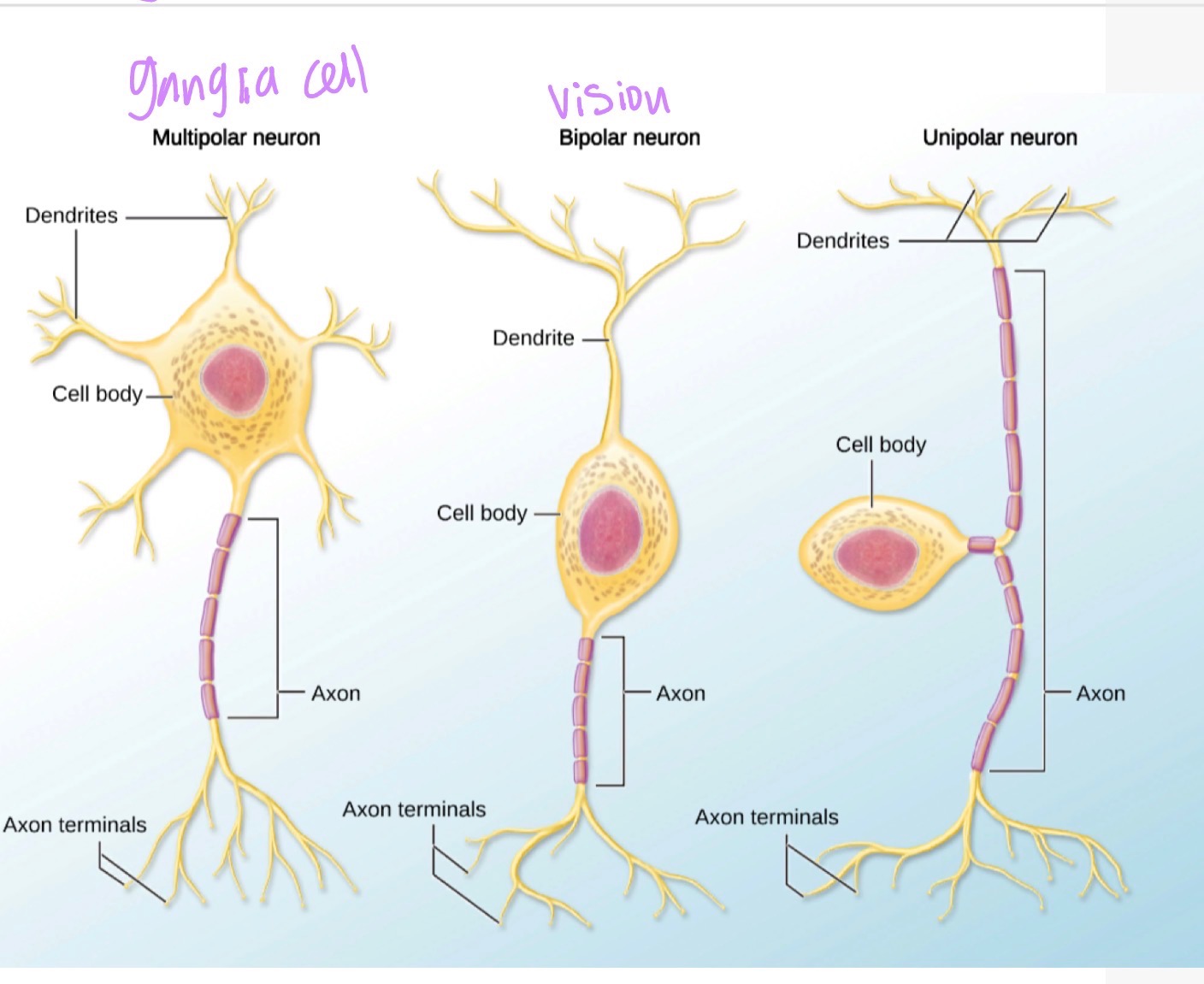
Structural Classifications of Neurons
Neuroglia of CNS
Astrocytes
Microglia
Oligodendrocytes
Ependymal cells
Astrocytes:
neuron support by:
- regulating the extracellular ion balance
- monitoring nerve impulses
- forming the blood brain barrier (along with capillary endothelial cells) and control transmission of solutes from the bloodstream into nervous tissue
- processes varying in size that facilitate exchanges between nutrient rich capillaries and neuronscomposed of: microfilaments and various organelles
Microglia
Neuron Support by:
- phagocytic cells
- ingest and break down waste products and pathogens
Oligodendrocytes
Neuron support by:
- wrapping the cell membrane around multiple CNS axons and forming a myelin sheath
- myelin sheath electrically insulates the cell
- Cytoplasm of cell provides support to maintain homeostatic environments for axons
- Prevent dissipation of neural impulse and crossing of neural impulses from adjacent neuronscomposed of: lipids and proteins
Ependymal Cells
Neuron cupport by:
- production of circulation of cerebrospinal fluid (CSF)
- Line the brain, ventricles, and central canal of the spinal cordcomposed of:
- cuboidal and columnar cells
- contain microvilli and cilia
Neuroglia of PNS (know one function for each cell)
Schwann cells
Satellite cells
Satelite Cells
Neuron support by:
- Form thin cellular sheaths around neuronal cell bodies in PNS ganglia
- maintain homeostatic environment
- regulate exchanges between the cell body and interstitial fluidComposed of:
- flat, uninucleate cells
Schwann Cells
Provide neuron support by:
- insulate myelinated PNS axons
- Support nerve regeneration
- allow saltatory conduction at the nodes of the Ranvier (part under the myelin sheath)Composed of: (myelin sheath)
- lipids and proteins
Neuron Function
Neuron os polarized with a net positive charge outside of the neuron and a net negative charge inside
The membrane depolarizes as the signal passes along the axon, repolarizing once the impulses has passed
At the axon terminal, the depolarization caused by the arriving signal triggers the release of neurotransmitters from the presynaptic cel
These chemicals travel across the synaptic cleft and bind to another neuron (thereby propagating the signal), or a terminal, effector, tissue
Resting Potential
Resting potential of neurons are -70mV
maintaining resting membrane
NA+/K+ ATPase
Action Potential
Axon Hillock
- must be depolrarized to the threshold value to trigger an action potentialDepolarization
- Na+ influxRepolarization
- K+ effluxHyperpolarization
- Some K+ channels are open
Synapse and Neurotransmitters
Synaptic cleft is where neurotransmitters (NTM) are secreted
NTM stored in the form of membrane-bound vesicles
Dopamine:
- excitatory and inhibitory
- inhibits unnecessary movements
- released from the substantia nigraAcetylcholine (Ach)
- excitatory in all cases except the heart (inhibitory)
- widespread neurotransmitter found in the CNS and at neuromuscualr junctions between peripheral nerves and musclesEpinephrine (E) and Norepinephrine (NE)
- excitatory
- function as both hormones and neurotransmitters, primarily in the CNS
- produced in the adrenal glands, brainstem, and hypothalamus ad released into the bloodstream
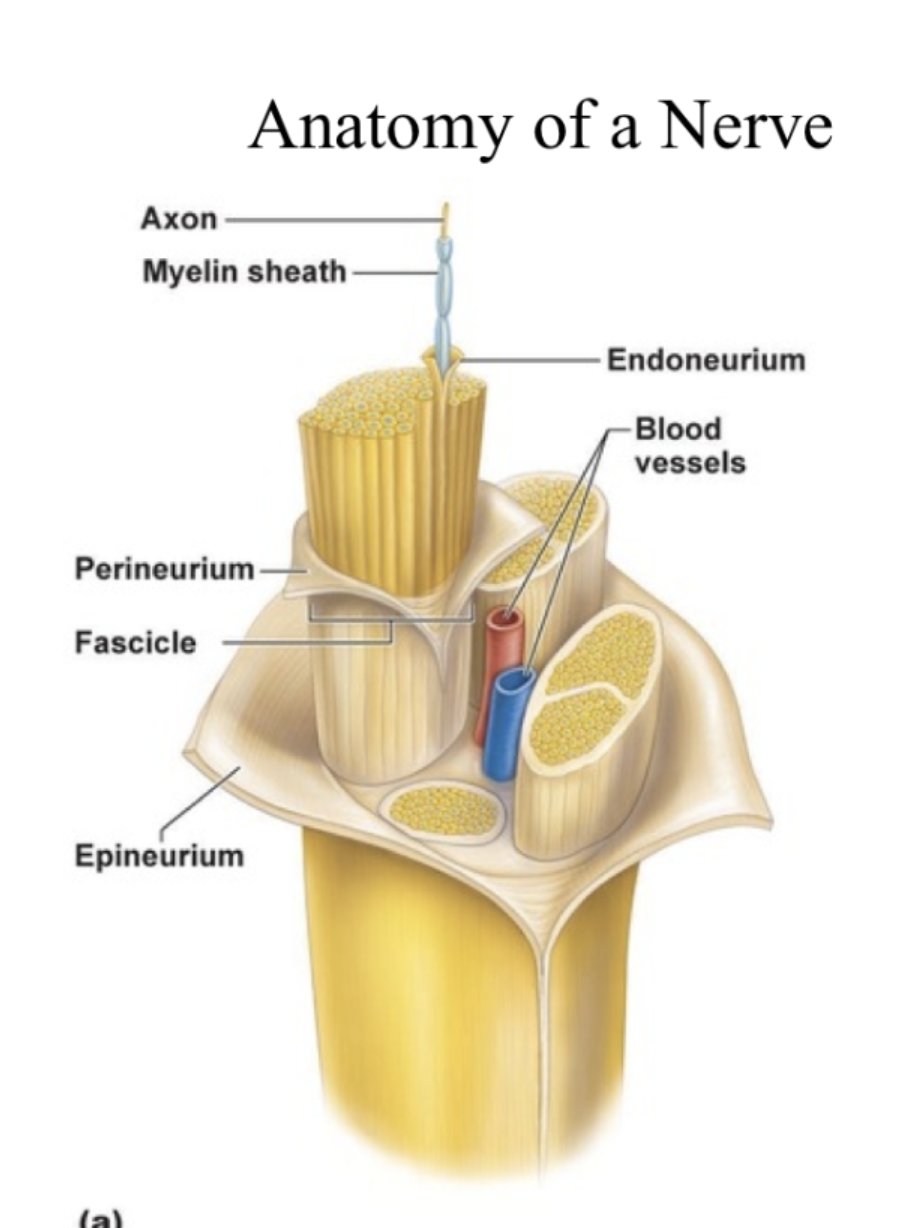
Anatomy of a Nerve
Nerve Fiber Types:
Sensory (afferent)
- somatic
- visceralMotor (efferent)
- somatic
- visceralmixed
Type of Nerve Fibers
Afferent (Sensory) Nerves
- from periphery to CNS
- Somatic sensory
- carrying information from the skin, skeletal muscle, and joints
- visceral sensory
- from visceral organs (liver or kidneys)Efferent (Motor) Nerves
- from CNS to periphery
- somatic motor
- provide innervation to the skeletal muscles
- visceral motor
- part of the ANS and they innervate smooth muscle, cardiac muscle and glandsMixed
- nerves that do both afferent and efferent fibers
- transmit messages in both directions
Pathologies
pinched nerve
carpal tunnel
sciatica
cause: excessive pressure on nerve by surrounding structures, overuse
Symptoms:
- tingling
- pain
- numbness
- weakness
- feeling of limb “falling asleep”