MRI Brain Anatomy
 Call Kai
 Learn
Learn Practice Test
 Spaced Repetition
 Match
Match Flashcards
 Knowt Play
1/161

There's no tags or description
Looks like no tags are added yet.
Name | Mastery | Learn | Test | Matching | Spaced | Call with Kai | Chat |
|---|
No analytics yet
Send a link to your students to track their progress
162 Terms
A- anterior cerebral artery
A

B- internal carotid artery
B

c- vertebral artery
C

D- basilar artery
D

E- posterior cerebral artery
E

F- middle cerebral artery
F

C- 7th cranial nerve, facial nerve
C

D. semi-circular canal
D
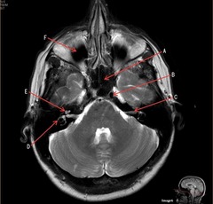
E- cochlea
E

F- maxillary sinus
F

T2 FLAIR; Sagittal
A FLAIR (Fluid Attenuated Inversion Recovery) sequence is utilized to suppress signal from cerebrospinal fluid (CSF).
What type of sequence?

T1, sagittal
What type of sequence?

Genu of Corpus Callosum
A- region

Splenium of Corpus Callosum
B- region

pons
C

cerebellum
D

lateral ventricle
A

corpus callosum
B
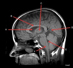
thalamus
C

tentorium cerebelli- cerebrum (occipital and temporal lobes) from brainstem and cerebellum
D- separates what

fourth ventricle
E

medulla oblongata
F

caudate nucleus
A

thalamus
B

third ventricle
C

lentiform nucleus
D

frontal sinus
E

basal ganglia region
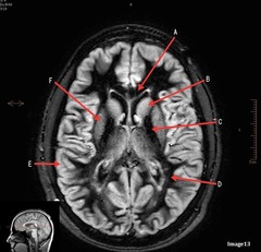
internal capsule
A (region)
dark band is called?

posterior horn of lateral ventricle
B

anterior horn of lateral ventricle
C

white matter
D

caudate nucleus
B

internal capsule
C

lentiform nucleus
F

gray matter
E

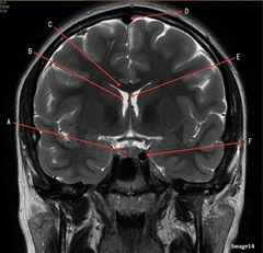
pituitary gland
A

lateral ventricle
B
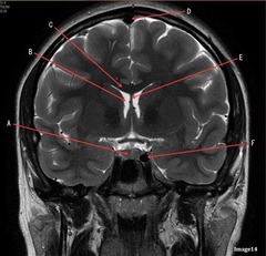
corpus
C

fornix
E

internal carotid artery
F
external carotid artery
face
A, where does it supply blood to?

internal carotid artery, anterior brain
B, where does it supply blood to?

Common carotid bifurcation
C

common carotid artery
D
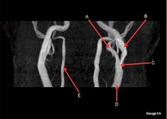
vertebral artery, posterior brain
E, where does it supply blood to?
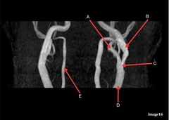
MRA extracranial circulation
what kind of image is this?

Torcular herophili
E

thalamus
B

A- mandibular condyle inside mandibular fossa of temporal bone
B- articular disc
C-articular tubercle
A
B
C

brachiocephalic trunk, innominate artery
G

R subclavian artery
H

internal carotid artery
K

posterior frontal vein
B

parietal veins
C

superior sagittal sinus
D

Torcular herophili
E

internal jugular vein
F
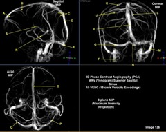
R transverse sinus
G

Superior sagittal sinus
H

R parietal veins
J

L parietal veins
L

Vein of trolard (superior anastomotic vein)
K

Left sigmoid sinus
M

Torcular herophili
N

Superior sagittal sinus
O

Left transverse sinus
P

Internal cerebral vein
S

Internal carotid artery
A

Caudate nucleus
B
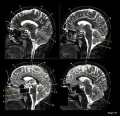
white matter
C

thalamus
D

straight sinus
F

lateral ventricle
G

lateral sulcus
H

optic chiasm
L

Splenium of corpus callosum
O

Cerebral peduncle
V

superior sagittal sinus
W

vein of galen
X

Inferior colliculus of midbrain,
just inferior to pineal gland / superior to the cerebral aqueduct
Y
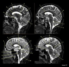
White matter
The corpus callosum is the only white matter tissue structure found in the midline sagittal slice of the brain.
Letter N in Image 137 is pointing to what type of tissue?

vertebral arteries
Which arteries join together to form the basilar artery?
anterior cerebral artery
letter A

7th cranial nerve- facial
8th cranial nerve vestibulocochlear on other side (posterior) of semicircular canal
letter C

medulla oblongata
letter F

internal capsule
letter C

white matter
letter D

lentiform nucleus
letter F
face
letter A responsible for blood supply to where
posterior brain
anterior brain
face
upper extremities

posterior frontal vein
B

parietal veins
C

superior sagittal sinus
D

Torcular herophili
E

anterior frontal vein
A

right transverse sinus
G

left parietal veins
L

Vein of Trolard
k

left sigmoid sinus
m

torcular Herophili
N

superior sagittal sinus
o
