Head and Neck Muscles
1/65
There's no tags or description
Looks like no tags are added yet.
Name | Mastery | Learn | Test | Matching | Spaced |
|---|
No study sessions yet.
66 Terms
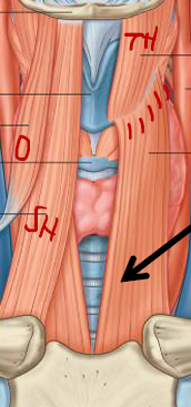
Describe the Neck
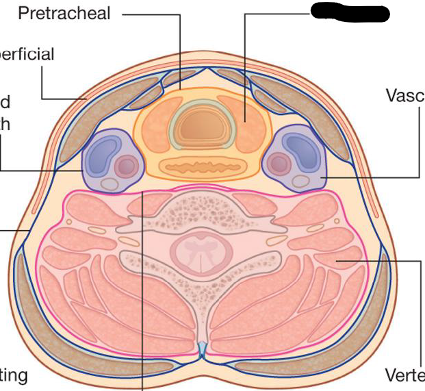
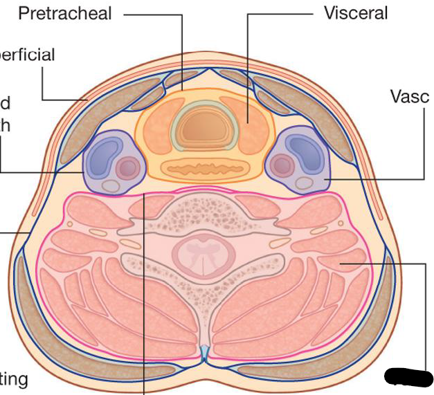
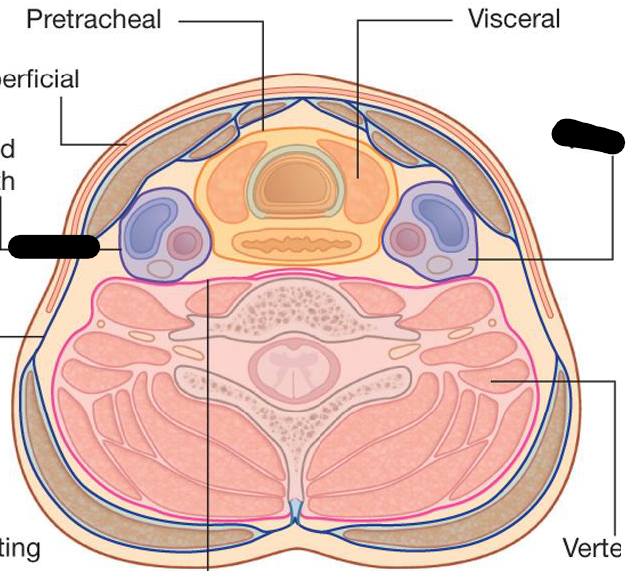
Extends from → lower border of mandible to manubrium / superior nuchal line of occipital bone to intervertebral disc of C7-T1
Divided into 4 compartments - Visceral, Vertebral, and Vascular(2)
What are the compartments of the Neck
Visceral Compartment:
Anterior, Organs of digestive and respiratory systems, and Endocrine glands
What are the compartments of the Neck
Vertebral compartment:
Posterior, Cervical vertebrae, Spinal cord, Cervical nerves, Muscles associated with vertebral column
What are the compartments of the Neck
Vascular Compartments (2)
Left and Right, Lateral, Contain major bv, vagus nerv
What are the components of the Neck
Anterior and Posterior Triangle
What are the boundaries of the Posterior Triangle
Posterior border of sternocleidomastoid, Anterior border of trapezius, Clavicle
What are the boundaries of the Anterior Triangle
Anterior border of sternocleidomastoid, Inferior border of mandible, Midline of neck
What does the Anterior Triangle muscle contain
Muscles - suprahyoid + infrahyoid muscles
BV - common carotid A and branches + jugular Vs
Cranial nerves
Thyroid and parathyroid glands
Pharynx and laryn
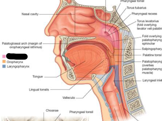
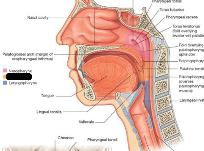
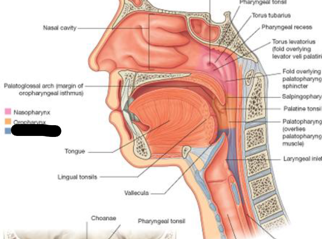
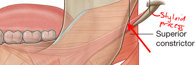
What does the Pharynx do and what is it divided into
Links oral and nasal cavity in head to larynx and esophagus in neck
Common pathway for air and food
Divided into → Nasopharynx, Oropharynx, and Laryngopharyn
What is this compartment of the Pharynx
Nasopharynx
From posterior apertures (choanae) of nasal cavities to soft palate
Continuous with oropharyn
What is this compartment of the Pharynx
Oropharynx
Posterior part of oral cavity
Inferior to soft palate, superior to epiglottis
Houses palatoglossal folds that house palatine tonsi
What is this compartment of the Pharynx
Laryngopharynx
Superior margin of epiglottis to top fo esophagus (C6)
What does the pharynx house
Houses tonsils
Pharyngeal tonsils → Adenoids and Nasopharynx
Palatine tonsils → Oropharynx
Describe the Larynx
Opens to pharynx
Continues with trachea
Valve to close lower respiratory tract
Instrument to produce sound
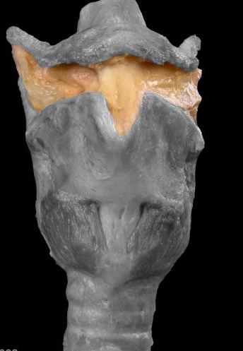
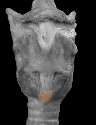
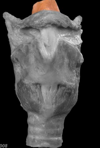
Describe the structures of the Larynx
Cartilages + Membranes/Ligaments
Suspended from Hyoid bone, attached to trachea
Lvl of C4 to C6
Keep airway open by routing food and drink into esophagus
Voice box - sound production
What does the Larynx do during swallowing
Larynx move up and forward to facilitate closing of the laryngeal inlet and open the esophagus
No food/drink in larynx
Describe the Thyroid Cartilage
Unpaired Cartilage
Lvl of C4 and C5
Has Laryngeal Prominence (Adam’s Apple)
Describe the Thyroid Notch
Superior to laryngeal prominence
Signifies superior margin of thyroid carilage
Describe the horns off the Hyoid
They come off of the Thyroid Cartilage
Superior - attachment for thyrohyoid ligament
Inferior - articulation with cricoid cartilage
What is this Membrane
Thyroid Membrane
B/T Hyoid bone and Thyroid cartilage
What is this Cartilage
Cricoid Cartilage
Lvl of C6
Below Thyroid Cartilage
Unpaired Cartilage
What is this ligament
Cricothyroid Ligament
B/T thyroid cartilage and cricoid cartilage
What is this
Epiglottis
Unpaired cartilage
Closes off trachea so food and drink does not enter
Attached by narrow stem by ligament to thyroid cartilage - Thyro-epiglottic ligament
What is this
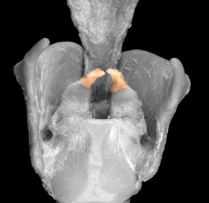
Corniculates
Paired cartilage
Small
Sit atop arytenoids
What is this
Arytenoids
Paired cartilage
Articulate inf with cricoid cartilage
Slide and rotate on the criocoid cartilage
Involved in sound production
Describe the Vocal Ligament
Sup margin of cricothyroid ligament thickened to from Vocal lig
Attached to internal surface of thyroid cartilage and vocal process of arytenoids
Vocal ligaments flank glottis
Arytenoids rotate and slide on cricoid cartilage, change how air passes out of glottis
Vocal lig enclosed in vocal folds
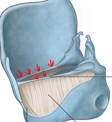
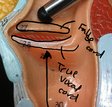
Describe the Larynx - Vocal Cords
Within larynx
2 vocal cords/folds
Vocal Fold/True Vocal cord (produces sound + inferiorly placed)
Vestibular Fold/False Vocal Cord (modifies sound produced by vocal fold)
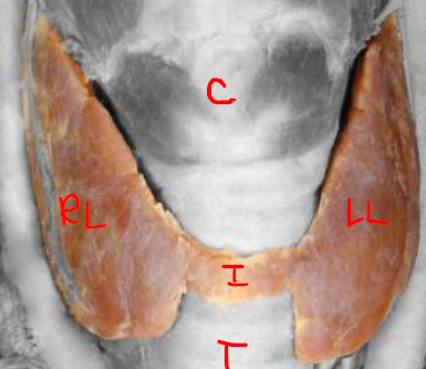
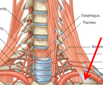
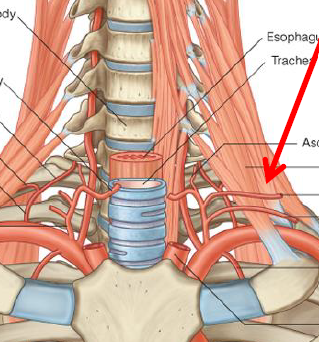
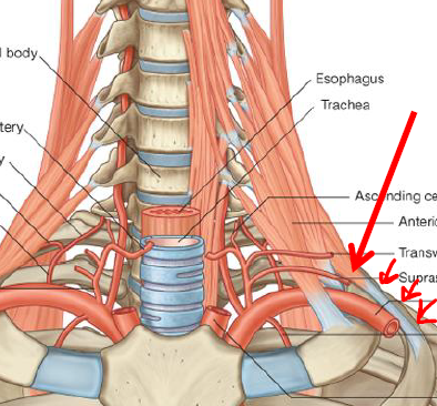
Describe the thyroid gland
Large gland with distinct anatomy → left and right lobes, connected by narrow Isthmus
Endocrine
Flanking trachea, inferior to larynx
How do you find the parts of the Thyroid Gland
R/L lobes best palpated by finding laryngeal prominence, move inferiorly to cricoid cartilage
Move posteriolateral to larynx
Isthmus lies on surface of trachea
Palpate isthmus by finding cricoid cartilage, move inferiorly to palpate along trachea
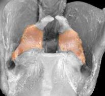
Describe the Parathyroid Glands
Small
Embedded in posterior surface of thyroid gland
Produces parathyroid hormone (Ca lvls in blood)
ID/Describe
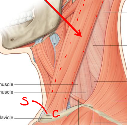
Sternocleidomastoid
Origin - Clavicular head-medial 1/3 of Clavicle / Sternal head-Manubrium
Insertion - Clavicular head-lateral ½ superior nuchal line / Sternal ehad-Mastoid process
Fxn - Unilateral contraction-ipsilateral lateral flecion / Unilateral contraction-contralateral head rotation / Bilateral-neck flexion
N - Accessory N
ID/Desciribe
Scalenus Anterior (anterior to brachial plexus and subclavian A
Origin - TP of C3-C6
Insertion - Upper surface of rib 1, scalene tubercle
Fxn - Elevate rib 1
N - Ant rami of C4-C7
ID/Describe
Scalenus Medius (posterior to brachial plexus and subclavian A
Origin - TP of vert C2-C7
Insertion - Upper surface of rib 1, posterior to groove for subclavian A
Fxn - Elevate rib 1
N - Ant rami of C3-C7
ID/Describe
Scalenus Posterior (posterior to brachial plexus and subclavian A)
Origin - TP of vert
Insertion - Upper surface of rib 2
Fxn - Elevate rib 2
N - Ant rami of C5-C7
What do the muscles of the face do
Move the skin, lips, nostrils, and eyelids in diff facial expressions
All innervated by the Facial N
ID/Describe
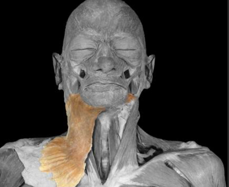
Platysma (large, thin, sheetlike muscle)
Origin - Clavicle
Insertion - Mandible
Fxn - Draws corners of mouth down / Grimace / Tenses skin of neck
N - Facial N
ID/Describe
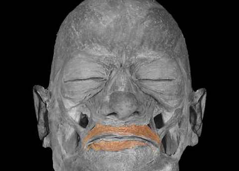
Orbicularis Oris
Origin - Facial muscles / Mandible / Maxilla
Insertion - Ellipse around mouth
Fxn - Closes lips / Protrudes lips / Presses lips against teeth / Pouting / Kissing
N - Facial N
ID/Describe
Zygomaticus Major
Origin - Zygomatic bone
Insertion - Corners of mouth
Fxn - Raises upper lip / smiling
N - Facial N
ID/Describe
Zygomaticus Minor
Origin - Zygomatic bone
Insertion - Upper lip medial to corner of mouth
Fxn - Raises upper lip
N - Facial N
ID/Describe
Levator Labii Superioris
Origin - Infraorbital margin of maxilla
Insertion - Upper lip
Fxn - Raises upper lip
N - Facial N
ID/Describe
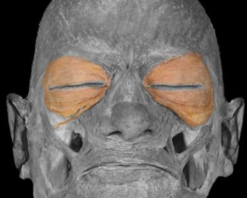
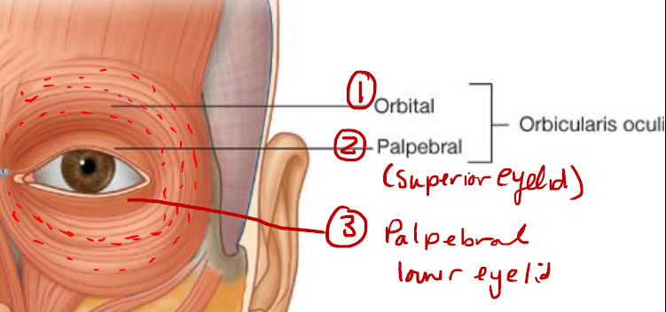
Orbicularis Oculi (3 portions)
Origin - Encircles orbit
Insertion - Encircles orbit
Fxn - Closes eyelids / Draws eyebrows medially and downward
N - Facial N
What are the 3 portions of the Orbicularis Oculi
1 Orbital portion
2 Palpebral portions (superior placed - upper eyelid)(inferior placed - lower eyelid)
ID/Describe
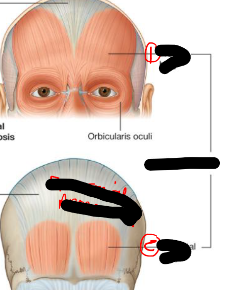
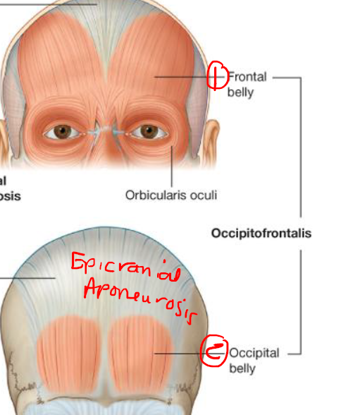
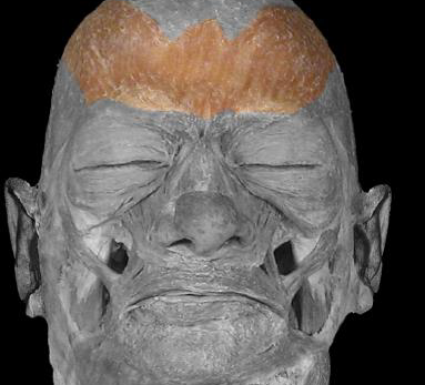
Occipitofrontalis
Two muscle bellies - Frontal belly (forehead) + Occipital belly
Connected by aponeurosis - Epicranial aponeurosis
ID/Describe
Frontal Belly
Origin - Epicranial Aponeurosis
Insertion - Skin of eyebrows
Fxn - Draws eyebrows upward / Wrinkles skin of forehead
N - Facial N
ID/Describe
Occipital Belly
Origin - Superior nuchal line of occipital bone / Mastoid process temporal bone
Insertion - Epicranial aponeurosis
Fxn - Draws scalp backward
N - Facial N
ID/Describe
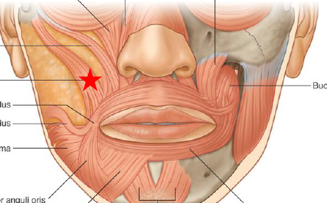
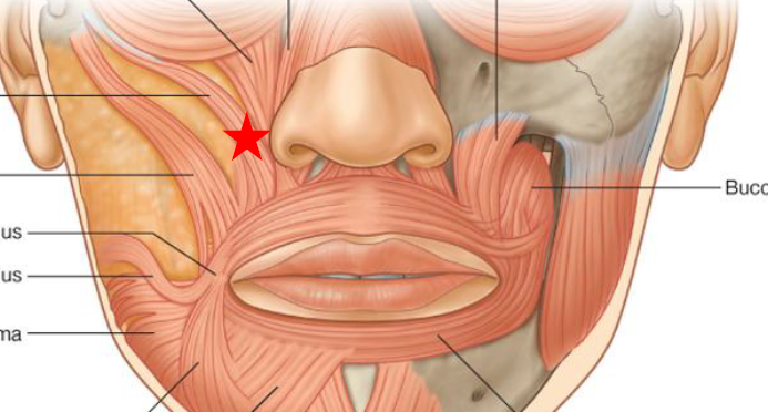
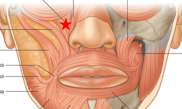
Buccinator (pierced by parotid duct)
Origin - Posterior parts of maxilla and mandible
Insertion - Blends with orbicularis oris and lips
Fxn - Presses cheek against teeth / Helps position food against teeth during chewing
N - Facial N
ID/Describe
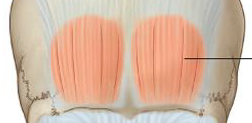
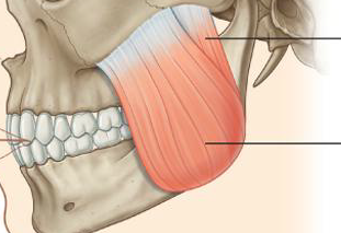
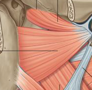
Masseter
Origin - Zygomatic arch / Maxillary process of zygomatic bone
Insertion - Lateral surface of ramus of mandible
Fxn - Elevate mandible
N - Mandibular branch of trigeminal N
ID/Describe
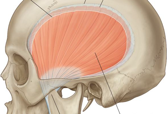
Temporalis
Origin - Temporal bone / Temporal fascia
Insertion - Coronoid process of mandible / Anterior marign of ramus of mandible
Fxn - Elevate mandible / Retract mandible
N - Mandibular branch of trigeminal N
ID/Describe
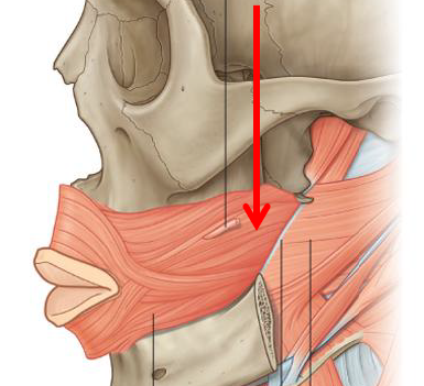
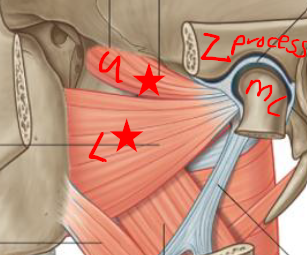
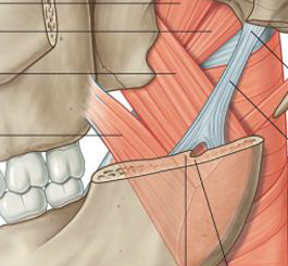
Lateral Pterygoid (2 heads - Upper + Lower)
Origin - U-infratemporal fossa / L-lateral side of lateral pterygoid plate
Insertion - Temporomandibular joint capsule / neck of mandible
Fxn - Protrude mandible / Move mandible side to side
N - Mandibular branch of trigeminal N
ID/Describe
Medial Pterygoid (2 heads - Superficial + Deep)
Origin - S-palatine bone + maxilla / D-medial surface of lateral pterygoid plate
Insertion - Medial surface of mandible near angle
Fxn - Elevate mandible / move mandible side to side
N - Mandibular branch of trigeminal N
ID/Describe
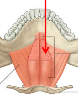
Genioglossus
Origin - Mental spines on back of mandible
Insertion - Forms bulk of tongue / Some fibers to hyoid
Fxn - Protrudes tongue
N - Hypoglossal N
ID/Describe
Hypoglossus
Origin - Hyoid bone
Insertion - Fibrous tissue of tongue near dorsum
Fxn - Flattens (depresses) tongue
N - Hypoglossal N
ID/Describe
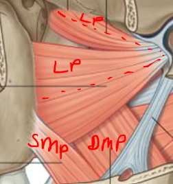
Styloglossus
Origin - Styloid process
Insertion - Lateral side of tongue / Body of hyoid bone
Fxn - Retracts tongue upward and backward
N - Hypoglossal N
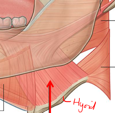
What do muscles of the hyoid move
Muscles that move the hyoid will move the larynx
b/c of membranous/muscular connection between the two
What are the 2 types of muscles that move the hyoid + larynx
Suprahyoid muscles → attach to superior margin of hyoid
Infrahyoid muscles → tatach to inferior marign of hyoid
ID/Describe
Stylohyoid (suprahyoid)
Origin - Styloid process
Insertion - Body of hyoid
Fxn - Elevates hyoid during swallowing
N - Facial N
ID/Describe
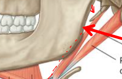
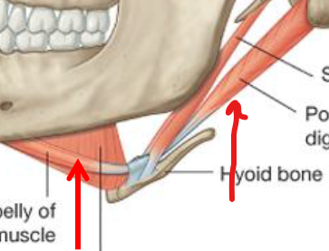
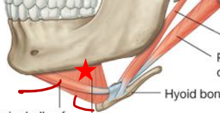
Digrastic (2 bellies - Ant + Post)(Suprahyoid)
Origin - A-lower margin of mandible / P-Mastoid process
Insertion - Tendon B/T two bellies to body of hyoid bone
Fxn - A-Elevates hyoid during swallowing and opening of mouth, depresses mandible / P-elevates hyoid during closing of mouth
N - A-Mylohyoid N / P-Facial N
ID/Describe
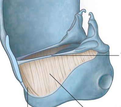
Mylohyoid (Suprachyoid)
Origin - Internal surface of mandible / Mylohyoid line of mandible
Insertion - Hyoid bone
Fxn - Helps form floor of oral cavity / Elevation of floor of mouth / Elevation of hyoid
N - Mylohyoid N (off branch of mandibular division of trigeminal N)
ID/Describe
Geniohyoid (Suprahyoid)
Origin - Mental spine of mandible
Insertion - Body of hyoid bone
Fxn - Supports and elevates floor of oral cavity / Depresses mandible when hyoid fixed / Elevates hyoid when mandible fixed
N - Ant ramus C1
ID/Describe
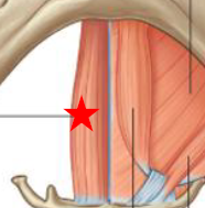
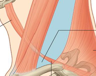
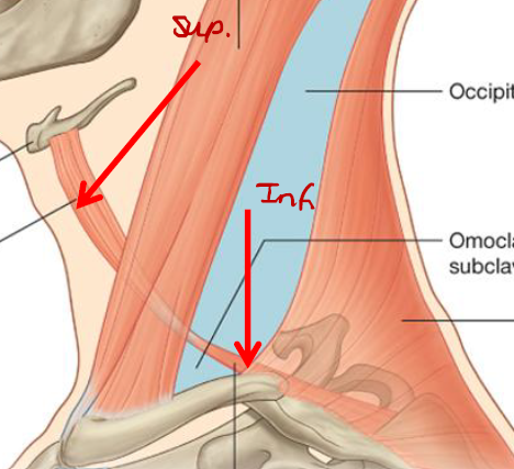
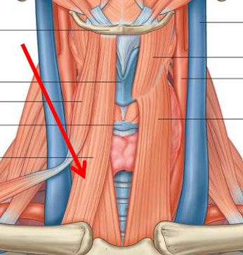
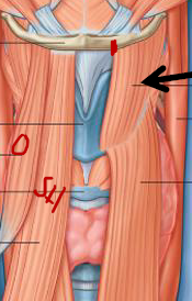
Omohyoid (2 bellies)(Infrahyoid)
Origin - Superior border of scapula
Insertion - Lower border of body of hyoid bone
Fxn - Depressing hyoid bone during vocalization and end of swallowing / Fixes hyoid bone
N - Ant rami of C1-C3
ID/Describe
Sternohyoid (Infrahyoid)
Origin - Manubrium / Clavicle
Insertion - Lower border of hyoid bone
Fxn - Depress hyoid bone during vocalization and end of swallowing / Fixes hyoid bone
N - Ant rami of C1-C3
ID/Describe
Thyrohyoid (Infrahyoid)
Origin - Thyroid cartilage
Insertion - Lower border of hyoid bone
Fxn - Depresses hyoid bone / Raise larynx while swallowing
N - Ant ramus of C1
ID/Describe
Sternothyroid
Origin - Manubrium / Medial part of first costal cartilage
Insertion - Front of thyroid cartilage
Fxn - Depresses larynx
N - Ant rami of C1-C3
Describe the Parotid Gland
Large salivary gland
Ant to ear, much of cheek region
Parotid duct comes off - Pierces buccinator to enter oral cavity
Describe the Submandibular Gland
Salivary gland below mandible
On L and R side
Drained by submandibular duct
Describe the Sublingal Gland
Salivary gland below tongue
On L and R side