DAS 105- panoramic xrays
1/76
Earn XP
Description and Tags
fuck yuo
Name | Mastery | Learn | Test | Matching | Spaced |
|---|
No study sessions yet.
77 Terms
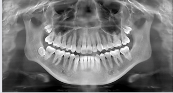
what does panoramic xray show
A wide view of the maxilla, mandible, and pts overall oral and facial structure
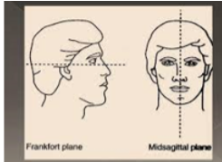
what is the frankfort plane
imaginary line that passes from bottom of the eye socket to top of ear canal
what is panoramic xray used to evaluate
dentition and supporting structures
impacted teeth
eruption patterns, growth, and development
disease lesions and conditions of the jaw
trauma
what is a pano not used to evalulate
caries
periodontal disease
periapical lesions
what are the fundamentals of pano
receptor and xray tube moves around pt
tube head moves in one direction and receptor rotates in opposite direction
what is tomography
allows imaging of one section of body while blurring the other sections
what is the rotation center
pivotal point where receptor and xray tube head rotate
what is the focal trough
three dimensional curved zone where structures are clearly viewed
what is the image resolution when image is inside the focal trough
well defined
image resolution when image is outside focal trough
blurry
how to make the focal trough narrower
get rotation closer to the teeth
what are resultant images
anatomic structures penetrated by xray twice, location of structures determine what type of image results
what is a real image
when structure lies between receptor and moving rotation center
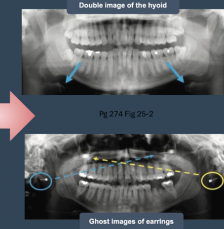
what is a double image
when structure is located behind the moving rotation center
what is the difference and similarity between a double and real image
double image is mirror but has the same proportions as a real image
what is a ghost image
structure located close to pt but outside the focal source ; appears blurred and magnified on opposite side of true angle
what are artifacts
motion
double images
ghost images
suboptimal positioning
what equipment is used in a pano
xray tube head
head positioner
exposure control
explain tube head
like intraoral tube head but collimator has opening in shape of narrow vertical slit rather than round circle
beam passes thru collimator as narrow band and exposes receptor thru slit
horizontal angulation fixed at -10 degrees
what makes up the head positioner
chin rest, notched bite block, forehead rest, lateral head support
what does the chin rest and bite block do
stabilizes pt dentition in anterior and posterior direction
what does the lateral head supporters do
stabilizes pt head in vertical and horizontal plane
what is unique about exposure factors in a pano
predetermined by manufacturer but radiography can select pt size and type of image being obtained
wat are the exposure steps
cover biteblock w barrier
select exposure settings
adjust machine height
explain procedure to pt, place lead apron, instruct them to remove jewelry/interfering objects
position spine and have pt stand straight and close to machine
pt position teeth on bite block
have pt heads midsagittal plane be perpendicular to floor and frankfort plane parallel to floor
what are common errors
ghost images
open lips
tongue not placed on hard palate
tipped chin
teeth not on biteblock
pt not centered
pt not standing straight
thyroid collar
what causes ghost images
glasses, jewelry, retainers, etc
why do open lips cause an error in a pano
makes dark radiolucent shadows in anterior region
why tongue not on hard palate matter
radiolucent shadows will appear around the apices of maxillary teeth
wai tipped chin bad
missing condyles
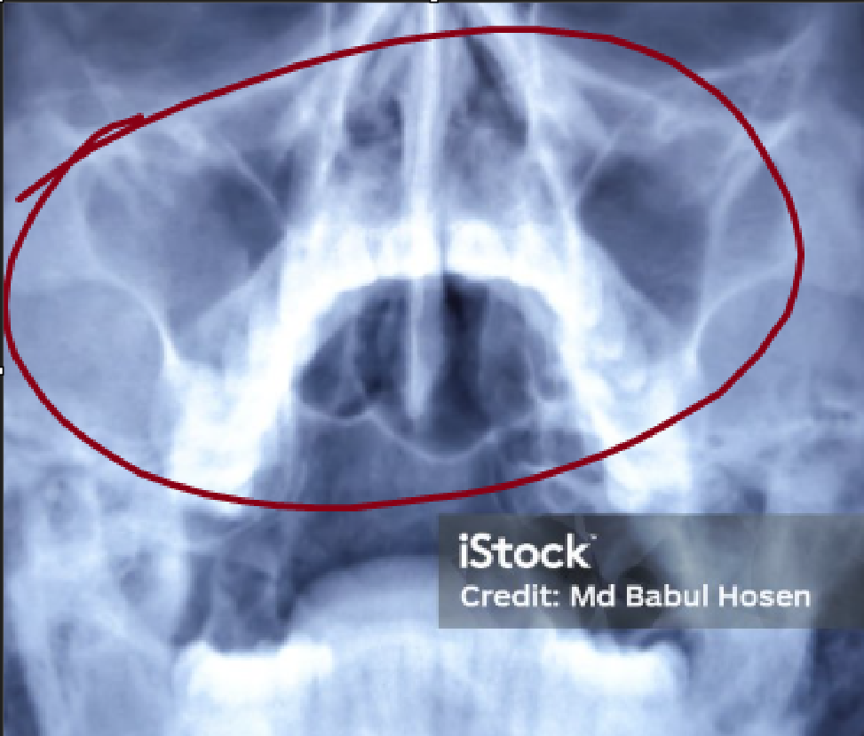
nasal cavity appears superimposed
maxillary incisors distorted
loss o detail
reverse smile line
wai teeth not on biteblock bad
distorts and overlaps the anterior and premolars
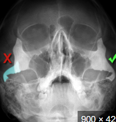
wai pt not centered bad
one side appears closer and other farther
wai pt not standing straight bad
beam will pass thru cervical spine causing radiopacity on image
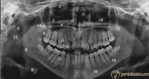
what are the normal anatomic landmarks
bony landmarks of maxilla, mandible, and surrounding strucutures
air spaces and soft tissues seen on pano images
what is this, its description and its appearance on images
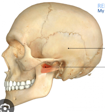
mastoid process
marked prominence of bone located posterior and inferior to TMJ
large rounded radiopacity located posterior and inferior, not seen on intraoral
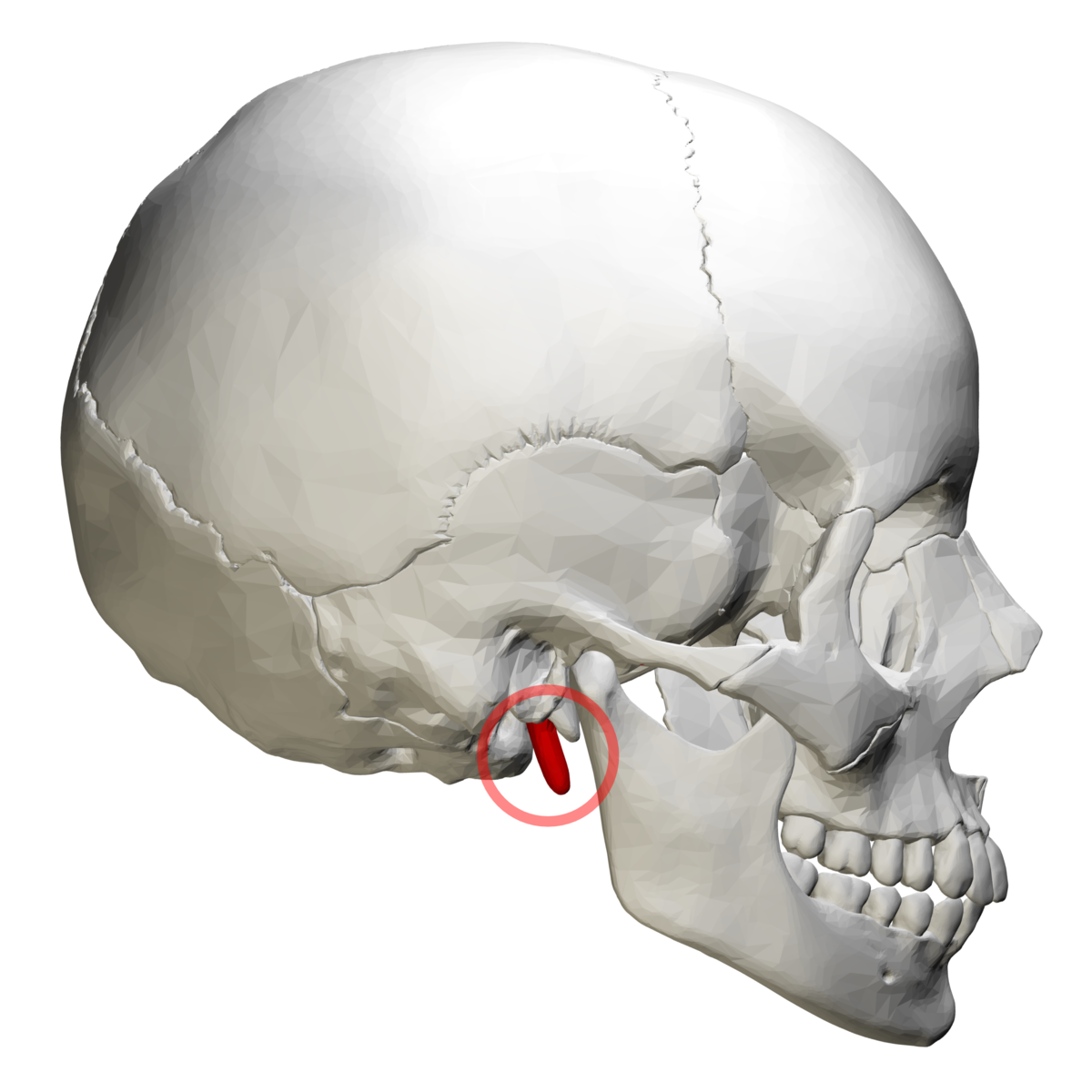
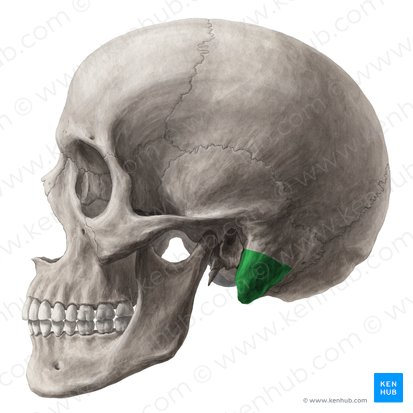
what is this, its description and its appearance on images
styloid process
long pointed projection of bone that extends downward for the inferior surface of temporal bone
long radiopaque spine that extends from temporal bone anterior to mastoid process, not seen intraoral
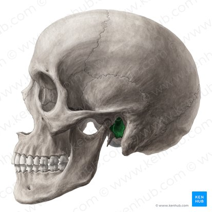
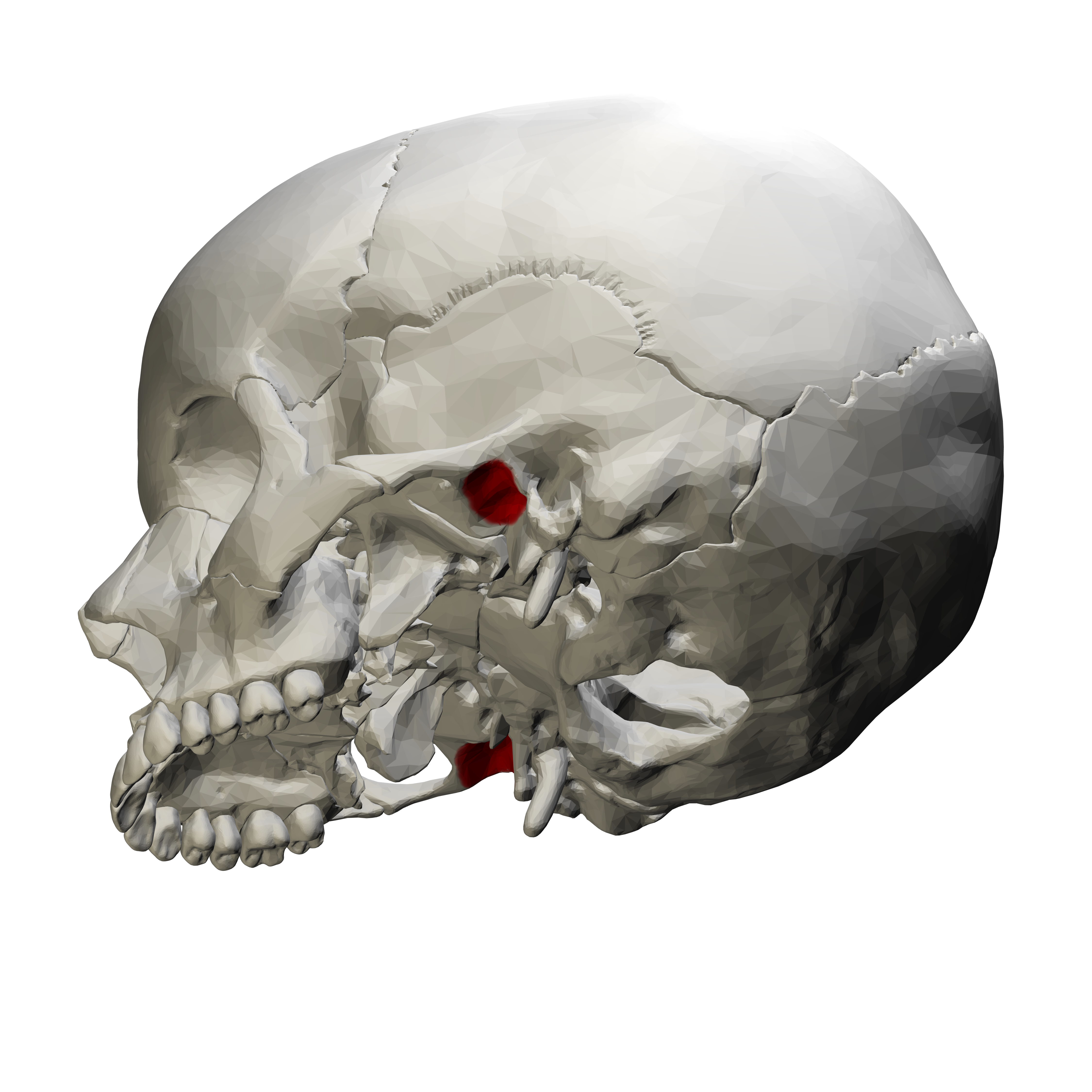
what is this, its description and its appearance on images
external auditory meatus
hole in temporal bone located superior and anterior to mastoid process
round radiolucency anterior and superior to mastoid process, not seen intraorally
what is this, its description and its appearance on images
glenoid fossa
concave depressed area of temporal bone
concave radiopacity superior to mandibular condyle, not seen intraorally
what is this, its description and its appearance on images
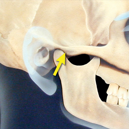
articular eminence
projection of temporal bone located anterior to glenoid fossa
rounded radiopaque projection of bone located anterior to glenoid fossa, not seen intraorally
what is this, its description and its appearance on images
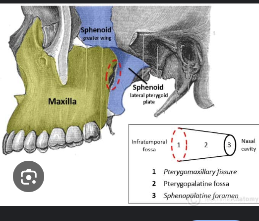
lateral pterygoid plate
thin, wing shaped extension of sphenoid located distal to maxillary tuberosity region
rounded radiopaque projection, not seen intraorally
what is this, its description and its appearance on images
pterygomaxillary fissure
narrow space that separates the lateral pterygoid plate and maxilla
rounded radiolucent area. not seen intraorally
what is this, its description and its appearance on images
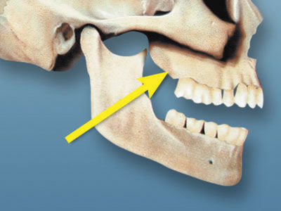
maxillary tuberosity
rounded prominence that extends posterior to third molar
rounded radiopaque bulge distal to third molar, seen on intraoral
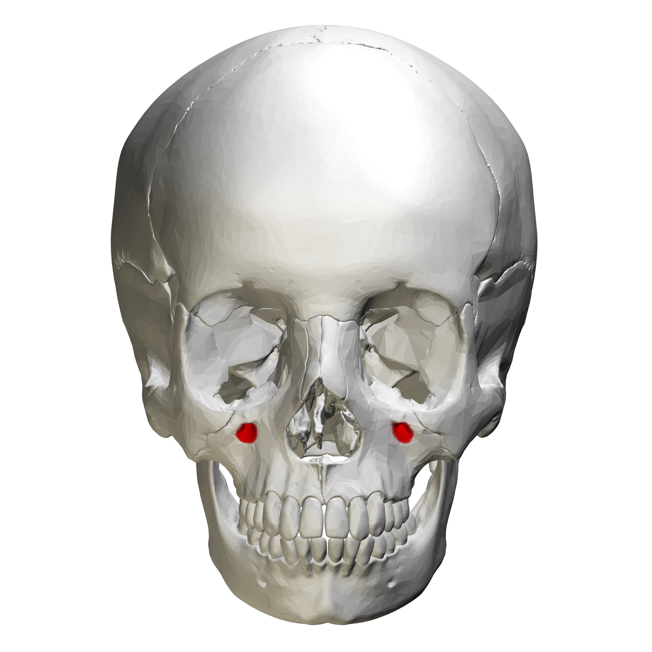
what is this, its description and its appearance on images
infraorbital foramen
hole in bone inferior to border or orbit
round radiolucency inferior to orbit, not seen intraoral
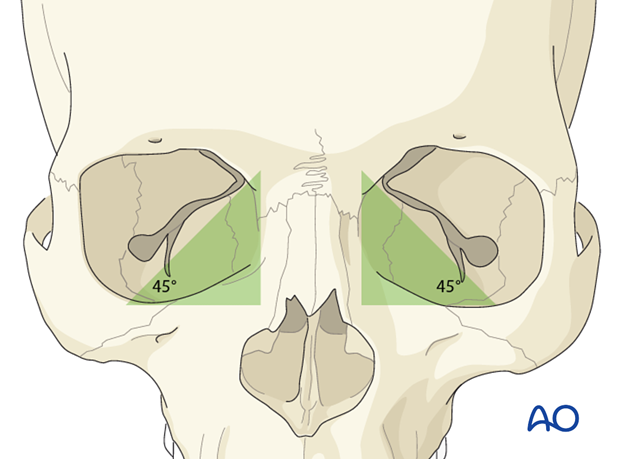
what is this, its description and its appearance on images
orbit
cavity that contains eyeball
round radiolucent compartment with radiopaque borders located superior to maxillary sinuses, not seen intraoral
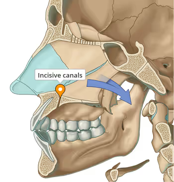
what is this, its description and its appearance on images
incisive canal
passageway thru bone that extends from superior foramina from incisive canal to foramen
tubelike radiolucent area with radiopaque borders, can be seen intraoral img
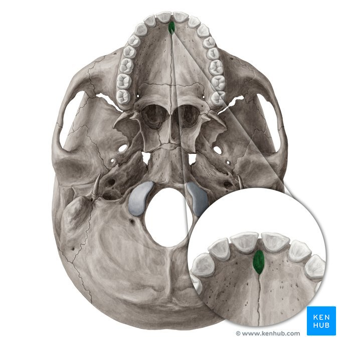
what is this, its description and its appearance on images
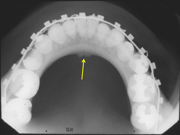
incisive foramen
hole in bone located at midline of anterior hard palate directly posterior to maxillary central incisors
round radiolucency located btween roots of maxillary central incisors, can be seen intraoral img
what is this, its description and its appearance on images
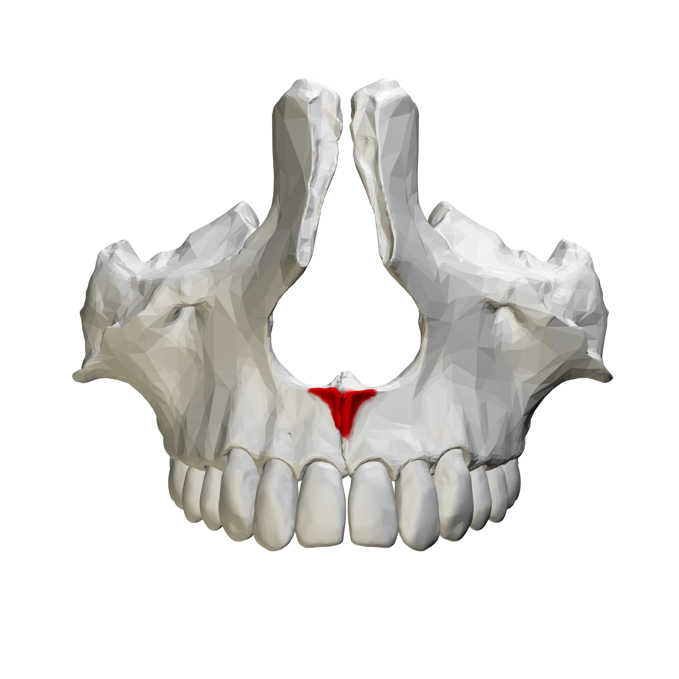
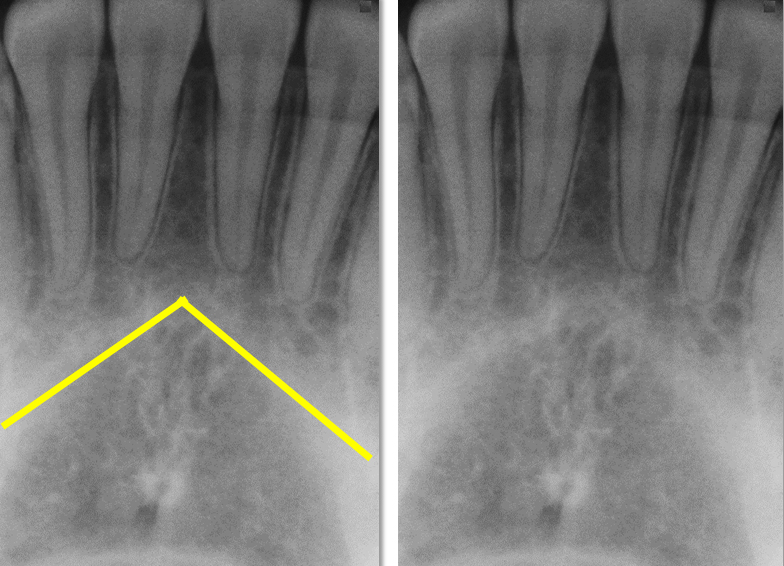
anterior nasal spine
sharp projection of maxilla located at anterior inferior portion of nasal cavity and septum
v shaped radiopaque area located at the intersection of floor nasal cav and septum, may be seen
what is this, its description and its appearance on images
nasal cavity
pear shaped compartment of bone located superior to maxilla
v shaped radiolucent area superior to maxillary incisors, may be seen
what is this, its description and its appearance on images
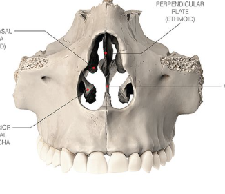
nasal septum
vertical bony wall that divides nasal cavity into left and right fossae
may be seen
what is this, its description and its appearance on images
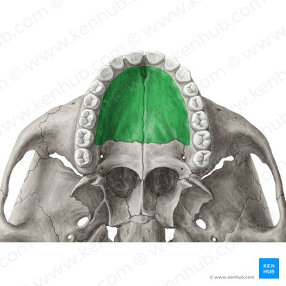
hard palate
wall that separates nasal cavity from oral
horizontal radiopaque band superior to apices of maxillary teeth, may be seen
what is this, its description and its appearance on images
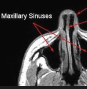
maxillary sinus
paired cavities within the maxilla superior to maxillary posterior teeth
radiolucent apreas superior to apices of maxillary premolars and molars, may been seen
what is this, its description and its appearance on images
zygomatic process of maxilla
projection of maxilla that articulates with zygoma
j or u shaped radiopacity located superior to maxillary first molar region, may be seen
what is this, its description and its appearance on images
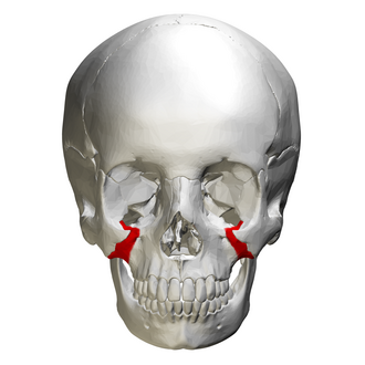
zygoma
cheekbone
diffuse radiopaque band that extends posteriorly from zygo process of maxilla, may be seen
what is this, its description and its appearance on images
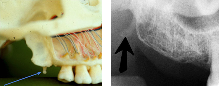
hamulus
small hooklike projection that extends from medial pterygoid plate
radiopaque, may be seen
what are the bony landmarks of the maxilla and surrounding structures
mastoid porcess
styloid process
external auditory meatus
glenoid fossa
articular eminence
lateral pterygoid plate
pterygomaxillary fissure
maxillary tuberosity
infraorbital foramen
orbit
incisive canal
incisive foramen
anterior nasal spine
nasal cavity
nasal septum
hard palate
maxillary sinus
floor of maxillary sinus
zygomatic process
zygoma
hamulus
what are the bony landmarks of the mandible and surrounding structures
mandibular condyle
coronoid notch
coronoid process
mandibular foramen
lingula
mental foramen
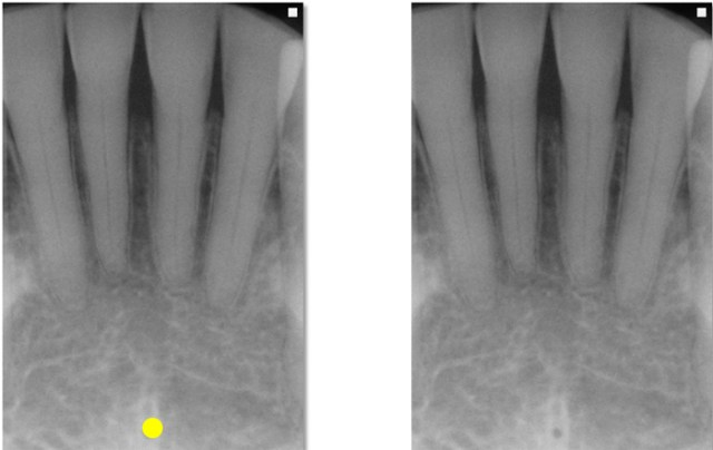
mental ridge
mental fossa
lingual foramen
genial tubercles
inferior border of the mandible
mylohyoid ridge
external oblique ridge
angle of the mandible
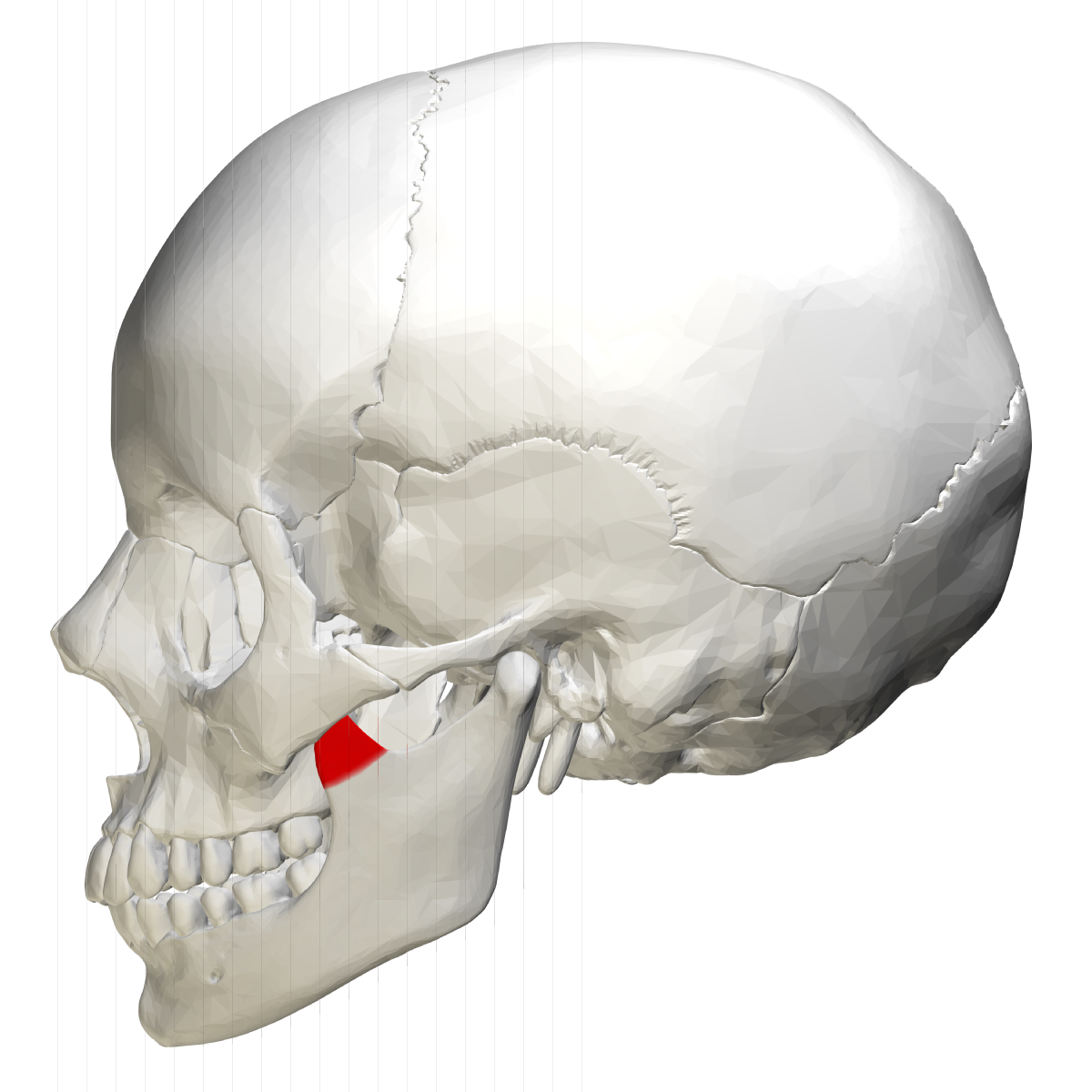
what is this, its description and its appearance on images
mandibular condyle
round projection extending from posterior superior border of ramus of mandible
bony rounded radiopaque projection, not seen intraoral
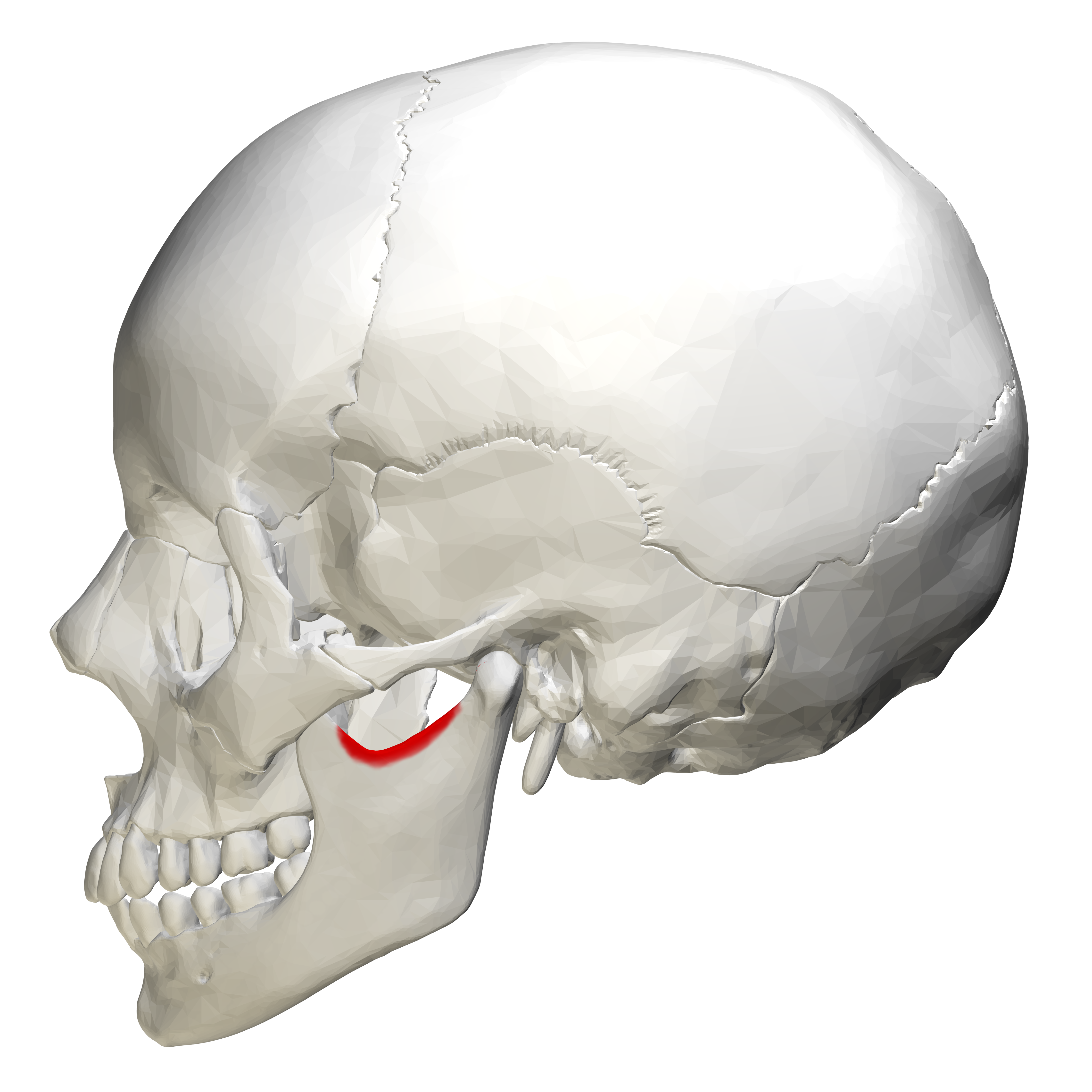
what is this, its description and its appearance on images
sigmoid notch
curved depression between mandibular condyle and coronoid process of mandible
radiopaque, not seen
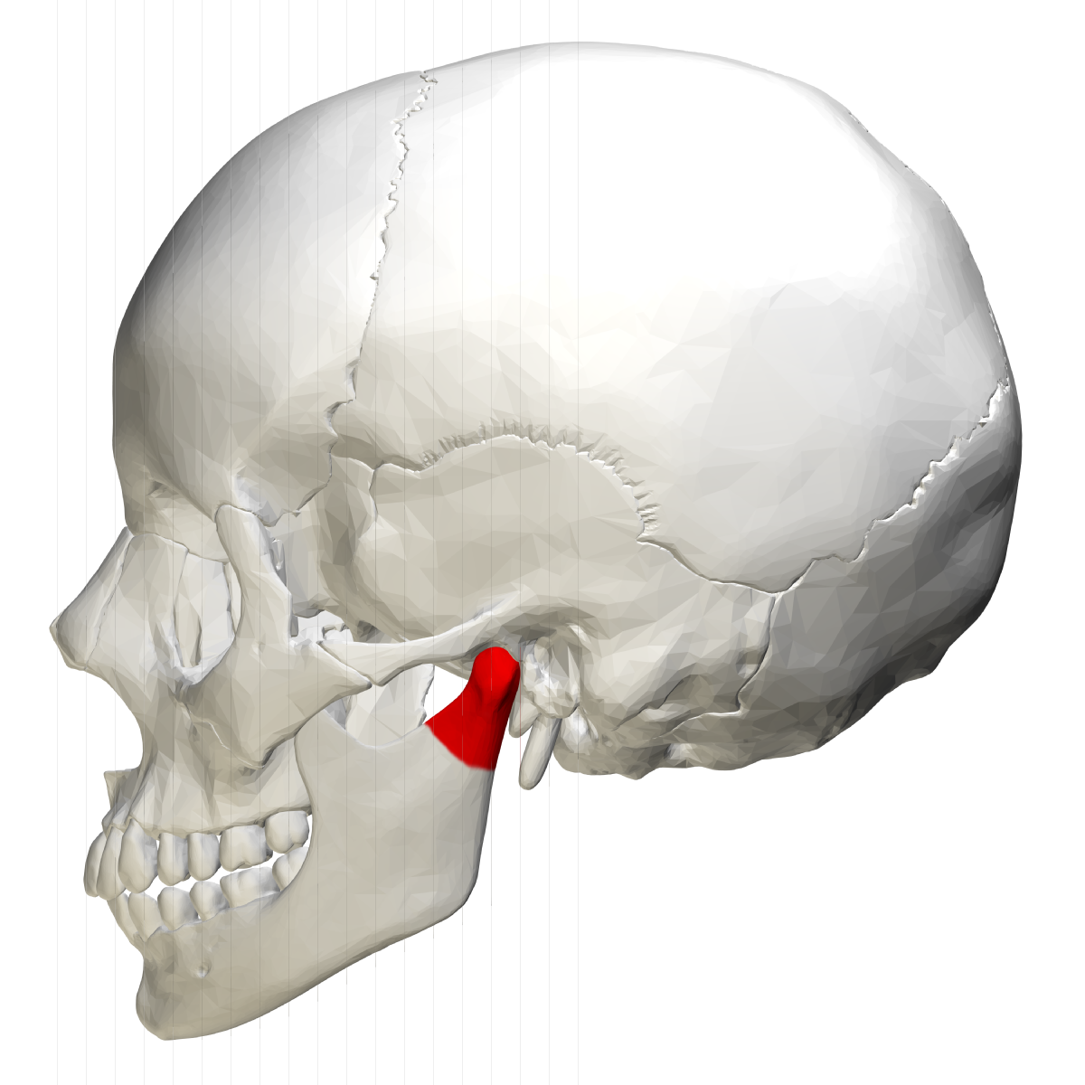
what is this, its description and its appearance on images
coronoid process
thin prominence shaped like crows beak
radiopacity, may be seen on maxillary molar periapical image
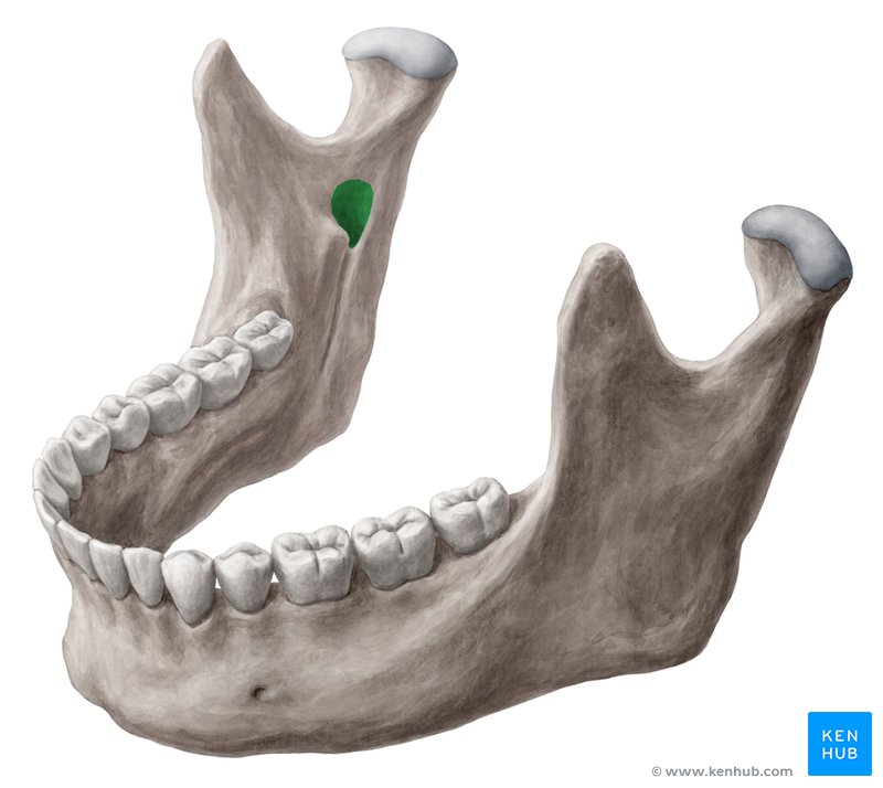
what is this, its description and its appearance on images
mandibular foramen
round hole on lingual aspect of ramus mandible
radiolucent, not seen intraoral
what is this, its description and its appearance on images- 1st line
lingula
small tongue shaped projection seen adjacent to mandibular foramen
radiopacity, not seen intraoral
what is this, its description and its appearance on images
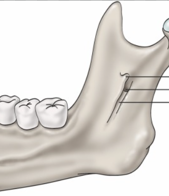
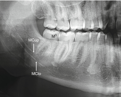
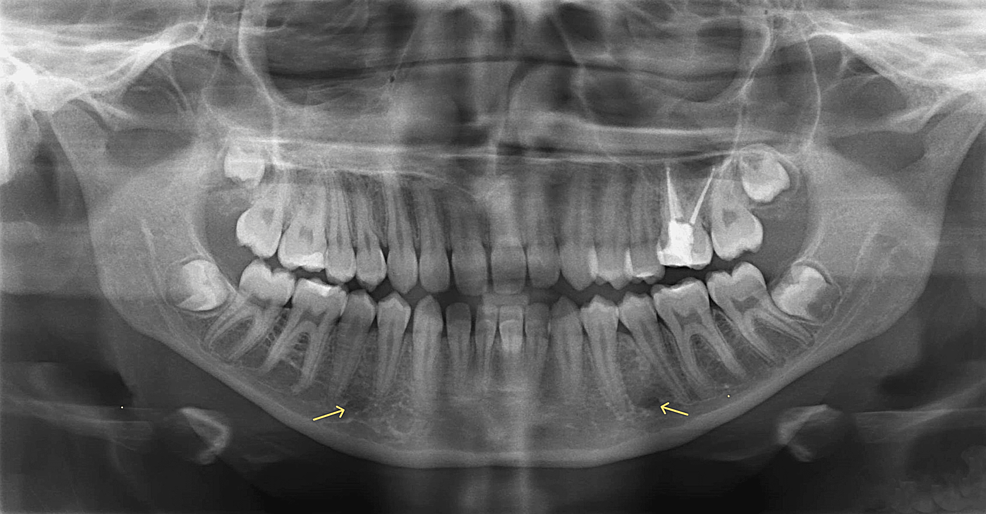
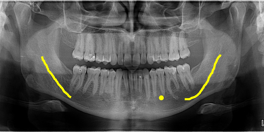
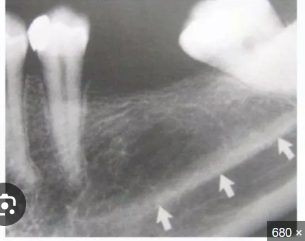
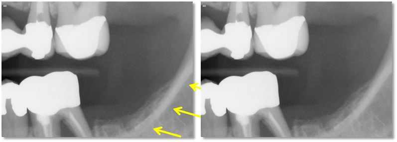
mandibular canal
tubelike passageway thru bone that travels within body or length of mandible
radiolucent band outlined by two thin radiopaque lines representing cortical walls of cancal, may be seen on intraoral
what is this, its description and its appearance on images
mental foramen
hole on external surface of mandible region
round radiolucency in apical region of mandibular premolars, may been seen intraoral
what is this, its description and its appearance on images- 9
hyoid bone
horseshoe shaped bone below mandible, between chin and thyroid cartilage
floating curved radiopacity, not seen intraoral
what is this, its description and its appearance on images
mental ridge
im not writing all that
radiopaque band that extends from mandibular premolar region to incisor region, may been seen
what is this, its description and its appearance on images
mental fossa
radiolucent area above mental ridge, may be seen
what is this, its description and its appearance on images
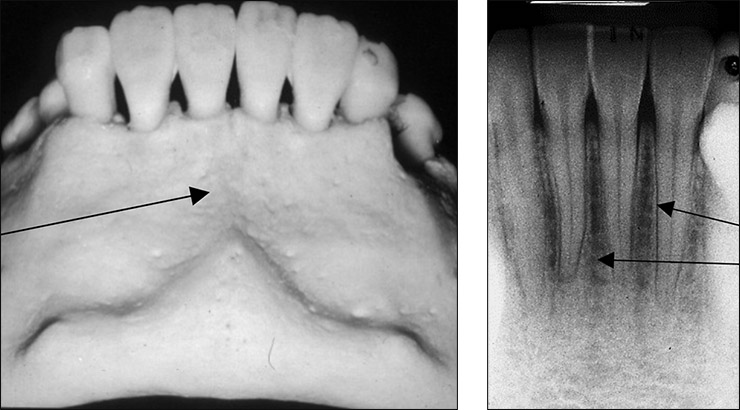
lingual foramen
tiny hole in internal surface of mandible near midline
small radiolucent dot located inferior to the apices of mandibular incisors, may be seen
what is this, its description and its appearance on images
genial tubercles
tiny bumps of bone located on lingual aspect of mandible
ringshaped radiopacity surrounding lingual foramen, may be viewed
what is this, its description and its appearance on images
inferior border of mandible
thick linear prominence of cortical bone that defines lower border of the mandible
radiopaque band outlines the lower border, may be seen
what is this
mylohyoid ridge
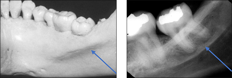
what is this
internal oblique ridge , may be seen
what is this
external oblique ridge, may be seen
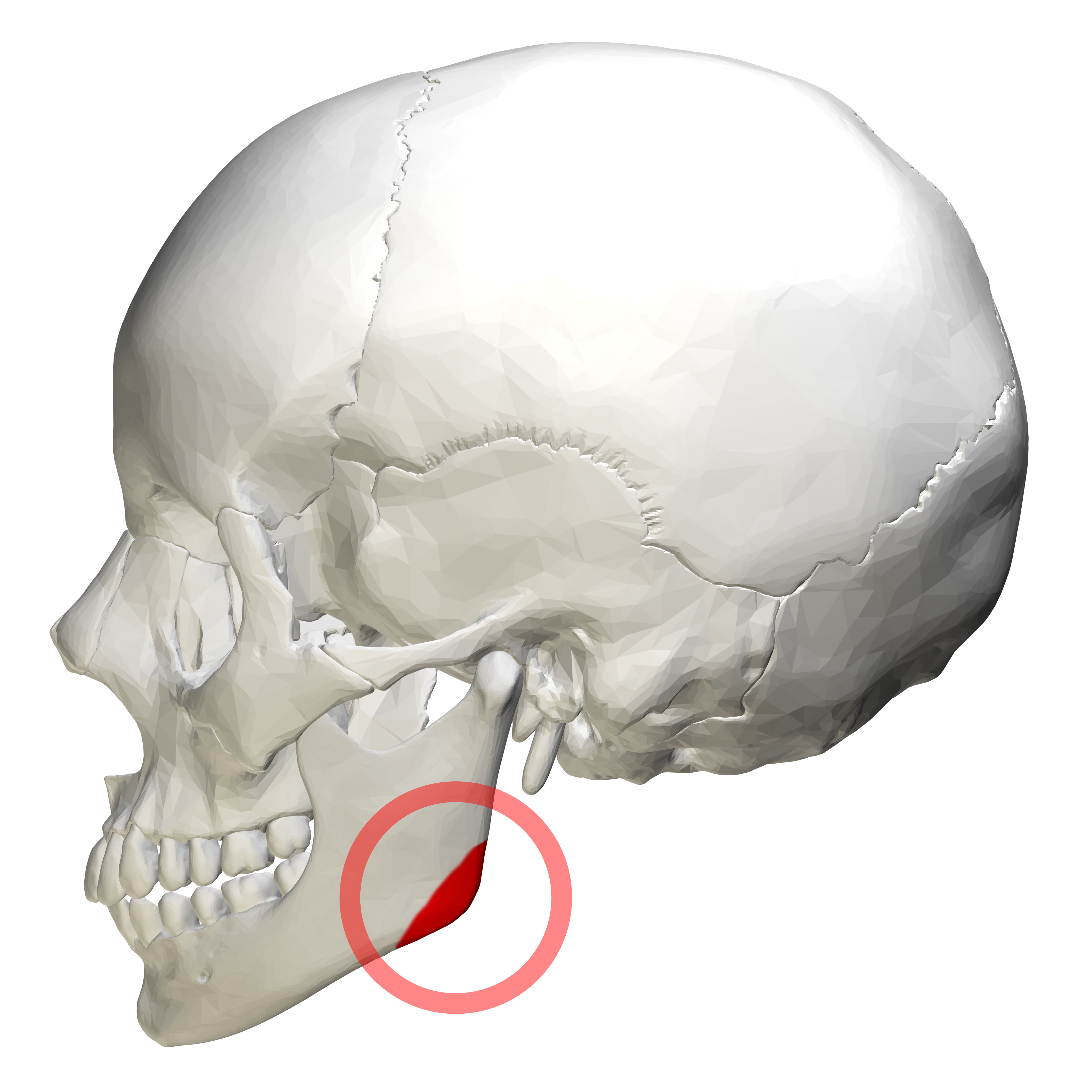
what is this,
angle of mandible , not seen
what are the air spaces seen on panoramic images
palatoglossal
nasopharyngeal
glossopharyngeal
where is the palatoglossal air space
between palate and tongue
where is the nasopharyngeal air space
pharynx posterior to cavity
where is the glossopharyngeal air space
pharynx located posterior to tongue and oral cavity
what soft tissues are seen on panoramic images
tongue
soft palate and uvula
lip line
ear