Histology Exam 3 - images
 Call Kai
 Learn
Learn Practice Test
 Spaced Repetition
 Match
Match Flashcards
 Knowt Play
1/64

There's no tags or description
Looks like no tags are added yet.
Name | Mastery | Learn | Test | Matching | Spaced | Call with Kai | Chat |
|---|
No analytics yet
Send a link to your students to track their progress
65 Terms
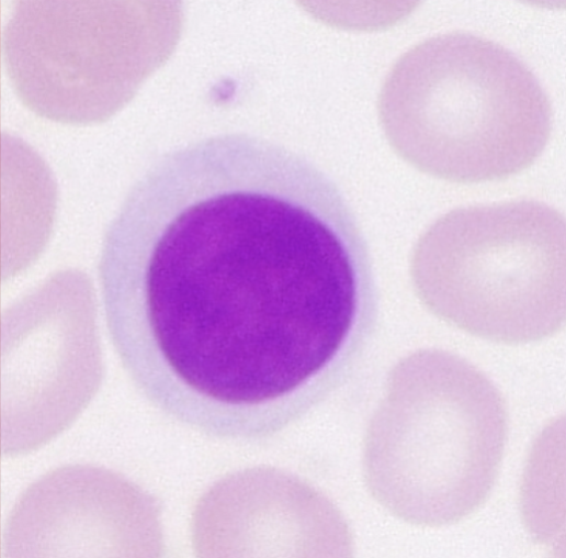
What is this?
Lymphocyte
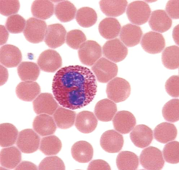
What is this?
eosinophil
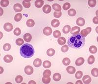
What three things can be seen in this image?
lymphocyte, neutrophil, and platelet
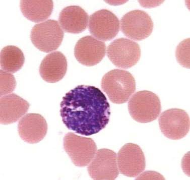
What is this?
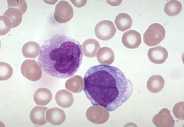
What are those two things in this image?
macrophages
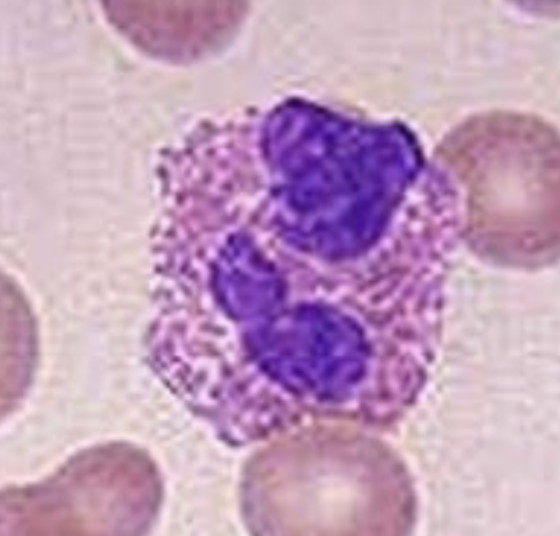
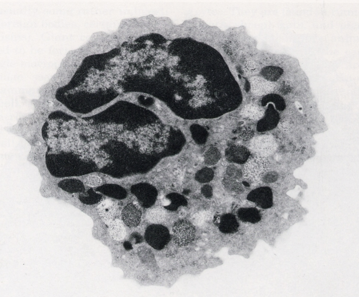
What is this?
basophil
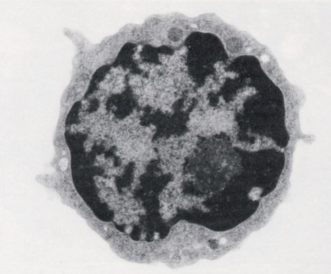
What is this?
lymphocytes
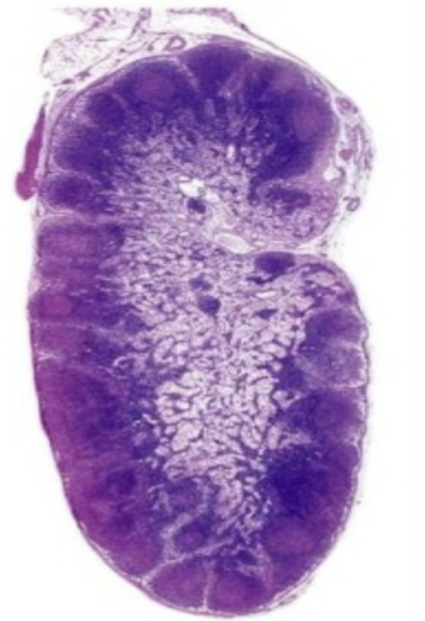
What is this?
Lymph node
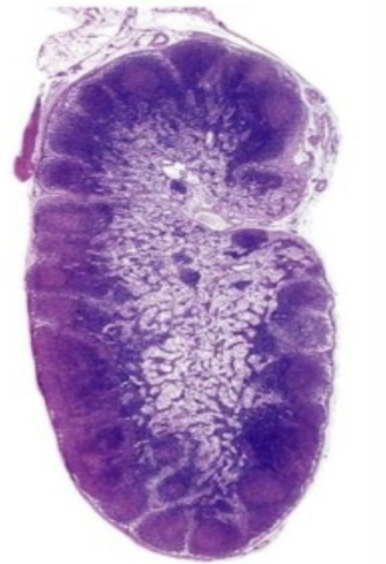
What is the inner lighter layer of this lymph node called?
Medulla
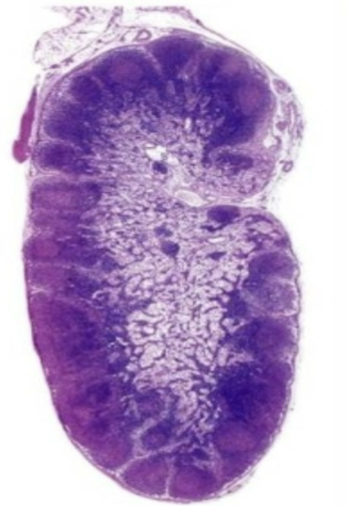
What is the outer layer of this lymph node called?
Capsule
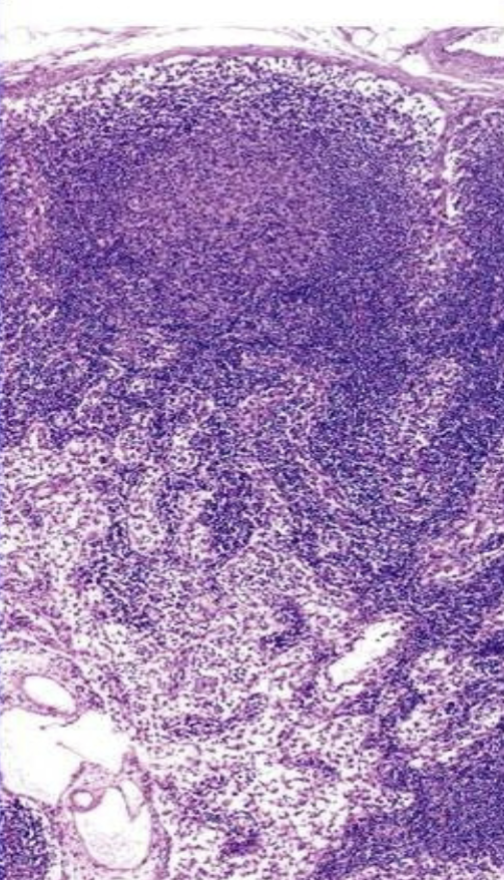
What is the light round area in the top area of this lymph node?
lymph node
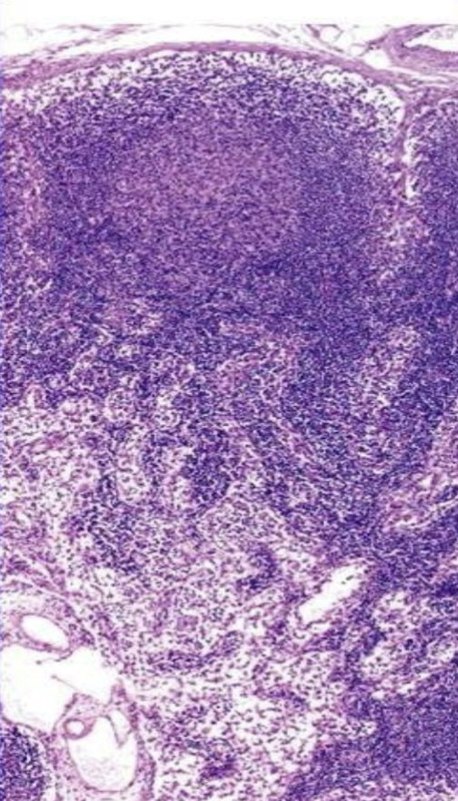
What is the thin eosinophilic layer outside of this lymph node?
capsule
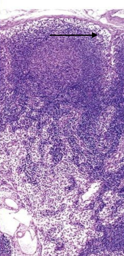
Where is this arrow pointing?
trabeculae
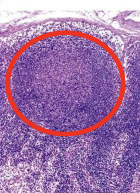
What is this?
Lymph Nodule
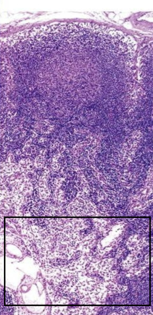
What is this boxed in area of the lymph node?
medullary sinus
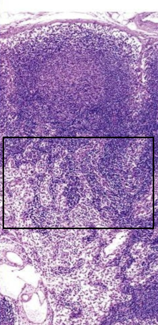
What is this boxed in area of the lymph node that houses a lot of t-cells?
Paracortical area
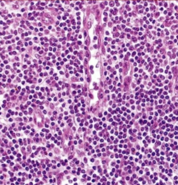
What is that gap in the cells in the lymph node is this?
High endothelial venules
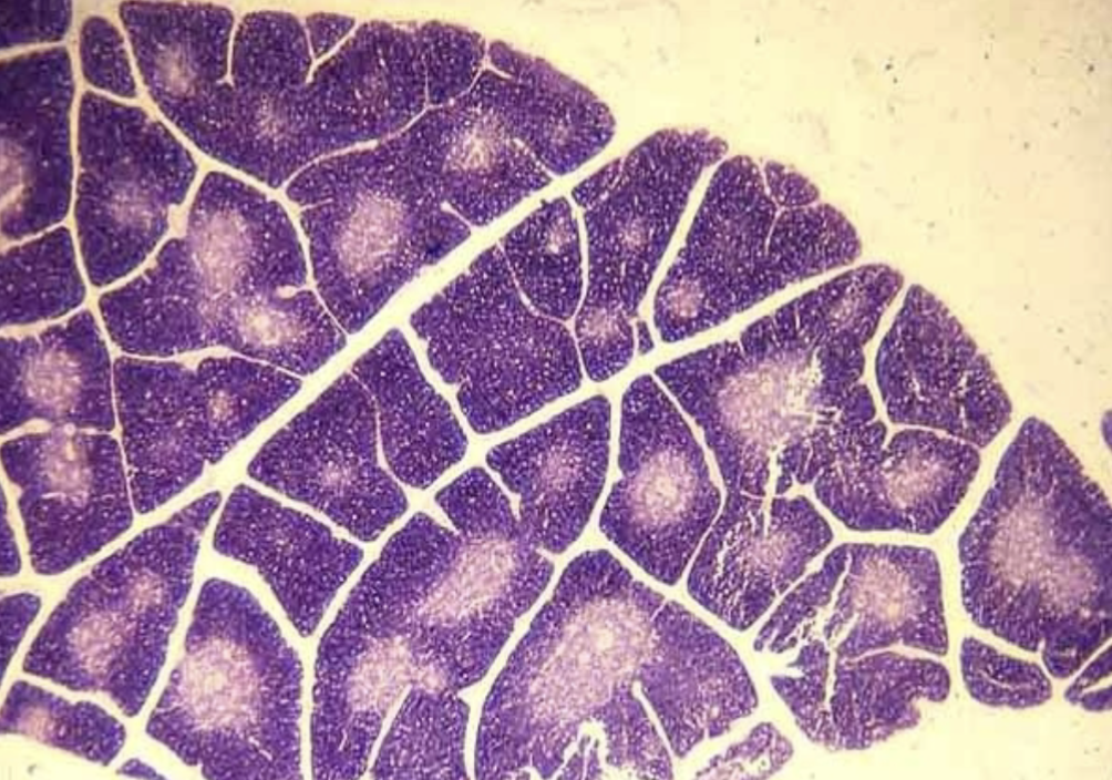
What is this?
thymus
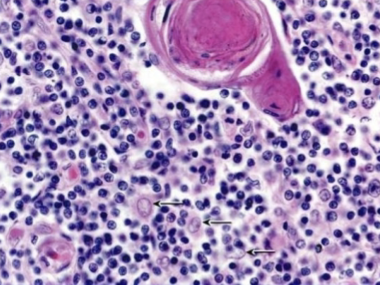
What is that big ol eosinophilic thing?
thymic (hassall’s) corpuscles
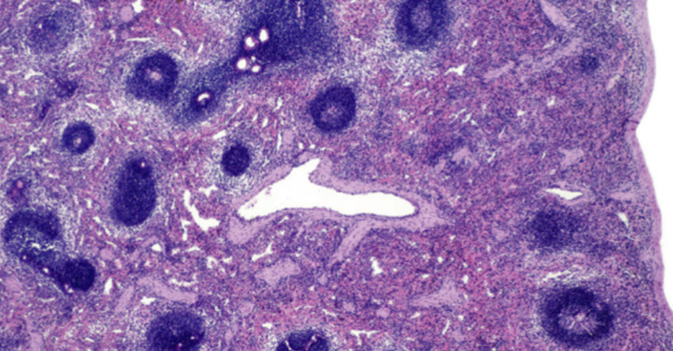
What is this?
Spleen
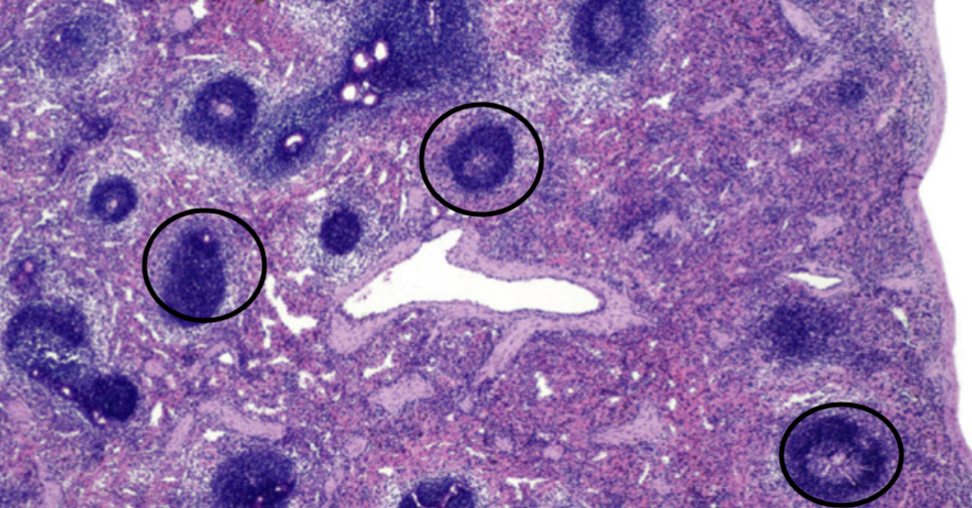
What are the circled areas?
lymphocytes
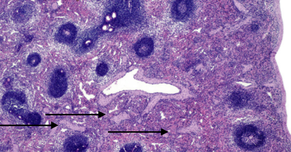
What are the light areas that the arrows are pointing to?
fibromuscular trabeculae
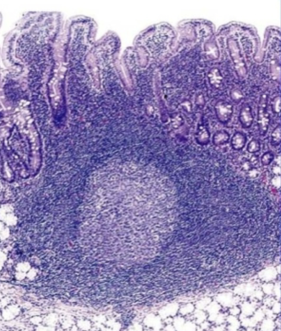
What is the general term for that lighter spot?
MALT -(mucosal associated lymphoid tissue)
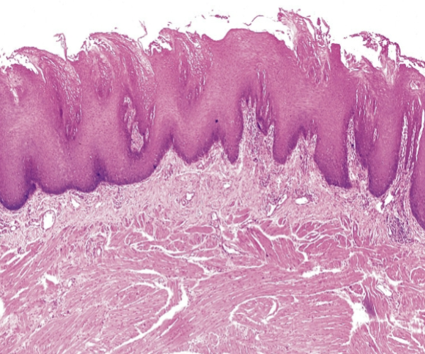
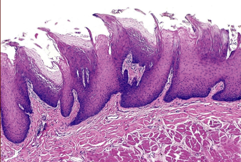
What is this?
tongue
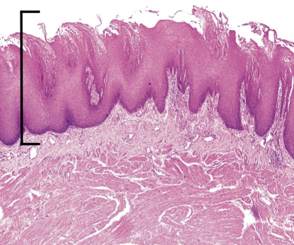
What is this top layer?
Papillae
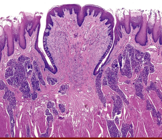
What is this tissue from?
tounge
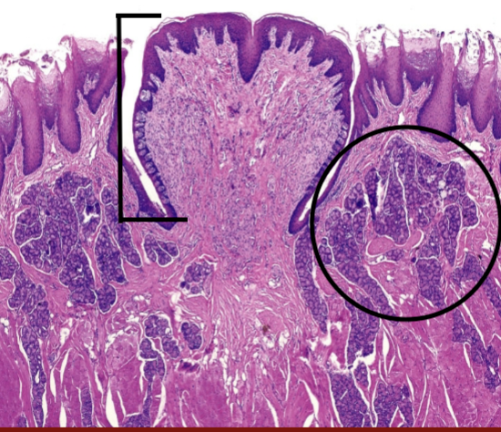
What part of the tongue area is barred out?
taste buds
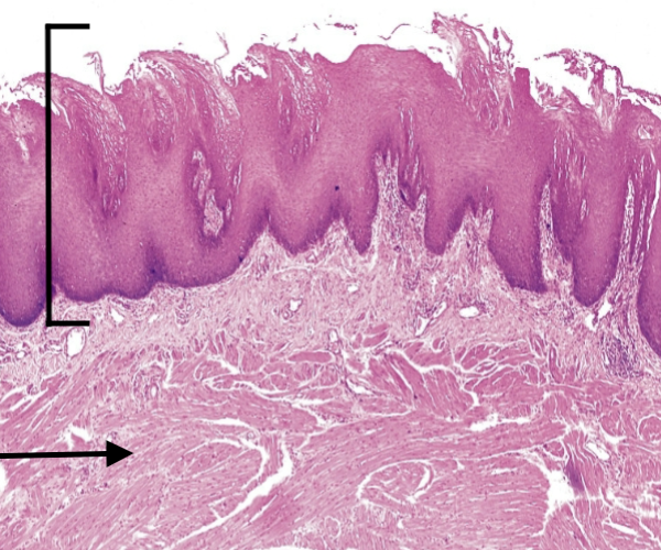
What type of muscle is haphazardly arranged in this image of tongue?
Skeletal muscle
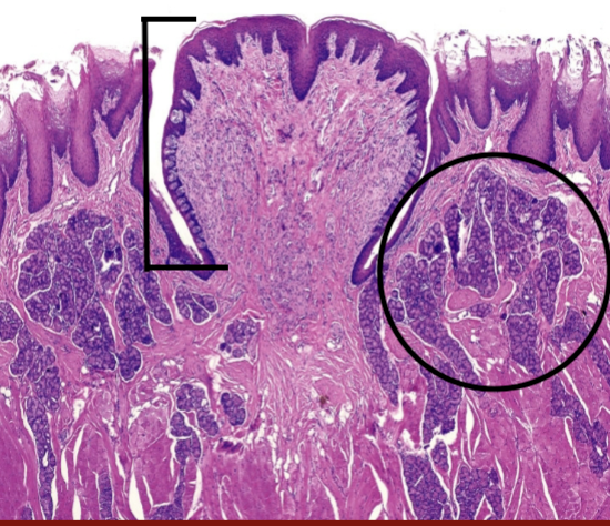
What is circled?
Sublingual glands
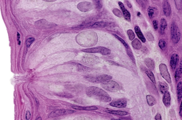
This is a tongue, what are those light colors things running to the surface?
nerves
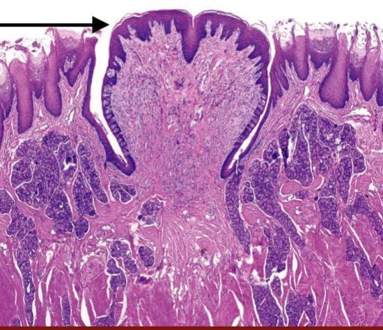
What type of epithelium is this arrow pointing to?
stratified squamous epithelium
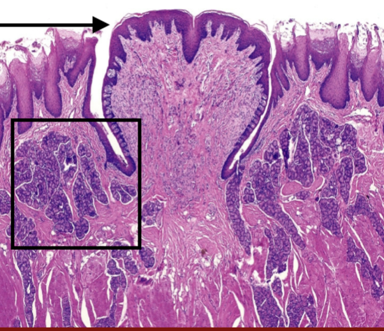
What is in the box of this image of tongue?
sublingual glands
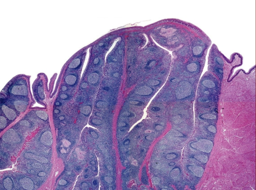
What is this?
Tonsils
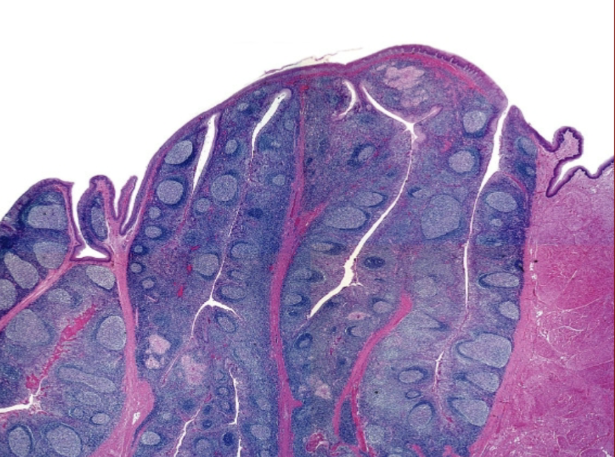
What kind of MALT is this?
GALT
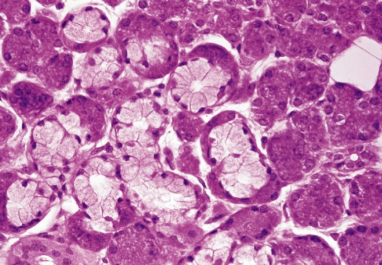
What type of gland is this?
Salivary glands
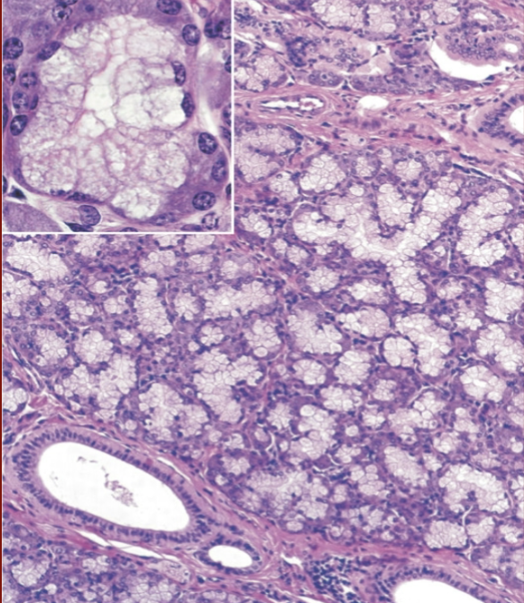
What is that white area in the bottom corner?
exocrine ducts
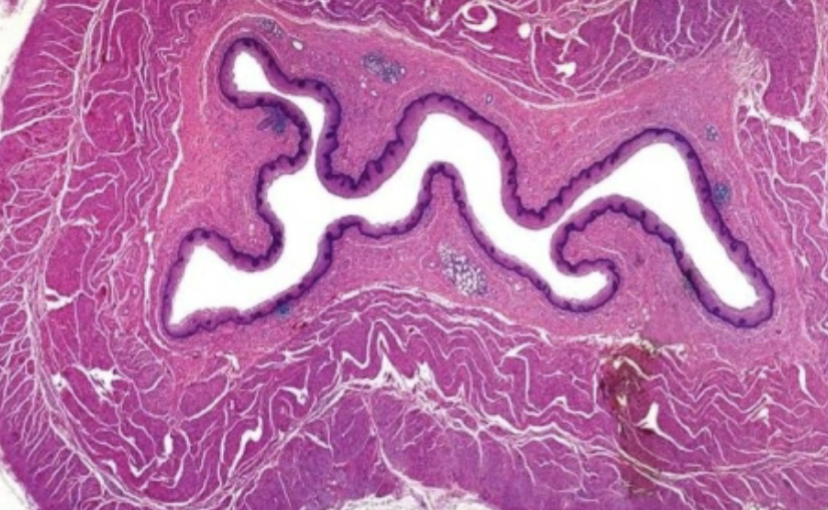
What is this an image of?
esophagus
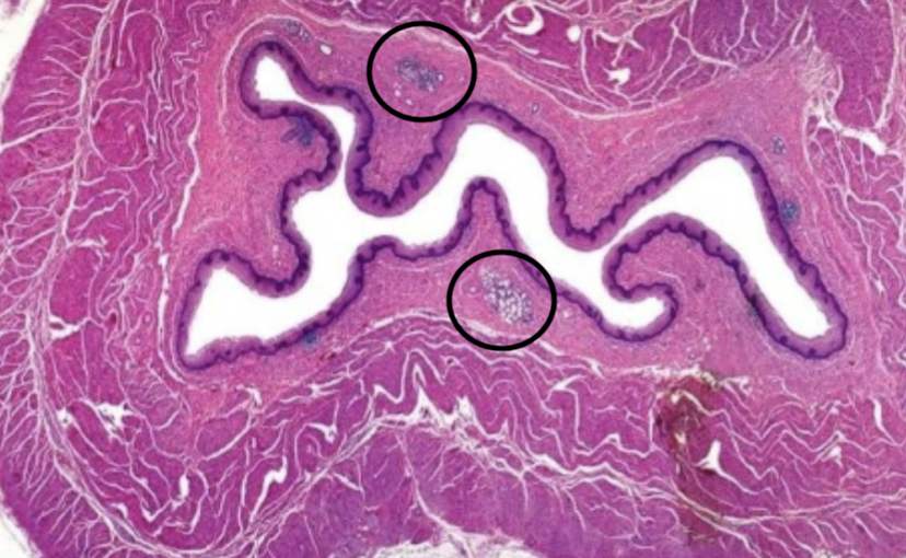
What is circuled?
submucosal glands
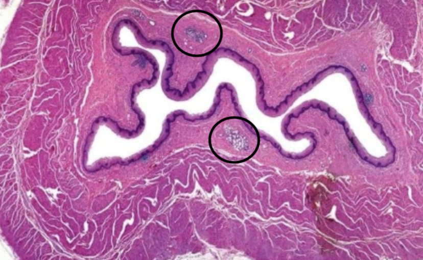
What epithelium does this have?
stratified squamous epithelium
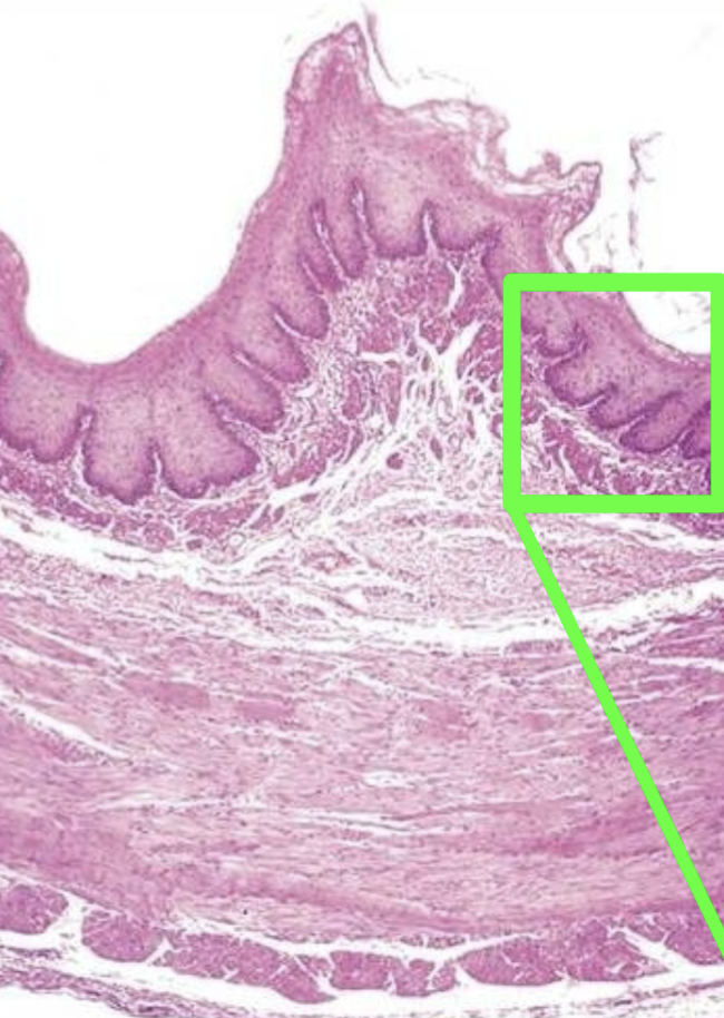
What layer is pointed out in green?
Mucosa
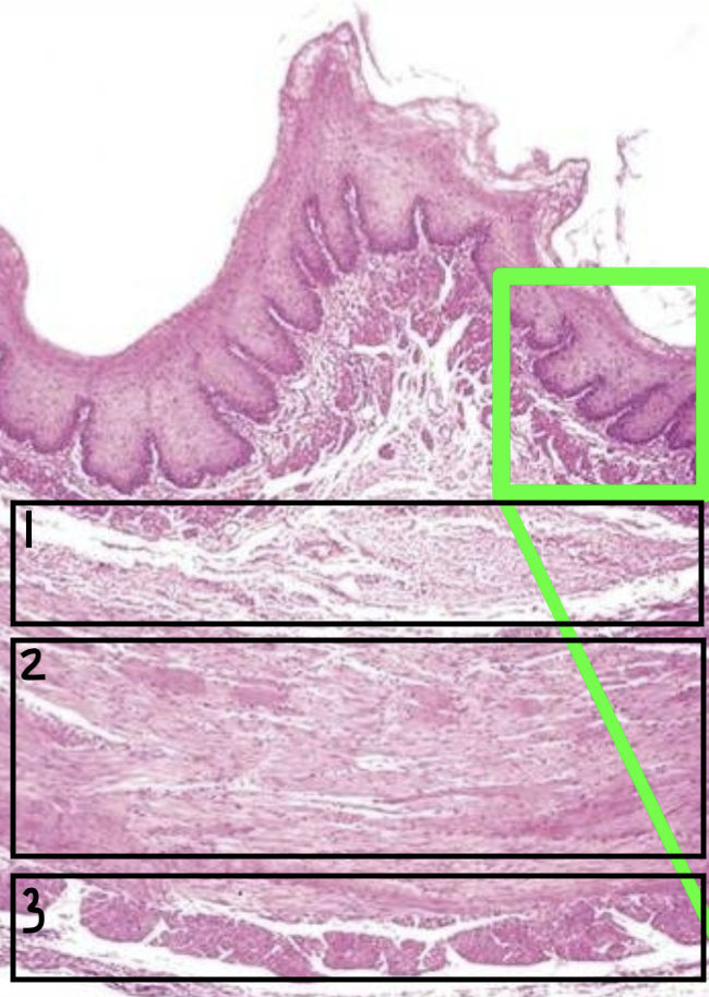
What layer is 1?
Submucosa
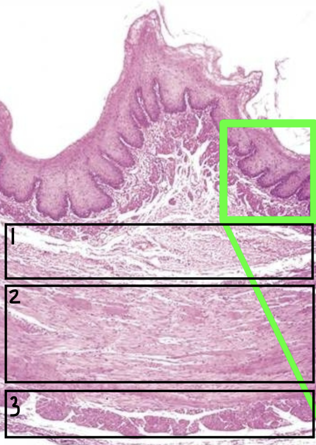
What layer is 2?
Muscularis
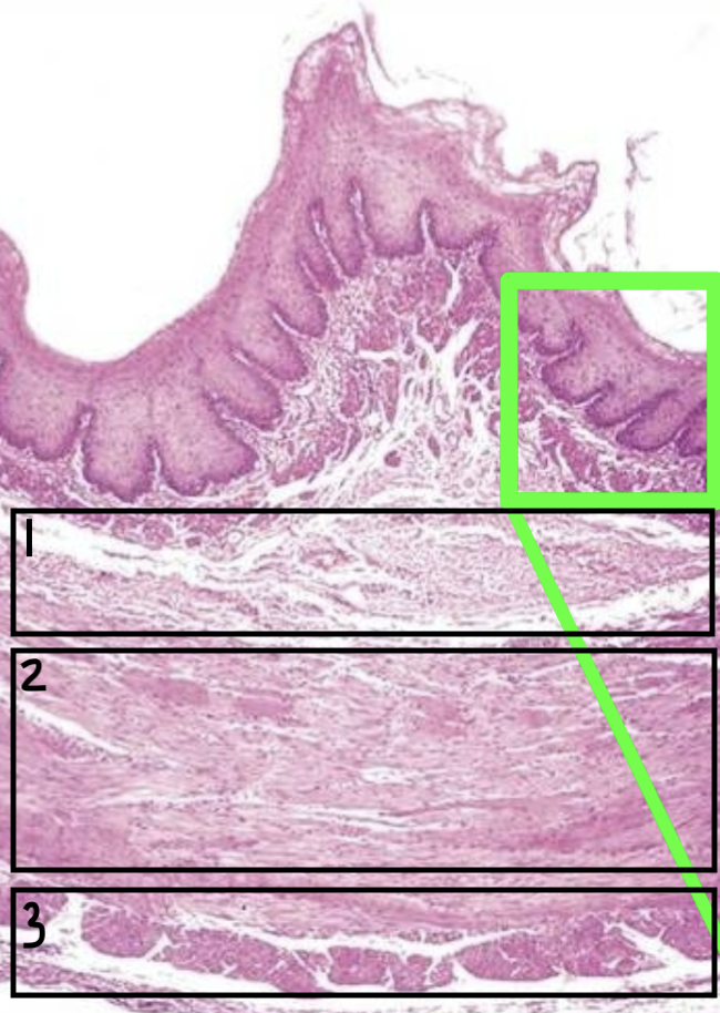
What layer is 3?
Serosa
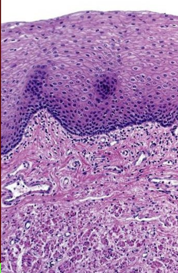
What type of epithelium is this?
Non-keratinized stratified squamous epithelium
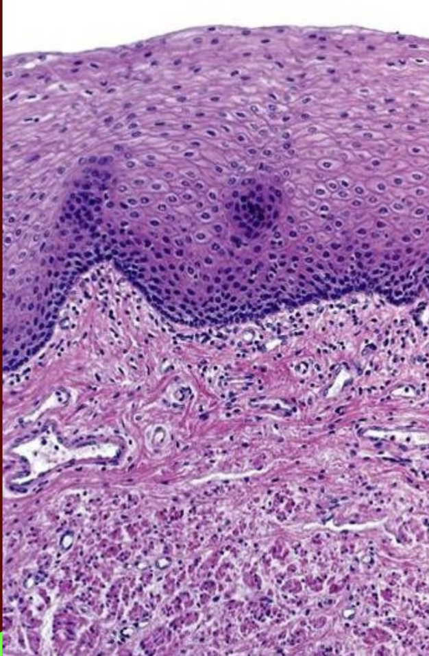
What type of muscle os this?
Smooth muscle