Evaluation and Emergency Management of the Trauma Patient - Abdominal Trauma
 Call Kai
 Learn
Learn Practice Test
 Spaced Repetition
 Match
Match Flashcards
 Knowt Play
1/20

There's no tags or description
Looks like no tags are added yet.
Name | Mastery | Learn | Test | Matching | Spaced | Call with Kai | Chat |
|---|
No analytics yet
Send a link to your students to track their progress
21 Terms
In cases of potential abdominal trauma, what exactly are we looking for and analyzing?
FLUID in the abdomen
Abdominocentesis
Fluid categorisation
Fluid analysis and Smear
What is the POCUS technique for viewing the abdomen?
A-FAST
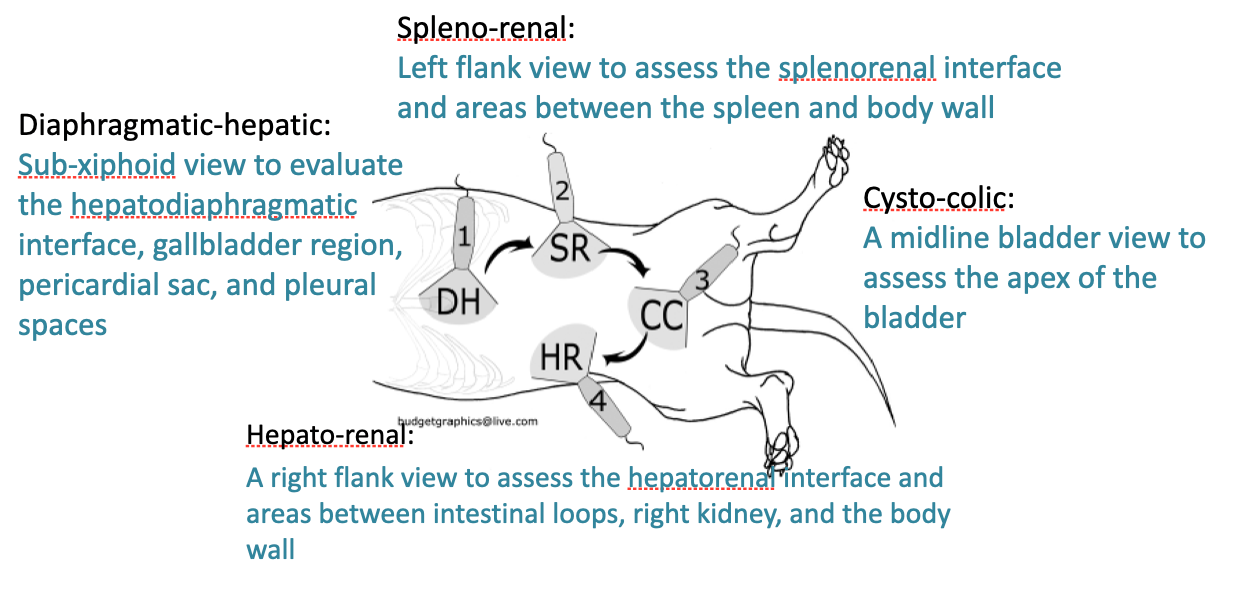
Has 4 major points that need to be assessed:
Spleno-renal
Left flank view to asses splenorenal interface and areas between the spleen and body wall
Cysto-colic
Midline bladder view to assess the apex of the bladder
Hepato-renal
Right flank view to asses the hepatorenal interface and areas between intestinal loops, right kidney, and body wall
Diaphragmatic-hepatic
Sub-xiphoid view to evaluate the hepatodiaphragmatic interface, gallbladder region, pericardial sac, and pleural spaces
When it comes to abdominal effusion, you will want to take a sample of the fluid and assess it via abdominocentesis. What are the 3 different classifications of fluid you could find in the abdomen?
Transudate - essentially very watery, not many proteins or cells
Modified Transudate - Increase of proteins and cells involved in inflammatory processes
Exudate - Very thick, mainly composed of neutrophils, RBCs and proteins

You will then perform a fluid analysis based on the appearance of the fluid. What are the 5 main kinds of abdominal effusion you could see?
1) Hemoabdomen - fluid is blood from an active bleed in the abdomen
To confirm, compare PCV of fluid to PCV of peripheral blood
If PCV of fluid is 10-25% that of the peripheral blood, you have an active bleed in the abdomen
SOMETIMES EXACERBATED BY SPLENIC CONTRACTION
2) Uroabdomen - urine in the abdomen
Will contain creatine or potassium, indicative of kidney damage / bladder rupture
3) Septic peritonitis - damage to the peritoneum → infection of the abdomen
Will be nasty fluid; containing degenerate neutrophils (inflamm.), intracellular bacteria, and lactate and glucose
4) Intestinal perforation - Feces / GI material in the abdomen
Intra/extracellular bacteria present in fluid
Particulate material
5) Bile peritonitis - bile free in the abdomen
Indicative of stomach, liver, or gallbladder damage
Will be BROWN in color, containing higher bilirubin than plasma levels
Neutrophils will be present due to inflammation from damaging bile
Further describe a hemoabdomen… what are the common causes? How do they present?
Presence of haemorrhagic effusion in the peritoneal cavity
Non traumatic (idiopathic)
Traumatic
Bleeding from kidneys, spleen and/or liver
Common in RTA
Presentation
Shocked/Collapsed patients
Secondary arrhythmias/ pulse deficits
** Can also be present in anaphylactic reactions
How would one diagnose a hemoabdomen ASIDE FROM PCV?
Coagulation profile
A-POCUS → blood looks shimmery on ultrasound instead of anechoic like normal fluid; due to RBCs and proteins (see image)

How would you approach initially treating and stabilising the patient? What are your options for treatment?
Goal → Manage shock and control source of bleeding
Treatment:
Start with goal directed fluid therapy BUT
Hypotensive or low volume resuscitation
Need to not exacerbate bleeding
Blood transfusions
Autotransfusions
Using the patient’s own blood from the abdomen and reintroducing it to the intravascular system
not the cleanest
If bleeding is from a cancerous mass, could spread cancer all through the body
If there’s other damage, could be bad blood
Need to inform O of risks before performing
Then of course put them on O2
What are the two options for management once the patient is stable?
If just a minor bleed → External pressure bandage wrap; monitor PCV
If a continuous bleed, NEEDS SURGICAL INTERVENTION
most will btw
Further describe a uroabdomen… what causes it? What are the two different classifications of a uroabdomen?
Leakage of urine to peritoneal or retroperitoneal
Loss of integrity of urinary system (Kidneys to intraabdominal urethra)
Classified by origin of leakage:
Uroperitoneum
Intraabdominal urethra
Bladder
Distal urether
Uroretroperitoneum
Kidneys
Proximal urether
What is one consequence of having urine leaking out into the abdomen? Think about what the kidney’s job is…
The body will try and maintain homeostasis as urine (which contains K+, Creat, Urea, and H2O) sits in the abdomen.
Results in intravascular disequilibrium:
Increased K → hyperkalemia
VERY NEGATIVE IMPACT ON THE KIDNEYS AND HEART
Kidneys → increased GFR, Aldosterone, Na/K ATPase → increased urine production → even MORE urine in abdomen
Heart → more potassium = increased action potentials → bradyarrhythmias
Increased Urea → azotemia
Increased abdominal H2O → DECREASED IV ELECTROLYTES
Will leave circulation to maintain osmolarity
Hyponatremia (decreased Na+)
Hypochloremia (decreased Cl-)

How would you diagnose a uroabdomen? Are there going to be clinical signs?
Non-specific Clinical signs
Bladder might or might NOT be palpable
+/- Urination
+/- haematuria before uroabdomen
Biochemistry
Azotaemia
Hyperkalaemia
Imaging - not the best either
APOCUS
Contrast radiography
Most surefire way is via fluid analysis
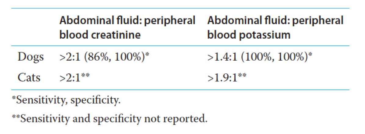
Test for creatinine and potassium in the free fluid
Still should ALWAYS compare the ratio between the creat in the fluid and the creat in blood
Also perform a smear of the fluid to rule out any bacteria
How would you then manage a uroabdomen and treat it?
Fluid resuscitation
Urinary catheterisation - need to get as much urine out as you can
Abdominal drainage - need to restore homeostasis
Electrolyte imbalance (Hyperkalaemia)
Medical vs. Surgical
Definitive surgical correction is usually required BUT medical stabilisation is essential before
What exactly is septic peritonitis? What are the three different classifications?
Peritonitis resulting from an identifiable source of intraperitoneal infection, typically bacterial in origin and most commonly a result of leakage of the GI tract
Classifications:
Primary - spontaneous (ex: cats with FIP)
Secondary - leakage of bacteria (foreign body caused)
Tertiary - recurrence (worse prognosis)
How would one diagnose septic peritonitis?
Haematology
Neutrophilia to neutropaenia
Biochemistry
Lactate - good indicator for how bad septic shock is
A-POCUS
MOST EFFECTIVE
Abdominal effusion
Cytology
intracellular bacteria
Degenerated neutrophils / toxic changes; will be pus-y
Peripheral glucose to abdominal
Difference of ≥ 38 mg/dl (2.1 mmol/L)
If glucose in abdomen is LOWER than glucose in blood, suggests septic peritonitis
Abdominal lactate to peripheral
> 2.5 mmol/L
If lactate in fluid is HIGHER than lactate in blood, suggests septic peritonitis
How would one then manage septic peritonitis?
NEEDS TO BE QUICK, CAN’T WAIT
Broad spectrum antibiotics
Fluid resuscitation - some cases may not be responsive due to septic shock causing vasodilation
Control hypotension
Vasopressors?
Infection source control !
Broad spectrum antibiotics
Ex-Lap
Peritoneal Lavage
Abdominal drain
Essentially, treat the infection and find the source ASAP
Further describe bile peritonitis… what can cause it? Why is it so damaging?
Is the leakage of bile into the peritoneal cavity. Caused by:
Gall bladder rupture after trauma
7-10 days post incident, so NEED TO KEEP AN EYE ON PATIENTS FOR THIS
Post-hepatic Biliary tract obstruction (gallbladder stones)
Further causes damage by:
Chemical/irritant effect on peritoneum
Severe inflammation
Coagulopathies (Vit K dependent) due to close association with the liver
How would one diagnose bile peritonitis?
Patient will be icteric
Hyperbilirubinaemia
Abdominal effusion
TBileffusion > TBilplasma (>2:1)
Bilirubin crystals
Abdominal cytology
Extracellular pigment
Rule out intracellular bacteria and concomitant sepsis
+/- culture and sensitivity
How would one manage bile peritonitis?
IS AN EMERGENCY
Emergency
Fluid resuscitation/Shock
Vitamin K/FFP if coagulopathy
Surgical
Ex-Lap
Resolution of the source
Peritoneal lavage
Abdominal drain
RANDOM TRAUMA NOW
What is traumatic pancreatitis, and what is unique about this in comparison to the other traumatic injuries in this lecture?
Can be damaged either DIRECTLY or INDIRECTLY
Is very sensitive to hemorrhages or hypovolemia; can become inflamed even if uninjured
Potential causes:
Subcapsular hematomas
Ischemia to pancreas
Causes the leakage of pancreatic enzymes, which can be dangerous to surrounding tissue
Autodigestion
Acute pancreatitis
VERY COMMON IN CATS
What happens during a diaphragmatic hernia? What can it cause, and how can you determine a potential hernia on physical exam?
Commonly secondary to blunt trauma
Varying severity of dyspnoea
Organs herniated:
Liver, stomach, small intestine, omentum, spleen
Physical exam
Borborygmi on thoracic auscultation to dull/absent lung sounds
”tucked in” abdomen
Diagnosis and management of diaphragmatic hernia?
Diagnosis
POCUS is diagnostic in 93% of cases
Radiographs
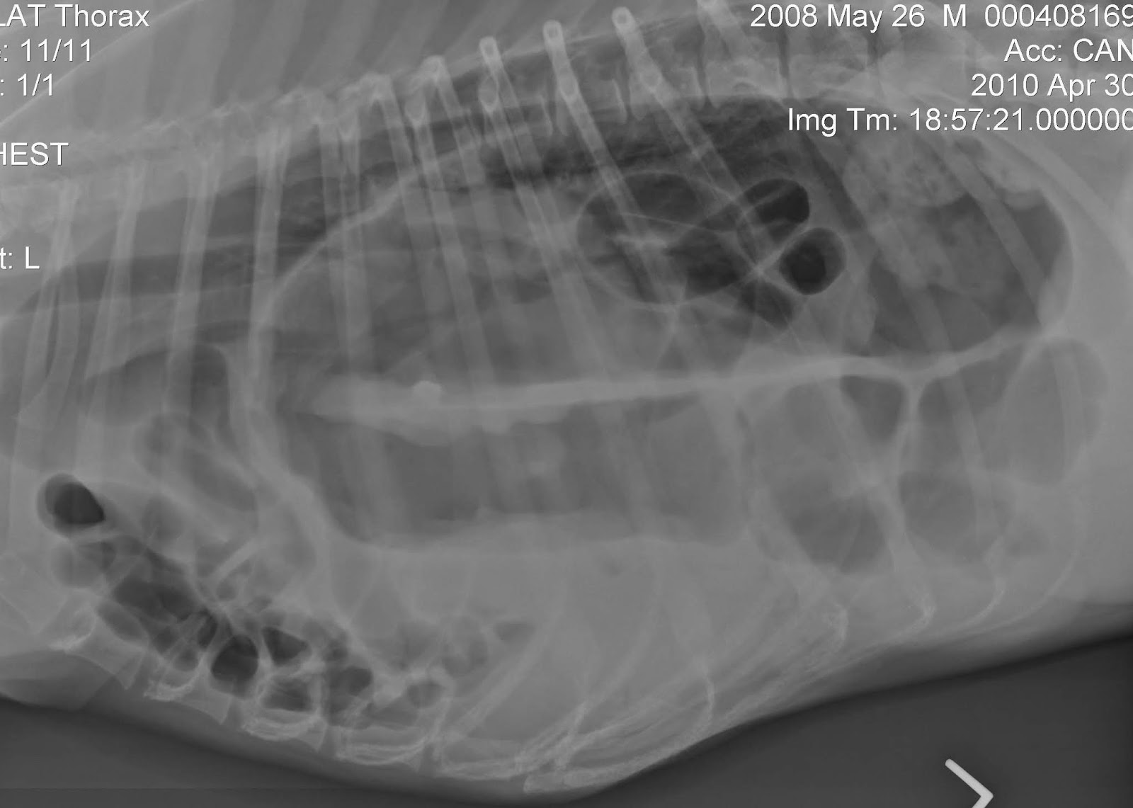
Presence of abdominal organs in the chest cavity
Loss of visualisation of the diaphragm
Management
Surgery +/- chest drain