PSY396 Lecture 5: Introduction to electroencephalography (EEG)
 Call Kai
 Learn
Learn Practice Test
 Spaced Repetition
 Match
Match Flashcards
 Knowt Play
1/31

There's no tags or description
Looks like no tags are added yet.
Name | Mastery | Learn | Test | Matching | Spaced | Call with Kai | Chat |
|---|
No analytics yet
Send a link to your students to track their progress
32 Terms
What is Electro-Encephalo-Gram?
Electrical Brain Picture
Electroencephalography
The recording of electrical changes at the scalp, which are caused by the synchronous activity of thousands of neurons that are parallel to each other
inventor of EEG
Hans Berger (1873-1941)
In 1924, first to measure electrical currents from human scalps
Coined the term electroencephalogram (“Elektroenkephalogramm”)
Described alpha-waves (Berger waves)
1924 succeeded in recording the first human electroencephalogram
1929 published his first paper entitled "recording the electrical activity of the human brain from the surface of the head".
in 1935, American roboticist William Grey Walter described “Berger was not regarded by his associates as in the front rank of German psychiatrists, having rather the reputation of being a crank. He seemed to me to be a modest and dignified person, full of good humour, and as unperturbed by lack of recognition as he was later by the fame it eventually brought upon him.” (W. Grey Walter, 1953, The Living Brain)
1937 importance of his discoveries in EEG were finally recognized at an international forum after British electrophysiologists Edgar Douglas Adrian and others confirmed Berger's basic observations in 1934
By 1938, electroencephalography had gained widespread recognition by eminent researchers in the field, leading to its practical use in diagnosis in the United States, England, and France.
Electroencephalography – The first
Richard Caton (1842-1926)
1875 First to measure electrical currents from brains of mammals
Registered change in electrical activity in the brain to sensory stimuli
Electroencephalography - Pioneers
Adolf Beck (1863-1942) Kraków
Ernst Fleischl von Marxow (1846-1891) Vienna
Vasili Yakovlevich Danilevsky (1852-1939) Charkov
Vladimir Právdicz-Neminski (1879-1952) Kiev
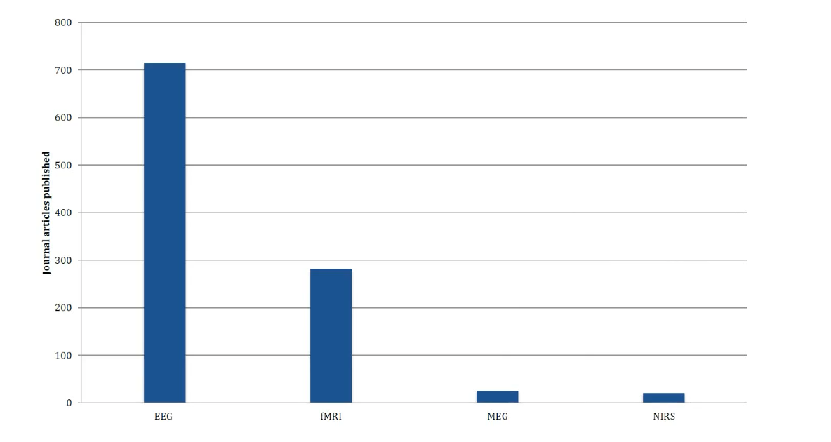
Documents by year for EEG, MRI and TMS

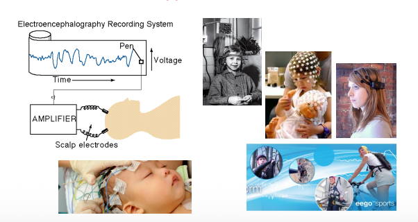
EEG - Apparatus
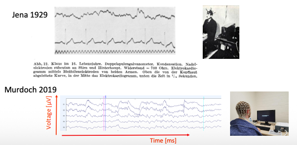
Electroencephalogram - now vs then
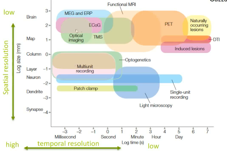
Methods in cognitive neuroscience
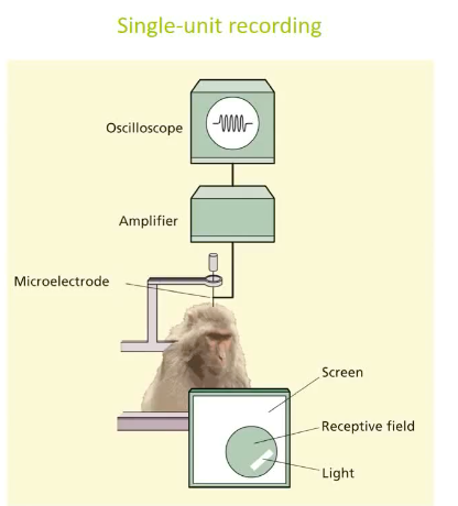
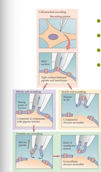
Patch clamp and single-/multi-unit recording
Directly measure neuronal activity
Invasive
Limited to few neurons
Unclear information code
How does EEG work?
A non-invasive technique that records electrical activity in the brain through electrodes placed on the scalp, used to study cognitive processes.
Electrical signals from the brain – Origin
In order to understand the origins of the voltage fluctuations that we measure on the scalp, we need to understand the origins of the voltage fluctuations in the brain
Captures post synaptic potentiation
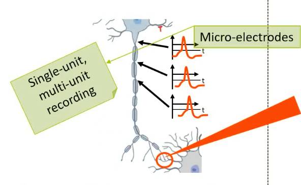
Neuronal activity - Action potentials
Discrete signal (voltage spike) that moves from the cell body of a neuron along its axon
At the end of the axon it enables the release of neurotransmitters
Postsynaptic potentials
Neurotransmitters bind to the membrane of the postsynaptic cell
Ion channels open and close leading to gradual changes in membrane potentials

Release of excitatory (inhibitory) neurotransmitter results in +ve (-ve) ions flow into the dendrites, and a net negativity (positivity) in the extracellular space
Meanwhile, when one end of neuron is positively charged, the other end of the neuron starts to be charged negatively (positive ions leave the cell body)
→ creates electrical dipole between different parts of the neurons
Scalp electrodes as neural activity detectors - Action potentials
Duration less than 1 ms (no/little temporal summation)
Random physical distribution of axons
Neurons fire in a different timing
Convoluted electrical activity
➥ not detectable
Scalp electrodes as neural activity detectors - Postsynaptic potentials
Duration of tens to hundreds of ms
Summation possible
➥ detectable
From postsynaptic potentials to EEG
Dipole of one pyramid cell is not sufficient → Summation of many required
Involving 103 - 106 Neurons
Occurring at about the same time
Neurons in the cerebral cortex are roughly aligned with each other → parallel
EEG recordings/set up
Are always bipolar
difference in voltage between an active electrode, i.e., an electrode of interest, and an electrode that is (relatively) electrically inactive, i.e., the reference electrode
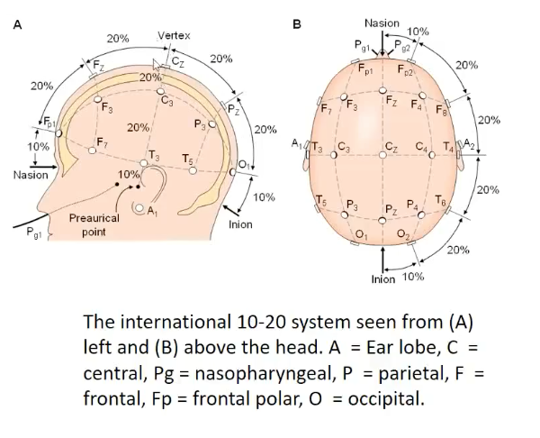
Mostly done from several electrodes that are placed at standardised positions
10-20 system and extension
Artifacts
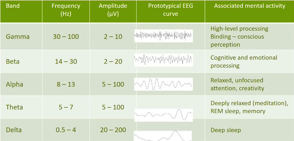
EEG – Spontaneous activity
EEG - Clinical applications
Neurological diagnoses
Detection & diagnosis → prognosis → therapy
Epilepsy (presence, epileptogenic zone, treatment outcome)
Creutzfeldt–Jakob disease (periodic sharp wave complexes)
Dementia (mild generalised background slowing)
Functional changes connected to head injuries, poisonings, or blood flow disorders (by conducting functional network analysis)
Sleep research (sleep disorder, brain development)
Neurofeedback:
EEG in cognitive neuroscience
Event-related potentials (ERPs):
Even-related synchronisation and desynchronization (ERS/ERD)
Coherence
Source localisation
Event-Related brain Potentials (ERPs)
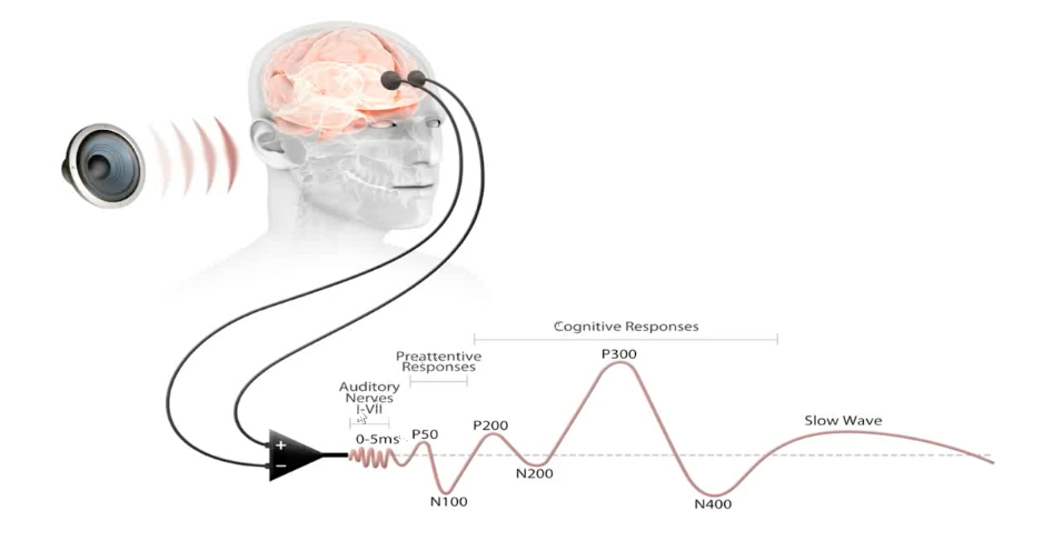
Brain responses time-locked to some event. This event may be:
a sensory stimulus (such as a flash or a sound),
a movement (such as response to a specified target stimulus),
or the omission of a stimulus (such as an increased time gap between stimuli).
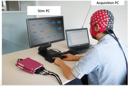
Doing an ERP experiment
During a study the participant listens to or watches some stimuli.
These stimuli are called events.
Electrical activity from the brain is recorded to a number of these events
Typical ERP set up
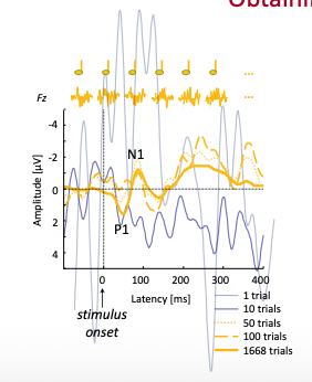
Obtaining ERPs
Consistent electrical changes can be seen by averaging together the electrical activity from a certain number of these events.
This activity is seen as a series of positive and negative-going deflections.
What do ERPs tell?
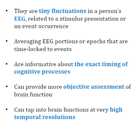
The timing and size of these deflections, and can be used to make inferences about
the time-course of processing in the brain
Why can we associate ERPs with specific cognitive processing?
EPRS: Example
Why do we get slower as we get older?
3 stages of sensorimotor information processing (e.g., Yordanova, et al., 2004)
1. Stimulus processing (P300)
2. Sensorimotor integration (LRP)
3. Motor-response generation (MRP)
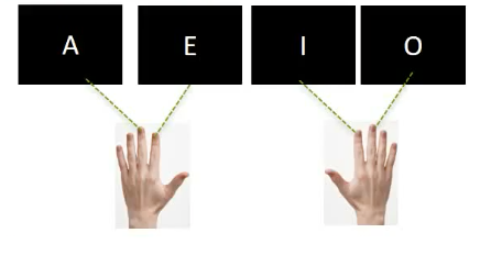
Four-choice-reaction task
Results:

EEG research@murdoch
“Exploring the Relationship between Mindfulness and Fear Extinction, and the Potential Implications of this Relationship for Posttraumatic Stress Symptoms (PTSS)” (DPsych Project: Ms Auretta Kumar)
“Entrainment of brain oscillations to improve inhibitory function in people with MCI”(Dementia Research Foundation funded project)
Advantages of ERP
ERP signal is directly related to neural activity and this electrical activity is conducted instantaneously to the scalp
→Therefore, ERP has an excellent temporal resolution
Cost effective, relatively mobile, tolerant of movement artefact, silent (advantageous for auditory research), no issue with claustrophobia, no magnetic field involved
Compared to behavioural paradigm, detect covert processing, does not require motor response, elucidate stage of processing
Disadvantages of ERP
The ERP signal is derived from different sources in the brain and it is not possible to infer exactly where these sources are from
→Therefore, ERP has a poor spatial resolution
Limited to cortical level
Takes long time to set up
high signal-to-noise ratio
→sophisticated data analysis tool and large number of trials and subject are needed