Head & Skull
 Call Kai
 Learn
Learn Practice Test
 Spaced Repetition
 Match
Match Flashcards
 Knowt Play
1/30

There's no tags or description
Looks like no tags are added yet.
Name | Mastery | Learn | Test | Matching | Spaced | Call with Kai | Chat |
|---|
No analytics yet
Send a link to your students to track their progress
31 Terms
Cranium
-anatomical position for head
2 parts
neurocranium
viscerocranium
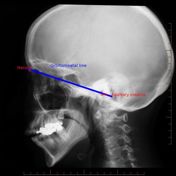
Orbitomeatal plane
defined by inferior margin of orbit & superior margin of
the external acoustic meatus (EAM)
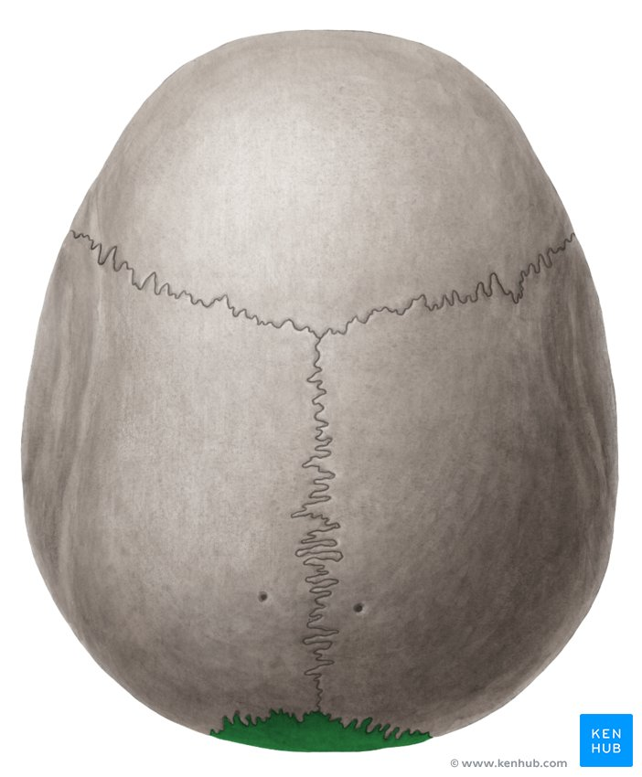
Calvaria
the roof of the neurocranium
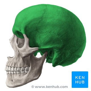
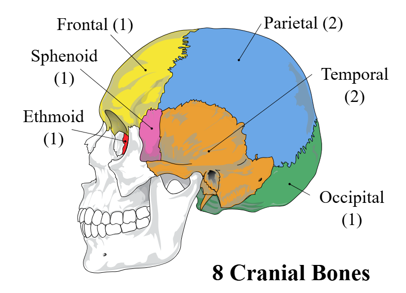
Neurocranium “the brain case”
formed by 8 bones:
4 singles- frontal, occipital, sphenoid, ethmoid
2 paired- 2 temporals and 2 parietals
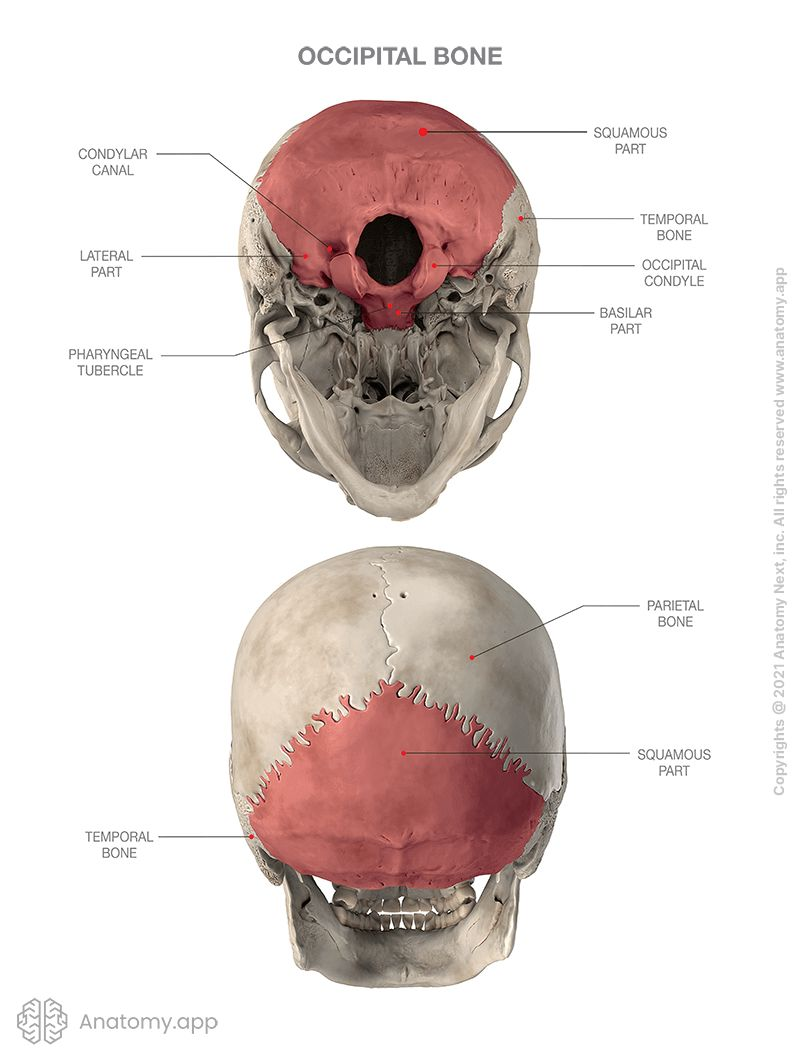
Occipital bone
Landmarks-
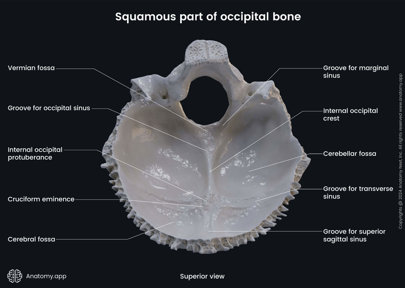
Internal surface-
Landmarks
▪ Foramen magnum
▪ Occipital condyles – bilateral convexities that
articulate with C1
▪ Superior and inferior nuchal lines
▪ External occipital protuberance
Internal surface:
▪ Internal occipital protuberance
▪ deep grooves for transverse sinuses
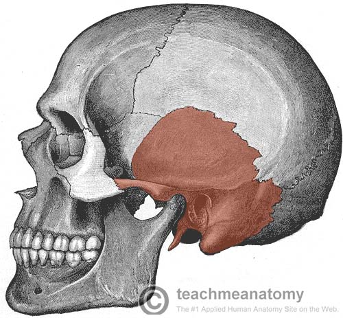
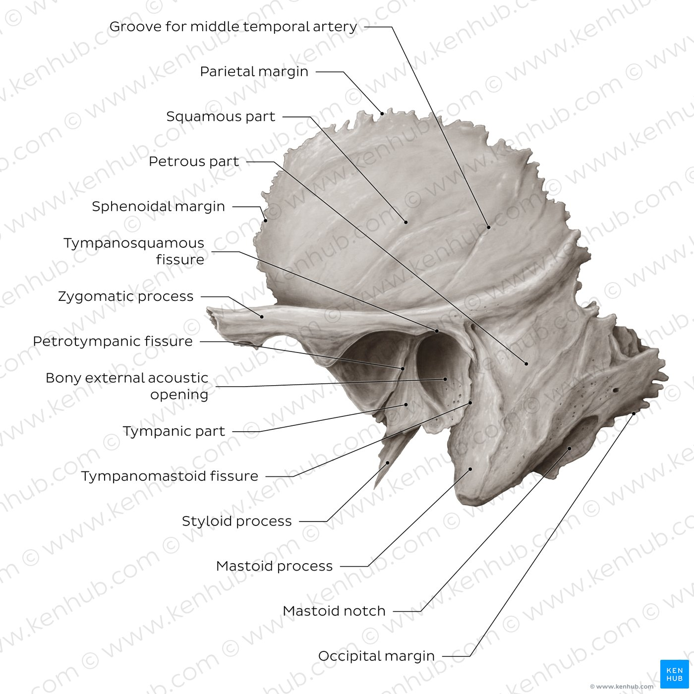
Temporal Bone
lateral view
medial view
Lateral view
▪ External acoustic meatus
▪ Styloid process
▪ Mastoid process
▪ Zygomatic process
▪ Squamous part
Medial view
▪ Petrous part - (L. stone-like)
▪ Internal acoustic meatus (CN VII & CN VIII)
▪ Styloid process - a sharp spine, about 2 - 2.5 cm. in length
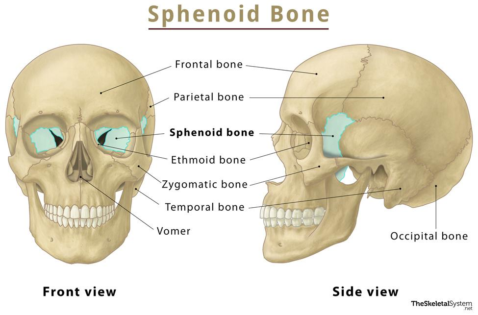
Sphenoid bone
A very complex, irregular bone
Forms : Part of orbit
- part of Ant. Cranial Fossa
- part of Middle Cranial Fossa
- part of the Temple, just anterior to the temporal bone
Contains:
- Ant. poles of frontal lobes
- Sella Turcica
- Multiple Neural foramina
- Pterygoid plates : muscle attachments
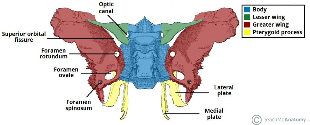
Sphenoid bone superior view
sella turcica “turkish saddle”
sella turcica contains the hypophysial fossa in which lies the pituitary gland (hypophysis)
Sphenoid also contains:
optic canal, foramen rotundum, foramen ovale
Sphenoid bone
Trans-sphenoidal surgical approach
Utilized for intracranial surgery, such as pituitary tumor
Instruments are inserted into part of the brain by passing through an incision made under the upper lip or at the bottom of the nose between the nostrils and then through the sphenoid bone
Notably less invasive than open procedure
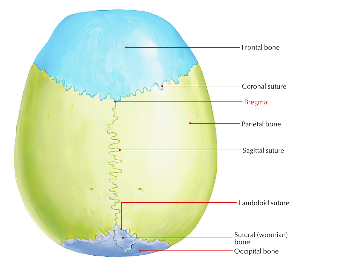
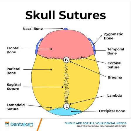
Cranial sutures
sagittal
bregma
lambda
Sagittal suture formed by joining of two parietal bones
Bregma junction of coronal and sagittal sutures
Lambda λ junction of sagittal and lambdoid sutures
Viscerocranium
Facial skeleton
upper and lower jaw
nose and nasal cavity
most of the orbits
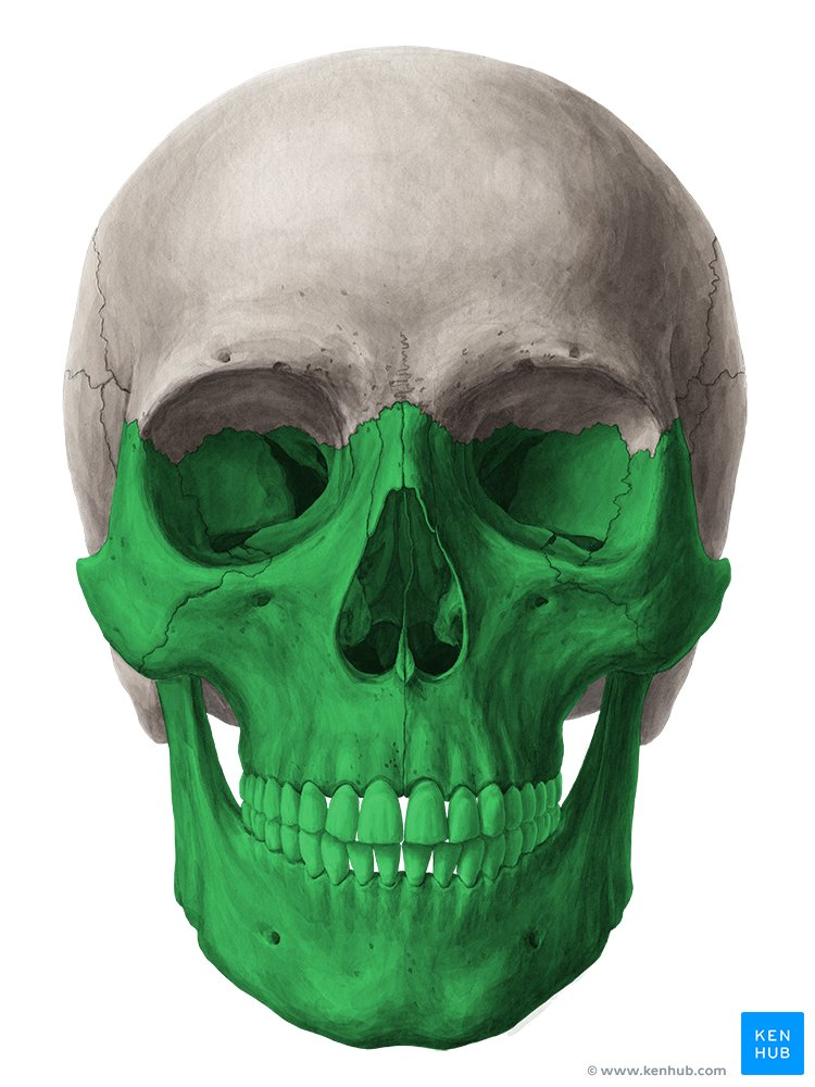
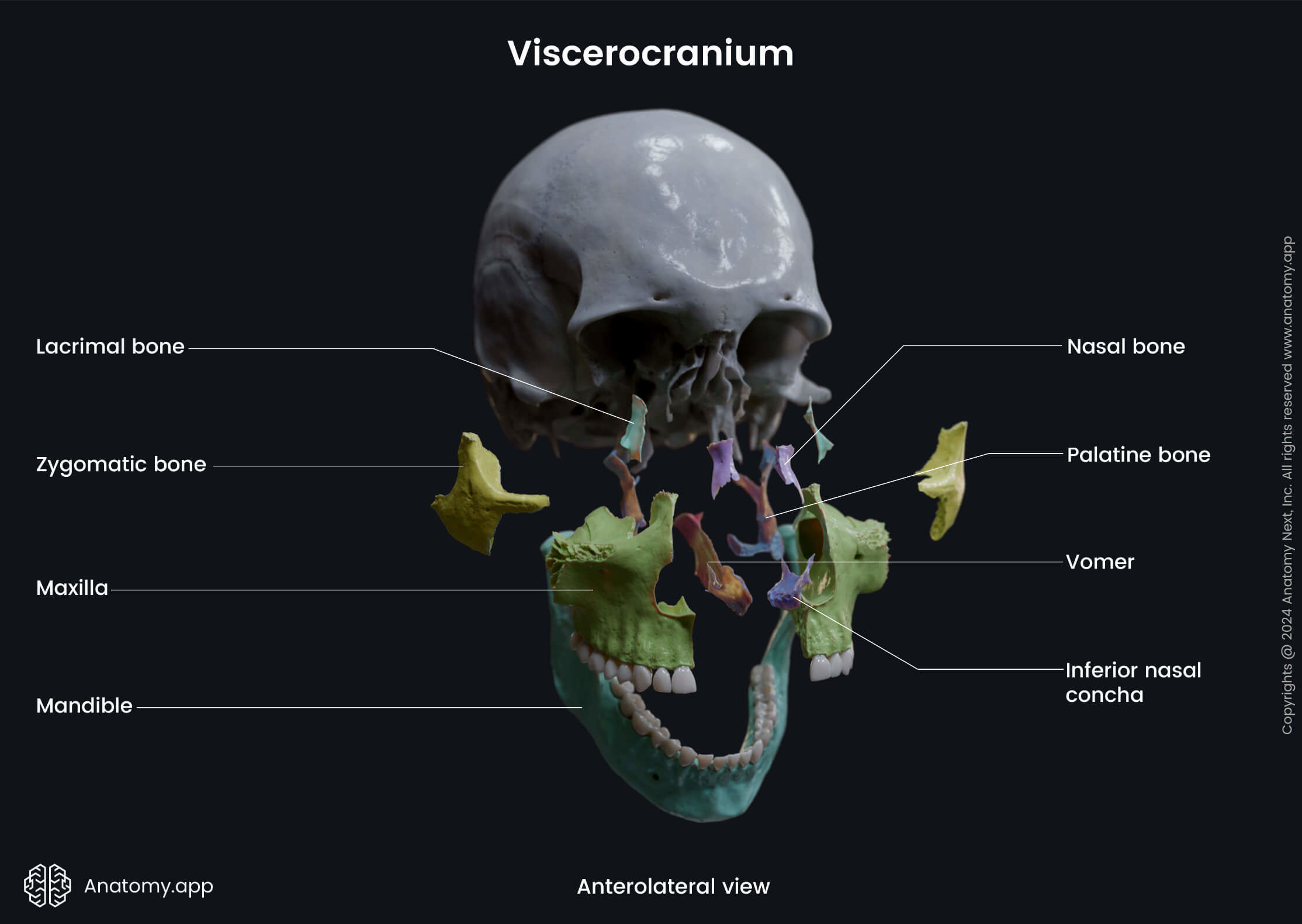
Viscerocranium is composed of 15 irregular bones
3 singular bones lying in midline: Mandible, ethmoid, vomer
6 bones as bilateral pairs: Maxillae, zygomatic, palatine, nasal,
Lacrimal , inferior nasal conchae
Viscerocranium
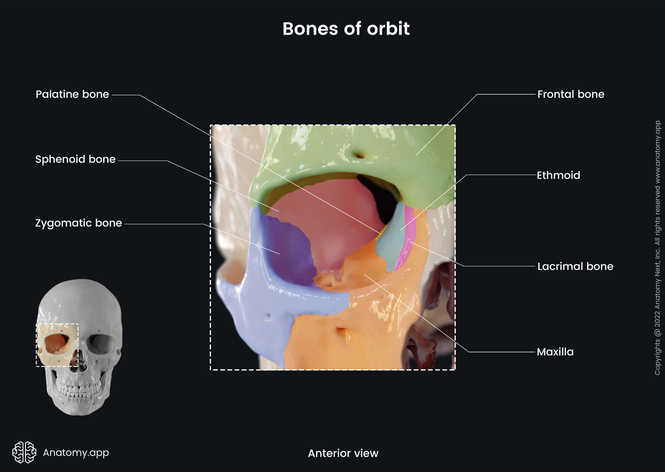
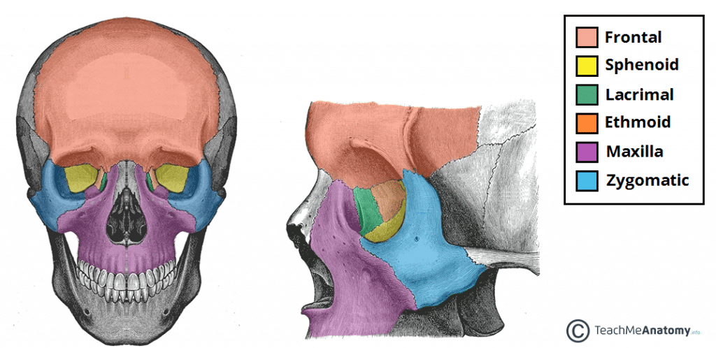
orbits (eye sockets)******
orbital rim formed by _,_, &_
remaining bones of orbit: _,_,_,&_
Orbit houses and protects…
Orbital rim formed by maxilla, zygomatic, and frontal bones
Remaining bones of orbit: Sphenoid, Lacrimal, Ethmoid, Palatine
Orbit houses and protects eyeball, extraocular muscles, related nerves & vessels, lacrimal gland, orbital fascia and fat
Cranium landmarks/features (“know these landmarks for the skull!”)
Temporal fossa
External occipital protuberance (EOP)
**Pterion**- 4 bones meet**- anterior branch of the middle meningeal artery passes beneath pterion
Zygomatic arch
Mastoid process
External acoustic meatus
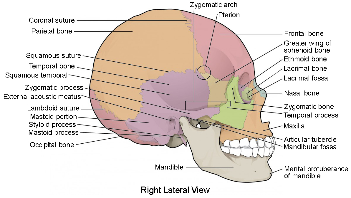
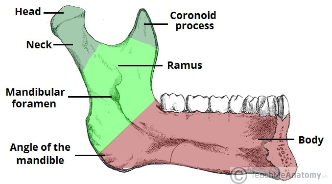
Mandible (“you should know these landmarks”)
▪ -Body
▪ -Ramus - the “vertical portion”
▪ -angle - intersection of Ramus & body
▪ -mental protuberance - prominent anterior region
▪ -condylar process - blunt, posterior knuckle-shaped
projection that forms convexity of the TMJ
▪ -coronoid process - more sharply angled projection- anterior to condylar process. Inferior attachment of temporalis
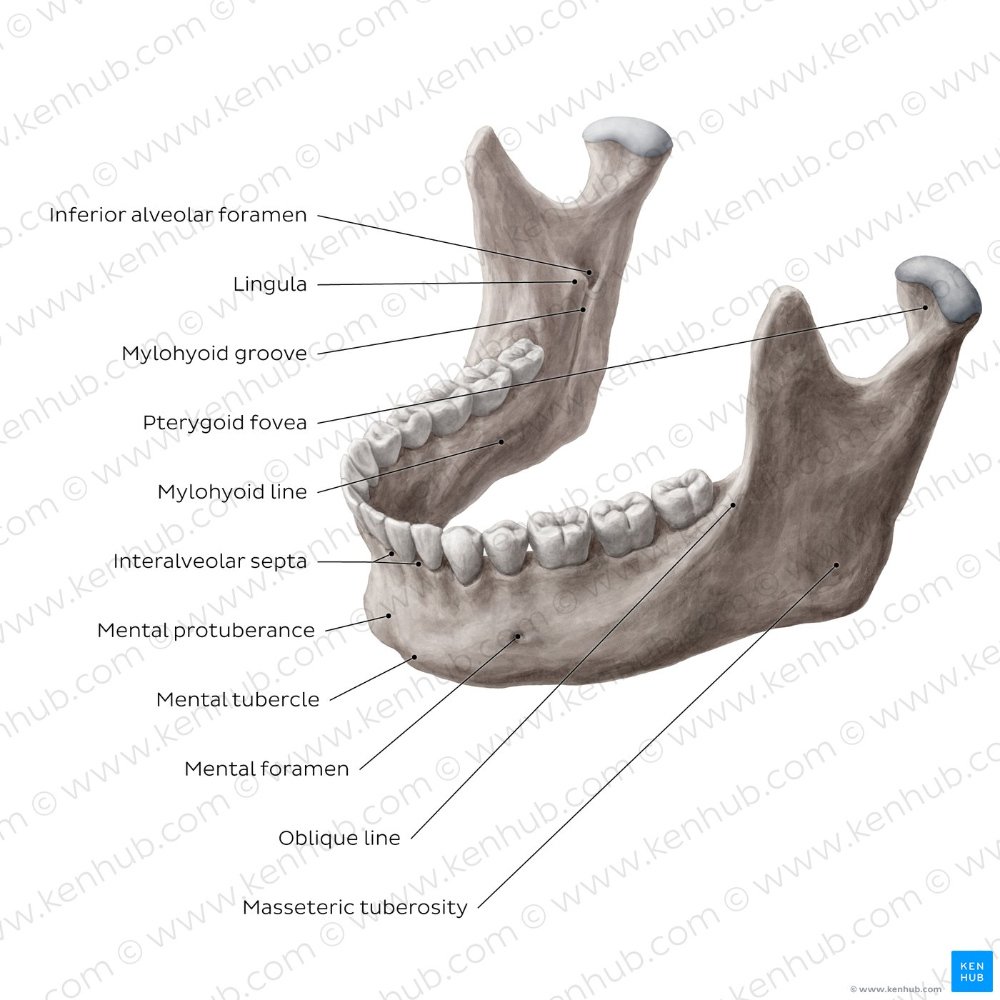
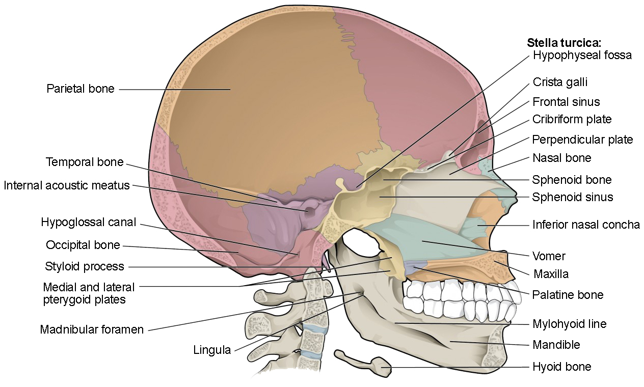
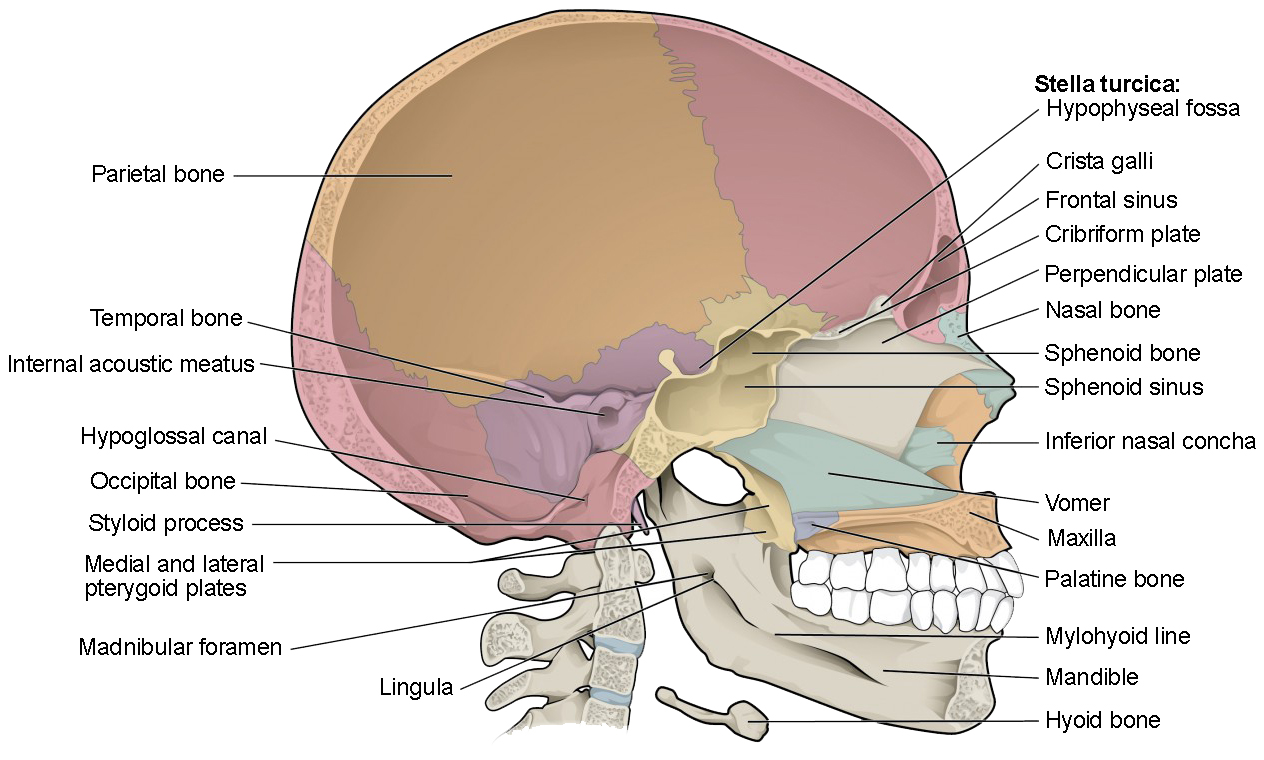
Skull (median section)
internal features
Cribiform plate (ethmoid bone)
Internal acoustic meatus
Basilar aspect of occipital bone
Foramen magnum
Sella turcica
Jugular foramen
Foramina of Cranial Base
inferior view
Mandibular fossa
Zygomatic process
Basilar part of Occiput
Foramen Magnum
Mastoid Process
Stylomastoid foramen (CN VII)
Carotid canal
External acoustic meatus
Foramina of Cranial base (will ask questions on superior )
Optic canal
CN II Optic nerve
Superior orbital fissure
CN V1 (5- the 1st part) Ophthalmic nerve
CN III Oculomotor nerve
CN IV Trochlear nerve
CN VI Abducent nerve
Foramen rotundum
CN V2 Maxillary nerve
Foramen Ovale
CN V3 Mandibular nerve
internal carotid artery
ICA enters inferior aspect of temporal bone. The artery takes a near 90 degree turn and travels anteromedially – emerges internally at foramen lacerum and turns superiorly to join Circle of Willis
Dura Mater
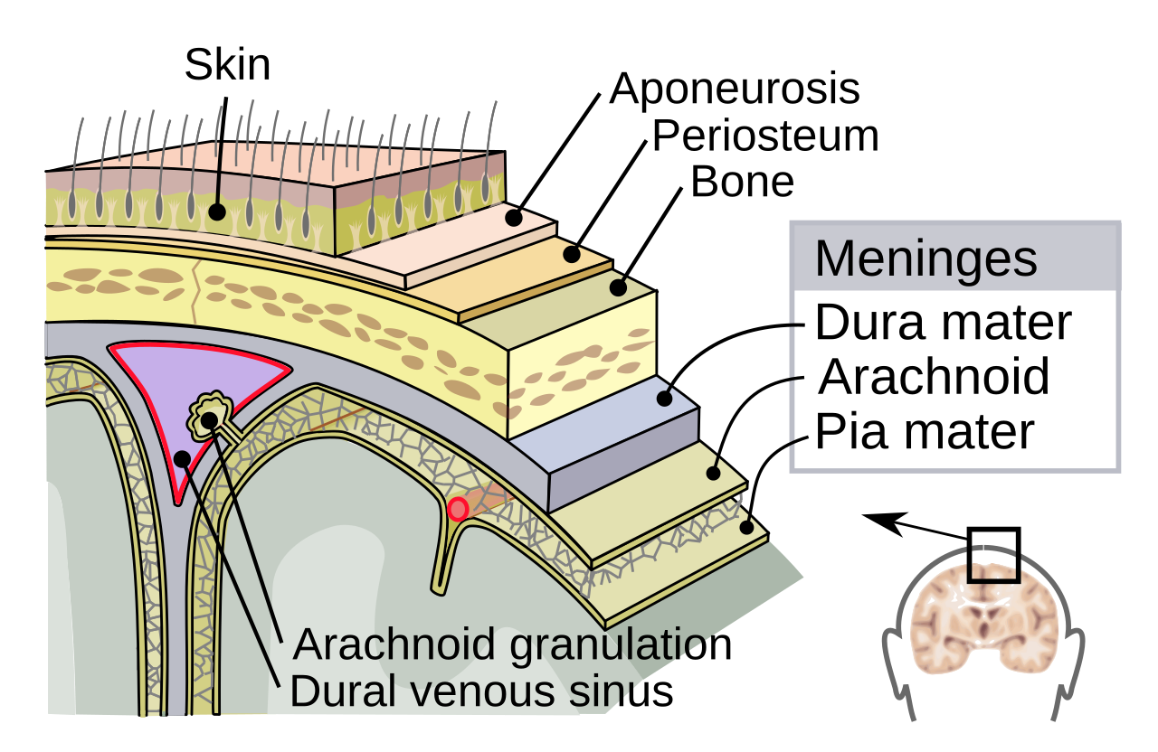
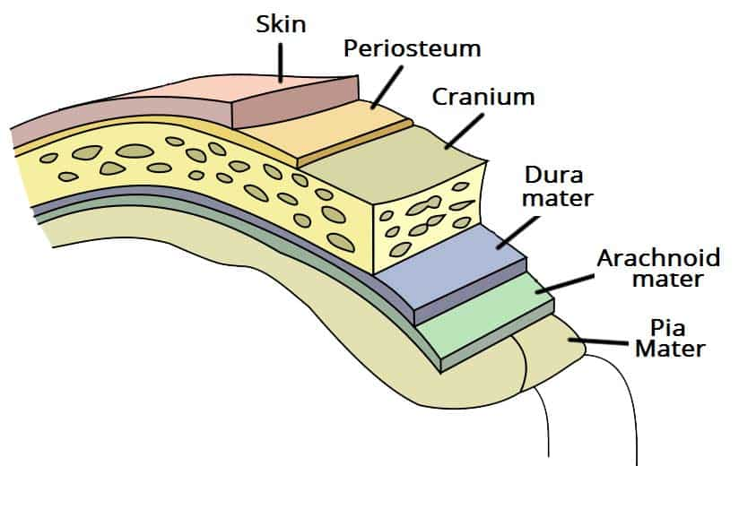
Dura mater adheres to internal aspect of calvaria (periosteal layer)
The deeper layer (meningeal layer), folds back from the periosteal layer to form infoldings that create the dural sinuses and as well as the following:
Falx cerebri
Tentorium cerebelli
Falx cerebelli
Falx cerebri
Large dural infolding between cerebral hemispheres
Runs anterior to posterior
Attaches anteriorly: frontal crest of frontal bone & crista galli of ethmoid
Posterior attachment: internal occipital protuberance
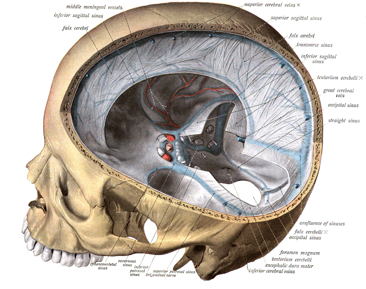
Tentorium cerebelli
is “tent-like” (L. tentorium, tent)
Dural infolding that separates the Occipital lobes of the cerebral hemispheres from cerebellum
Compartments formed by tentorium are known as:
Supratentorial
Infratentorial
The Falx cerebelli is a dural infolding that partially separates the cerebellar hemispheres
Pia mater
deepest meningeal layer
Very thin, highly vascularized
Pia adheres to brain surface and follows contours into sulci of cerebral cortex
Also covers spinal cord and its fissures
“NO QUESTIONS ON DURAL VENOUS SINUSES ON FINAL”
Epidural hemorrhage
occurs in the potential space between the dura and the cranium
Laceration of the middle meningeal artery and its accompanying dural sinuses is the most common etiology.
Subdural hematoma
Bleeding within the dural-arachnoid junction creates a “pathological space”
Shift of midline structures and is visible with imaging
Usually results from head trauma
- 50% mortality rate
will not ask function of the lobes of cerebrum, may reference them in a question referring to an injury
will not ask about sulci
will not need to know cerebrum medial stem
will not ask about ventricles
WILL ASK ABOUT THREE PARTS OF BRAINSTEM
Need to know three parts of brain stem***
Brainstem
Midbrain (Mesencephalon) is the most superior aspect of
brainstem
Pons (L. bridge ) lies between midbrain and medulla oblongata
Pons lies in anterior aspect of posterior cranial fossa
Medulla oblongata is the most inferior division of the brainstem
Medulla continuous with spinal cord
3 parts of brain stem:
Brainstem
Midbrain (Mesencephalon) is the most superior aspect of
brainstem
3 parts of brain stem
Pons
Pons (L. bridge ) lies between midbrain and medulla oblongata
Pons lies in anterior aspect of posterior cranial fossa
3 parts of brain stem
Medulla oblongata
is the most inferior division of the brainstem
- Medulla continuous with spinal cord