Innate and Adaptive Immunity Vaccines - Slide Deck
 Call Kai
 Learn
Learn Practice Test
 Spaced Repetition
 Match
Match Flashcards
 Knowt Play
1/29

Earn XP
Description and Tags
Immuno Exam 1 - Basic Science Core
Name | Mastery | Learn | Test | Matching | Spaced | Call with Kai | Chat |
|---|
No analytics yet
Send a link to your students to track their progress
30 Terms
Innate Immunity
First line of defense against pathogens
We are born with innate immunity and it develops based on our genetic make-up
Rapid response: within minutes of an infection
Not specific: same molecules/cells respond to a range of pathogens
Has no memory: same response after repeated exposure
The mechanisms involved in innate immunity might not be sufficient to overcome and defeat the infection, either in a “timely fashion” or at all
Is the “Complement system”
Innate Immunity - Physical barriers & chemical defenses
1) Skin
2) Mucous membranes
3) Acid, enzymes, mucus, others
Innate Immunity - Cellular defenses
1) WBCs
2) Cytokines
Layers of the Skin (3)
Epidermis (outer layer)
Dermis (inner layer)
Hypodermis (Deepest Layer)
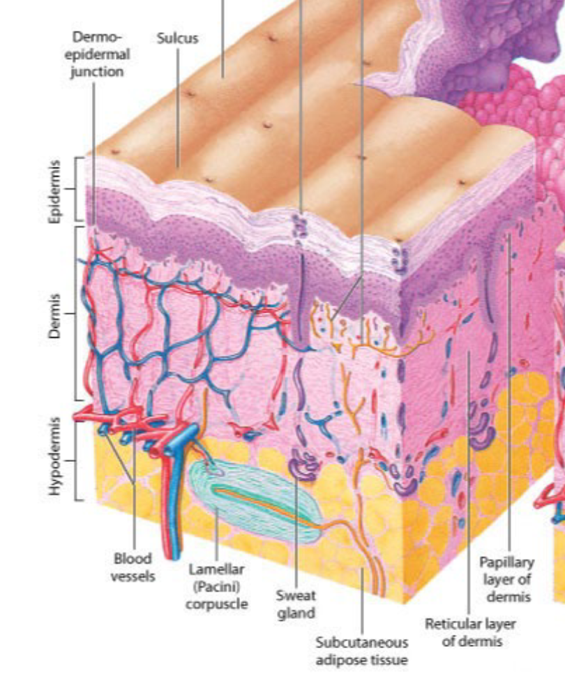
Epidermis
The epidermis contains no blood vessels and is entirely dependent on the underlying dermis for nutrient delivery and waste disposal via diffusion through the dermo-epidermal junction
The epidermis is a stratified squamous epithelium
consists primarily of keratinocytes in progressive 5 layers
Tightly packed together
Mnemonic for the 5 Layers of Epidermis
Come
Lets
Get
Sun
Burnt
5 Layers of Epidermis
1) Stratum Corneum
2) Stratum Lucidum
3) Stratum Granulosum
4) Stratum Spinosum
5) Stratum Basale
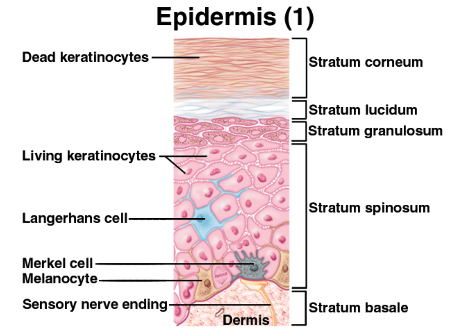
Stratum Basale
Contains the “Stratum germinativum” which:
a single layer of cells that divides constantly (“stem cells of the skin”) – as new cells form, older ones are pushed toward the skin surface
Stratum Corneum
the largest and most abundant of the epidermis.
This layer ranges in thickness from 15-100 or more cells depending on anatomic location and is the primary protective barrier from the external environment.
Contain “Dead Keratinocytes”
Provide elasticity
Protect underlying (living) cells from drying out
Desquamation
Sloughing off of skin cells
Keratinocytes → keratin (Main cells sloughed of) → Corneocytes (keratin-filled sacs)
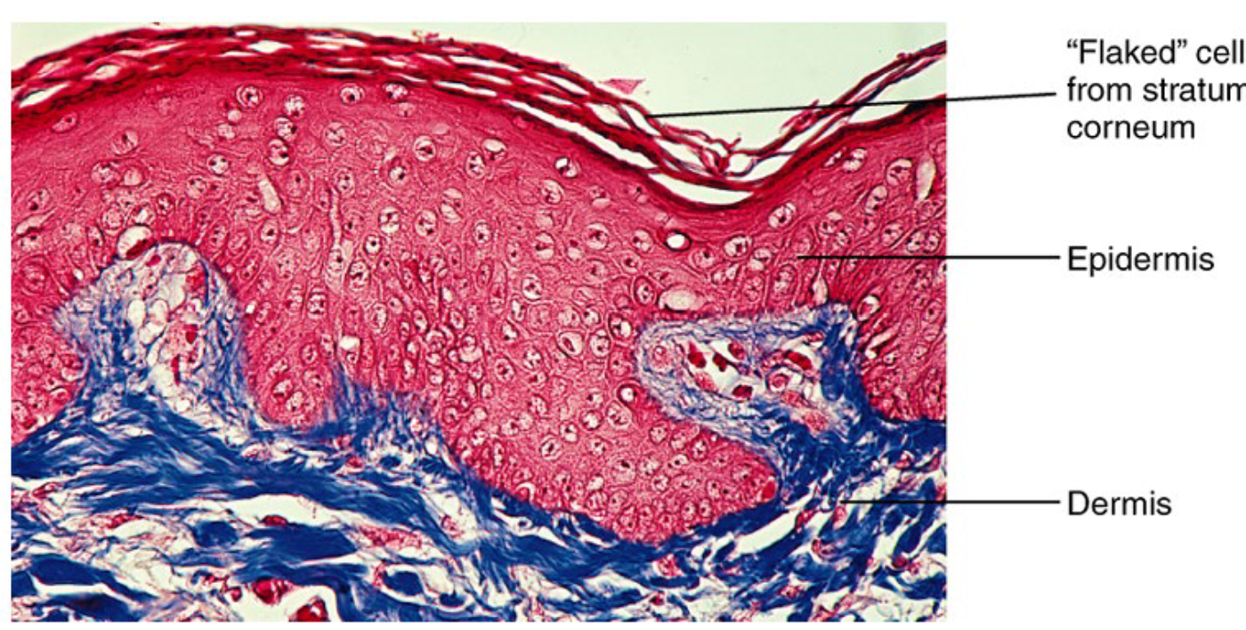
Four types of Cells of the Epidermis
1) Keratinocytes
2) Melanocytes
3) Dendritic cells (Langerhans cells)
4) Nerve endings
Keratinocytes (Squamous Cells) /Corneocytes
Produce keratin (strength, flexibility, waterproof)
Melanocytes
Produce melanin (pigment that absorbs harmful UV radiation from sunlight before it reaches tissues below the skin)
Ratio of melanocytes to keratinocytes
Exposed to the sun → 1:4
Not exposed to the sun → 1:30
Dendritic cells (Langerhans cells)
Identifies bacteria and other invaders and presents them to the immune system for recognition and destruction
Sensory Cells
Nerve Endings
Allow for sensory perception
Dermis
Deeper to and thicker than epidermis
Thickness = 0.3-4.0 mm
Mainly fibroblasts are scattered rather than packed, plus they are interwoven with fibers
Some of the fibers are tough & strong (collagen), others are thin and elastic (become wrinkles when degraded)
Has Dermal papillae
Dermal Papillae
Found in the dermis
Upward projections that create the ridges & grooves of the fingerprints
Dermis Components (5)
1) Nerve endings
2) Hair follicles
3) Sweat (eccrine) glands
4) Sebaceous glands (pilosebaceous follicles)
5) Blood vessels
Sebaceous Glands
Found over the entire surface of the body except the palms, soles, and dorsum of the feet.
They are largest and most concentrated in the face and scalp where they are the sites of origin of acne.
The normal function of sebaceous glands is to produce and secrete sebum, a group of complex oils that include triglycerides and FATTY ACIDS, wax esters, squalene, cholesterol esters, and cholesterol.
Sebum (4 fatty acid)
Lubricates the skin to protect it against friction and makes the skin more impervious to moisture.
Contains 4 fatty acid types:
1) Sapienic acid
2) Lauric acid
3) Oleic acid
4) Palmitoleic acid
These Fatty acids can disrupt the structure and
function of bacterial cell membranes
Palmitoleic acid
Found in Sebum
Also effective in blocking the adherence of Candida albicans to the skin
Salivary Glands (3 Types)
Secretions are important for the hygiene and comfort of the mouth and teeth!
Three Main Glands are:
Parotid
Submandibular
Sublingual
The daily secretion of saliva normally ranges between 800-1500 ml.
Secretions aid in lubrication and digestion of food, and enhance speech, taste, and swallowing.
It also dissolves and washes out food particles from between the teeth
Anti-Pathogen Components in Saliva (4)
1) Lysozymes
2) Lactoferrin
3) Histatins
4) Immunoglobulin A (IgA)
Lysozymes
Enzymes that can attack bacterial cell walls
In Saliva
Lactoferrin
In Saliva
Which chelates iron, preventing the multiplication of
organisms that require it for growth
Histatins
In Saliva
Which are a group of proteins that can disrupt bacterial
& fungal cell membranes
Immunoglobulin A (IgA)
In Saliva
Which prevents bacteria from adhering to
surfaces in the mouth
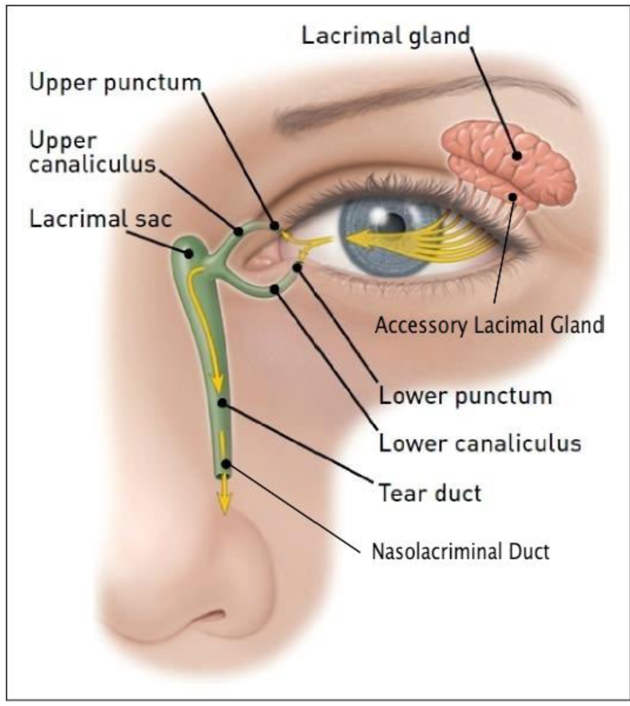
Lacrimal Glands & Lacrimation → Anti-Pathogen Components of Tears
1) Lysozymes
2) Lactoferrin
3) Lipocalins, which are proteins that also chelates iron
4) Immunoglobulin A (IgA)
Gastric Pits
Deep channels called gastric pits, which are lined with columns of specialized cells called gastric glands