Module 4 MCB
1/16
There's no tags or description
Looks like no tags are added yet.
Name | Mastery | Learn | Test | Matching | Spaced |
|---|
No study sessions yet.
17 Terms

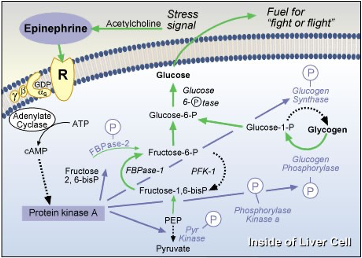
Identify the ligand, second messenger, what type of receptor is this?
ligand = epinephrine
second messenger = cAMP
type of receptor = G-protein coupled receptor
Even if the ligand is present, there are several ways that this pathway can be turned off. What are these steps?
ATP + NADH act as feedback inhibitors, high conc. of ATP + NADH -> inhibit key enzymes -> stop cycle
Recycling = deactivate = hydrolysis of GTP from a triphosphate to diphosphate

Western blot using b arrestin antibody. Lane M contains the markers with sizes of bands indicated on the left. Lanes 1 and 2 contain lysates made from cells that did not have the inhibitor for bARK. Lanes 3 and 4 contain lysates made from cells that did have the inhibitor for bARK. Underneath the lane indicates the number of minutes the cells were incubated with epinephrine.
During the immunoprecipitation step an antibody against the epinephrine receptor is first used, and then on the Western blot an antibody against b arrestin is used. Why?
Immunoprecipitation isolates epinephrine receptor + bound contents from lysate using an antibody specific to the receptor
-> pulls down the receptor + associated proteins (B-arrestin) out of the mixture
= Western blot uses B-arrestin antibody to detect whether B-arrestin was bound to the receptor
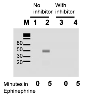
What do the results in lanes 1 and 2 tell you regarding what first needs to happen before b arrestin can bind to the receptor.
Lane 1 = (0 mins of epinephrine) = little/no B-arrestin detected
Lane 2 = (5 mins of epinephrine) = strong B-arrestin signal
ligand binding/exposure of epinephrine is required for B-arrestin to associate with receptor = receptor must be activated by epinephrine for B-arrestin to bind

What information do the kinase inhibitor studies provide about b arrestin binding to the receptor?
Lane 3 = (0 mins of inhibitor) = no B-arrestin
Lane 4 = (5 mins of inhibitor) = no B-arrestin
even after 5 mins of exposure to epinephrine, when kinase bARK is inhibited B-arrestin binding does not occur
receptor phosphorylation by bARK is necessary for B-arrestin binding

What does the following mean with respect to a signal pathway?
X inhibits Y, therefore Y cannot inhibit Z, Z is active
A. Is the following pathway active or not active? (- indicates an inhibitor, + indicates an activator). Why?
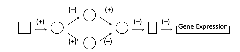
Not active, inhibitor is blocking an upstream step needed to activate downstream signalling. Both pathways are inhibited at different points
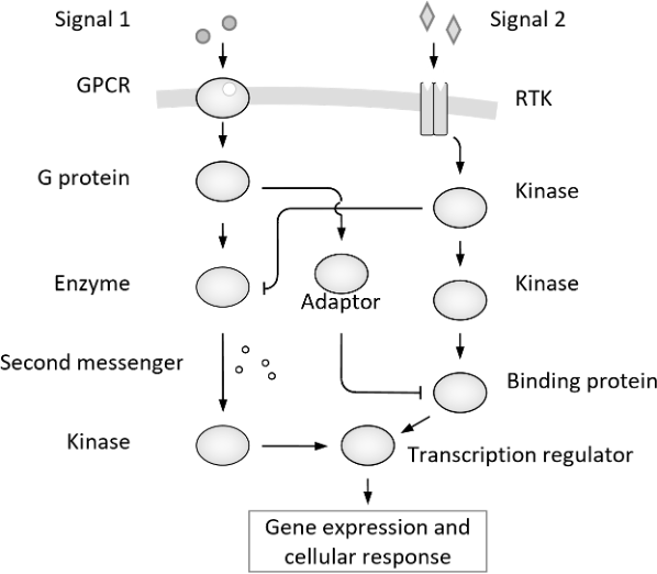
Under which of the following conditions is target-gene expression induced? (there may be more than 1 answer)
A. When only Signal 1 is added
B. When only Signal 2 is added
C. When no signal is added
D. When both signals are added together
= Both A and B, each pathway activated inhibitory step of other pathways
When Signal 1 is added, signal 1 pathway inhibits the signal 2 pathway. Gene expression is still induced but only from signal 1
When signal 2 is added, signal 2 pathway inhibits signal 1 pathway. gene expression is only induced from signal 2
i) Work out which of the protein kinases 1 or 2 is upstream of the other by observing the data in the table below
ii) If you make a cell with a constitutively active PK2 and an inactive PK1 what cell response would you observe and why?
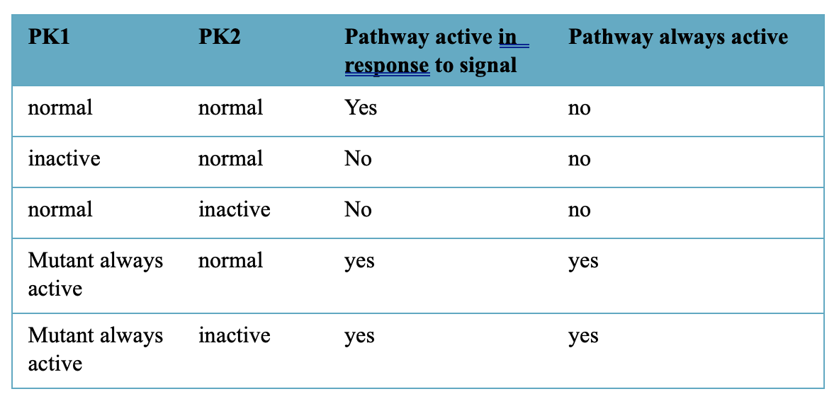
i) PK2 is upstream of PK1
Constitutively active PK1 can drive the pathway even when PK2 is inactive = PK2 activates PK1
ii) Activation of pathway, constitutively active PK2 can still drive pathway with inactive PK1
Genes encoding mutant forms of a receptor tyrosine kinase were expressed in a cell. For each of (i) and (ii) specify if they can bind ligand and if they can become phosphorylated. Then, if the mutant genes are expressed at considerably higher levels than the normal receptor in the cell, decide if each mutant can act as a competitive inhibitor– this means will they affect the function of the cell to respond to signal, ie compete with the normal receptor
A) A mutant receptor tyrosine kinase that lacks its extracellular domain
B) A mutant receptor tyrosine kinase that lacks its intracellular domain
RTK Autophosphorylates tyrosine residues in INTRAcellular domain upon LIGAND BINDING at EXTRAcellular domain
A) = NO ligand binding, NO Phosphorylation as ligand binding triggers AUTOphosphorylation, no ligand binding
B) YES ligand binding, NO phosphorylation, autophosphorylation occurs in intracellular domain, no intracellular domain
What does autophosphorylation mean?
Whole molecule can phosphorylate itself, doesn’t need something to add phosphate, is acting on itself to phosphorylate
Why do you run a protein gel first before adding radioactive ATP?
run protein gel to register presence of ALL 3 receptor types, LOADING CONTROL
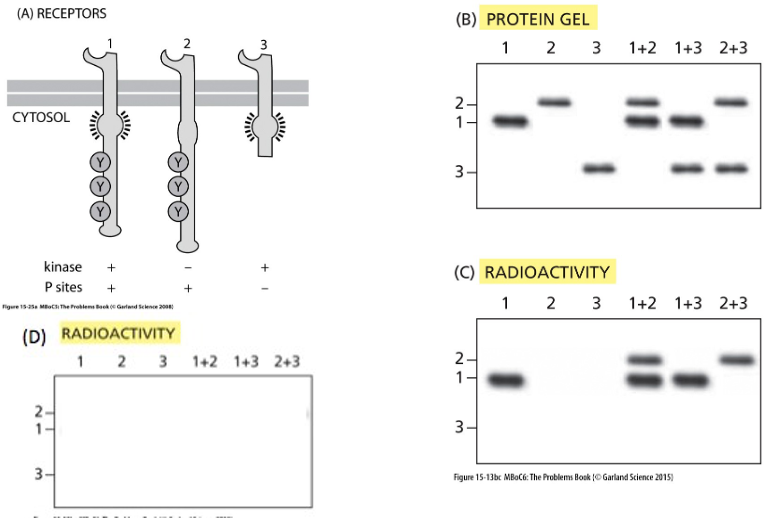
When a receptor tyrosine kinase binds its ligand and forms a dimer, do the individual receptor molecules only phosphorylate themselves or does one receptor cross-phosphorylate the other and vice versa? To investigate this question, you have constructed genes for three forms of a RTK as shown in figure A:
Number 1 is the normal form with an active kinase domain and three sites for phosphorylation,
Number 2 is a large form that carries an inactivating point mutation in the kinase domain but retains the three phosphorylation sites, and
Number 3 is a short version that has an active kinase domain but is lacking the sites of phosphorylation.
You express the genes on their own or in combination in a cell line that lacks this receptor and then break open the cells and add the ligand for the receptor in the presence of radioactive ATP. You immunoprecipitate the receptors and analyze them for expression levels by staining for protein (Fig. B) and for phosphorylation by autoradiography (Fig. C).
(i) Does the data support a model where the individual receptor molecules can only phosphorylate themselves or does one receptor cross-phosphorylate the other and vice versa?
(ii) What results would you have expected on the autoradiograph if the other model was the correct answer? (If each receptor could phosphorylate itself)
i)
Lane 1 (normal) + 2 (no kinase) = Receptor 1 + 2 phosphorylated, receptor 1 phosphorylates itself + 2
Lane 1 (normal) + 3 (no sites) = only receptor 1 phosphorylated, receptor 3 cant be phosphorylated (has no sites) but can activate receptor 1 via dimerization
Lane 2 (no kinase) + 3 (no sites) = no phosphorylation, receptor 2 cannot autophosphorylate due to no kinase, receptor 3 has no sites so cant be phosphorylated
(ii)
- Lane 1 = YES phosphorylation (active kinase + sites)
- Lane 2 = NO phosphorylation (no kinase)
- Lane 3 = NO phosphorylation (no sites)
- Lane 1 + 2 = Receptor 1 can phosphorylate, NOT receptor 2
- Lane 1 + 3 = Receptor 1 can phosphorylate, NOT receptor 3
- Lane 2 + 3 = NO phosphorylation (neither can phosphorylate itself)
In cells with the G12V mutation in Ras, what happens to cell proliferation? Briefly explain why and include whether ligand binding is required.
Ras acts like a switch -> is active when bound to GTP and inactive when it hydrolyses GTP to GDP
G12V mutation prevents Ras from hydrolysing GTP, prevents Ras inactivating
-> Ras stays permanently active
-> stuck in on state
-> as in on state, Ras activates mAP kinase pathway which promotes cell division and growth
-> cell proliferation increases dramatically and becomes uncontrolled
Ligand binding = Normal Ras activation depends on ligand binding to receptor tyrosine kinase
-> activates Ras downstream
With G12V mutation, Ras is already constantly active
-> therefore Ligand binding is not required to activate Ras
How is the Ras protein usually activated and deactivated?
Activated = when ligand binds to receptor tyrosine (RTK) on cell surface
-> RTK dimerizes + autophosphorylates = activates Ras
-> Ras swaps GDP to GTP
-> Ras is bound to GTP, is activated and can trigger downstream signalling
Deactivation = Ras hydrolyses GTP into GDP -> returns to inactive state
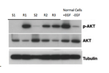
AKT is transcription factor that is phosphorylated by Erk (a MAPK) when the EPF (epidermal growth factor) pathway is activated. Proteins were extracted from normal cells that were either untreated (-EGF) or treated (+EGF) with EGF. Proteins were also extracted from 5 patient tumours (labelled S1, S2, R1, R2, R3), which have the ras mutation, but also have been treated with a ras inhibitor. Proteins were run on a SDS PAGE and then western blotted with 3 different antibodies- one recognizes AKT, one recognizes the phosphorylated form of EGF (pEGF) and one that recognizes tubulin.
(i) From looking at this Western blot how can you tell if the pathway is active or not?
(ii) What does this experiment tell you about the EGF pathway in the different tumours, and for which ones is the ras inhibitor a possible treatment?
i) If p-AKT is present = pathway is ACTIVE. phosphorylation of AKT indicates EGF pathway through Ras and MAPK is active
ii) S1 + S2 = inactive as little to no pAKT, therefore inhibitor IS effective,
R1, R2, R3 = active as high amounts of pAKT, therefore inhibitor IS NOT effective
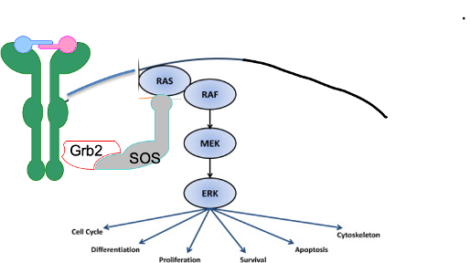
Imagine you are working in a cancer clinic and encounter a patient with a cancer that has the Raf-V600E mutation. You treat with a Raf inhibitor but the cancer does not respond. You decide you need to add a new inhibitor to your treatment plan. Which of the following would work?
a. An inhibitor of Ras-GEF
b. An inhibitor of Mek
c. An inhibitor of Ras
d. An inhibitor of the Receptor tyrosine kinase
e. An inhibitor of Erk
f. An inhibitor of Stat
Cancerous process starts RAS onwards, best to inhibit something downstream of mutation, i.e. Mek and Erk,
= Erk in case there was a mutation in Mek, ERK is the molecule that directly activates the pathway