Chapter 11: Functional Organization of Nervous Tissue
0.0(0)
Card Sorting
1/71
Earn XP
Study Analytics
Name | Mastery | Learn | Test | Matching | Spaced |
|---|
No study sessions yet.
72 Terms
1
New cards
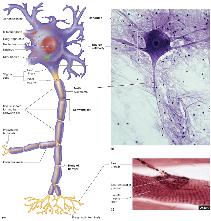
Two main cell types in nervous tissue
neurons and glial cells
2
New cards
Nerve
bundle of axons outside the brain and spinal cord
3
New cards
Cranial nerves
orginate from the brain; 12 pairs
4
New cards
Spinal Nerves
originate from the spinal cord; 31 pairs
5
New cards
Ganglion
collection of neuron cell bodies (holds nucleus) outside the brain and spinal cord (dorsal root ganglion)
6
New cards
Plexus
extensive network of axons and sometimes neuron cell bodies; located outside the CNS
\
ex. brachial plexus
\
ex. brachial plexus
7
New cards
Glial Cells
Supportive cells with many functions
8
New cards
Functions of the nervous system
maintain homeostasis
receive sensory input (monitor internal/external stimuli)
Integrating information (CNS)
controlling muscles and glands (effector organs)
establishing and maintaining mental activity
receive sensory input (monitor internal/external stimuli)
Integrating information (CNS)
controlling muscles and glands (effector organs)
establishing and maintaining mental activity
9
New cards
Central nervous system
brain and spinal cord
10
New cards
peripheral nervous system
sensory receptors and nerves
11
New cards
Sensory division of PNS
transmits AP from receptors to CNS (afferent)
12
New cards
Sensory receptors
can be neuron endings or specialized cells that detect external and internal stimuli; send input along nerves to brain or spinal cord
13
New cards
motor division of PNS
transmits AP from CNS to effectors (efferent)
14
New cards
somatic nervous system
from CNS to skeletal muscles
voluntary; single neuron system
voluntary; single neuron system
15
New cards
autonomic nervous system
from CNS to smooth muscle, cardiac muscle or glands
subconscious or involuntary control
two neuron system
* first from CNS to ganglion
* second from ganglion to effector
\
subconscious or involuntary control
two neuron system
* first from CNS to ganglion
* second from ganglion to effector
\
16
New cards
divisions of ANS
sympathetic, parasympathetic, or enteric
17
New cards
sympathetic ANS
prepares body for physical activity
18
New cards
Parasympathetic ANS
regulates resting functions
19
New cards
Enteric ANS
plexuses within the wall of the digestive tract
20
New cards
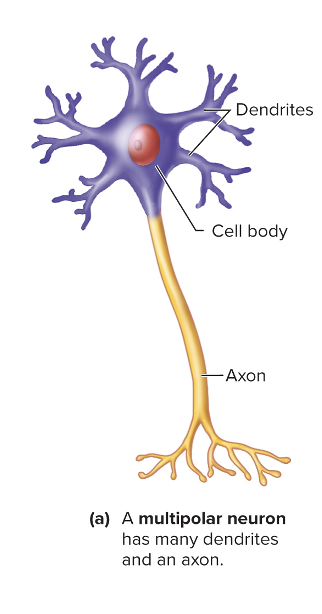
multipolar/motor neuron
receive information through dendrites
integrate information in cell body
send signals to cause action in effectors through axon
integrate information in cell body
send signals to cause action in effectors through axon
21
New cards
Interneurons
within the CNS, connect 1 neuron to another
\*sometimes not seen in very simple neural pathways
\*sometimes not seen in very simple neural pathways
22
New cards
Multipolar
most neurons in CNS; motor neurons
23
New cards
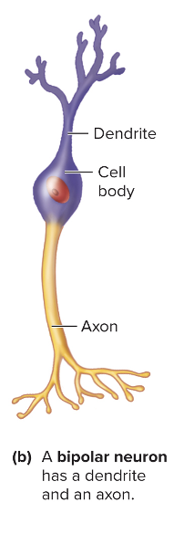
Bipolar Neurons
sensory in retina of the retina and nasal cavity
24
New cards
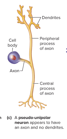
psuedo-unipolar
single process that divides into two branches.
part that extends into the periphery has dendrite-like sensory receptors
part that extends into the periphery has dendrite-like sensory receptors
25
New cards
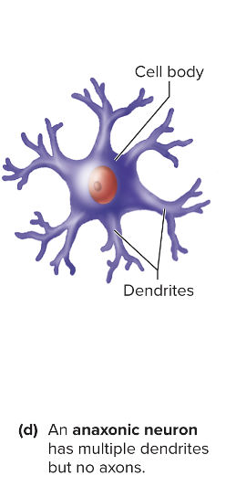
anaxonic
no axons, only dendrites
found in brain and retina where they only communicate using graded potentials
found in brain and retina where they only communicate using graded potentials
26
New cards
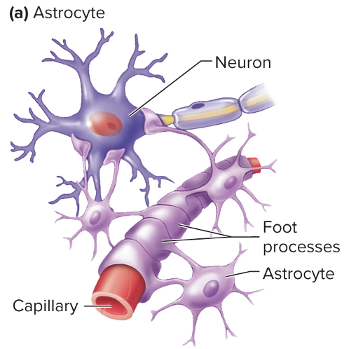
Astrocytes
glial cell
prevents blood composition fluctuations from affecting the brain function
forms blood-brain barrier (keep some thing in and some things out)
promote development of synapses
prevents blood composition fluctuations from affecting the brain function
forms blood-brain barrier (keep some thing in and some things out)
promote development of synapses
27
New cards
reactive astrocytosis
CNS injury sites are walled off to limit inflammation
can have scar forming astrocytes which limits axon regeneration in damaged neurons
can have scar forming astrocytes which limits axon regeneration in damaged neurons
28
New cards
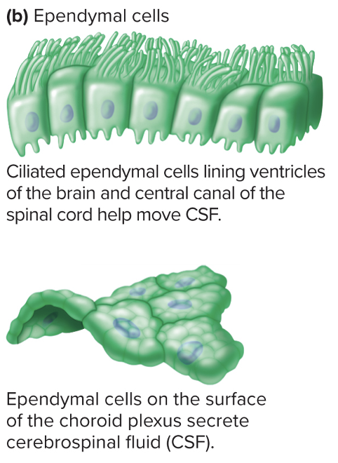
Ependymal cells
glial cell of CNS
line brain ventricles and central canal of spinal cord
specialized versions form choroid plexus
**forms cerebrospinal fluid**
has some astrocyte-like functions
line brain ventricles and central canal of spinal cord
specialized versions form choroid plexus
**forms cerebrospinal fluid**
has some astrocyte-like functions
29
New cards
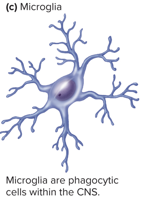
Microglia
glial cell in CNS
specialized CNS macrophage
respond to inflammation, phagocytize necrotic tissue, microorganisms, and foreign substances that invade the CNS
can be used in autopsy to detect CNS damage
specialized CNS macrophage
respond to inflammation, phagocytize necrotic tissue, microorganisms, and foreign substances that invade the CNS
can be used in autopsy to detect CNS damage
30
New cards
Oligodendrocytes
glial cell of CNS
form myelin sheaths by wrapping cytoplasmic extensions around axons
can form myelin sheaths around portions of several axons
form myelin sheaths by wrapping cytoplasmic extensions around axons
can form myelin sheaths around portions of several axons
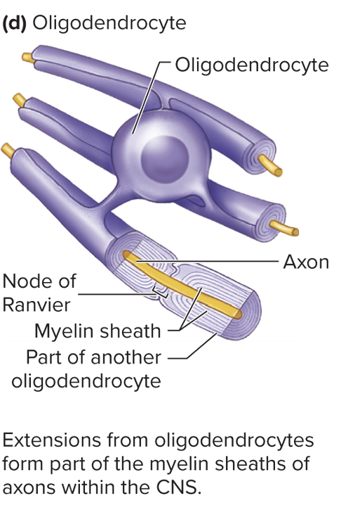
31
New cards
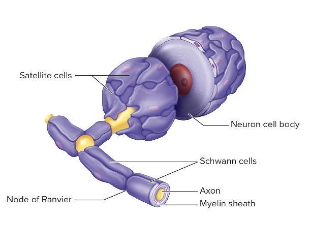
Shwann Cells
glial cell of PNS
wrap only one axon to form myelin sheath
outer layer is neurilemma (most cytoplasm, nucleus, and organelles)
wrap only one axon to form myelin sheath
outer layer is neurilemma (most cytoplasm, nucleus, and organelles)

32
New cards
Satellite cells
glial cell of PNS
surround neuron cell bodies in sensory and autonomic ganglia
provide support, nutrients, and protection from heavy metal poisons
surround neuron cell bodies in sensory and autonomic ganglia
provide support, nutrients, and protection from heavy metal poisons
33
New cards
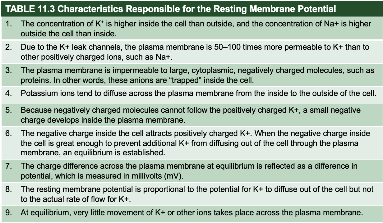
Characteristics responsible for the resting membrane potential
34
New cards
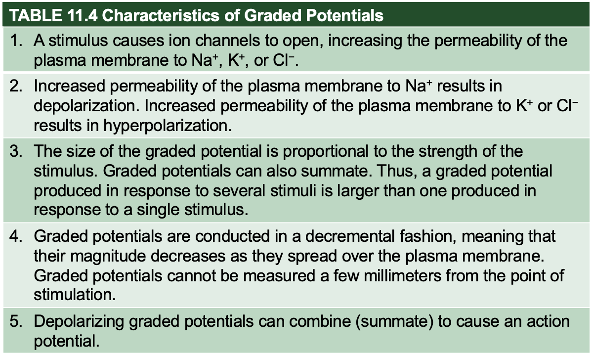
graded potentials result from
ligands bind to receptors
change in charge across membrane
mechanical stimulation
temp change
spontaneous change in permeability
change in charge across membrane
mechanical stimulation
temp change
spontaneous change in permeability
35
New cards
graded potential occurs in ….
areas of synaptic contact
cell body and dendrites
cell body and dendrites
36
New cards
action potential occurs in ….
Axons
37
New cards
graded potential
small change in membrane potential in a localized area
magnitude varies based on stimuli and frequency
hyperpolarizing (Cl+ in/K+ out) or depolarizing
can summate/add to reach threshold
conducted over the plasma membrane in a decremental fashion (decrease in magnitude as the spread)
magnitude varies based on stimuli and frequency
hyperpolarizing (Cl+ in/K+ out) or depolarizing
can summate/add to reach threshold
conducted over the plasma membrane in a decremental fashion (decrease in magnitude as the spread)
38
New cards
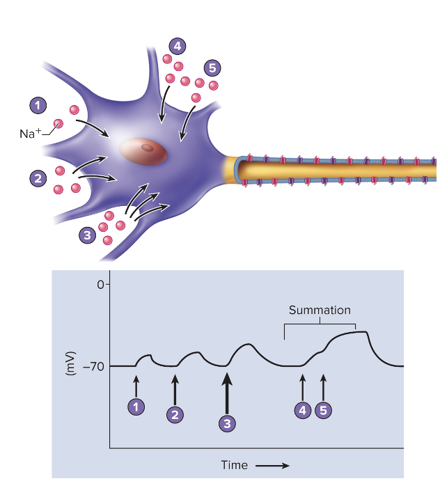
graded potential steps
1. small act of Na+ entering → slight depolarization
2. and 3. more Na+ enters → greater depolarization
4. stimulus is applied to cell causing a small depolarization
5. summation → larger depolarization
39
New cards
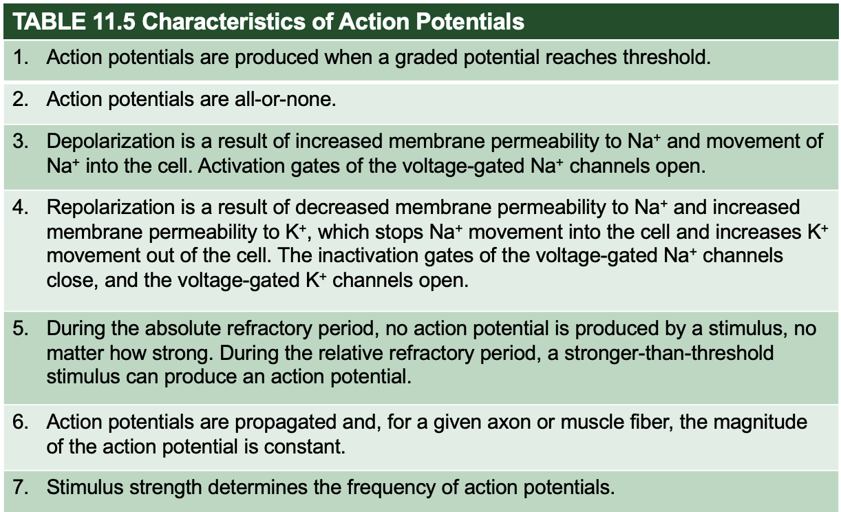
Characteristic of action potentials
produced when graded potential reaches a threshold
all or none response
this is how neurons communicate with an effector
all or none response
this is how neurons communicate with an effector
40
New cards
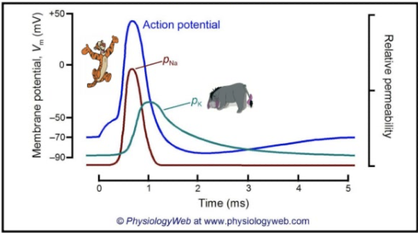
1st step of an action potential and a voltage-gated ion channel
resting membrane potential
voltage-gated = closed, inactivation gate = open
outside is + charged
voltage-gated = closed, inactivation gate = open
outside is + charged
41
New cards
2nd step of an action potential and a voltage-gated ion channel
Na+ activation channel = open quickly (graded potential)
Depolarization when threshold is reached
K+ start to open slowly
Inside becomes more +
open voltage channels increase open voltage channels until all open
positive feedback cycle
Depolarization when threshold is reached
K+ start to open slowly
Inside becomes more +
open voltage channels increase open voltage channels until all open
positive feedback cycle
42
New cards
3rd step of an action potential and a voltage-gated ion channel
repolarization
inactivation gates of Na+ voltage channels close
More k+ channels open
\*no more Na in and increased k+ out →repolarization
inactivation gates of Na+ voltage channels close
More k+ channels open
\*no more Na in and increased k+ out →repolarization
43
New cards
4th step of an action potential and a voltage-gated ion channel
End of repolarization
reestablishes resting condition
reestablishes resting condition
44
New cards
Activation v inactivation in Na+ and K+ channels
1. rest: Na+ activated closed, Na+ inactivated open
2. graded: Na+ activated open quickly, Na+ inactivated open, K+ open slowly
3. depolarization: Na+ open until all are open, K+ opening
4. Repolarization: Na+ inactivated close, K+ still open
5. End of repol: Na+ activated close, inactivated open, k+ still open
6. Afterpotential: Na+ all closed, K+ still open
1. Rest: K+ and Na+ closed
45
New cards
Afterpotential
Hyperpolarization
allows k+ to leave cell
allows k+ to leave cell
46
New cards
Refractory Period
sensitivity of area to further stimulation decreases for a time
absolute (complete insensitivity, beginning go AP to \~end of repol.)
relative (stronger than threshold stimulus can initiate another AP)
absolute (complete insensitivity, beginning go AP to \~end of repol.)
relative (stronger than threshold stimulus can initiate another AP)
47
New cards
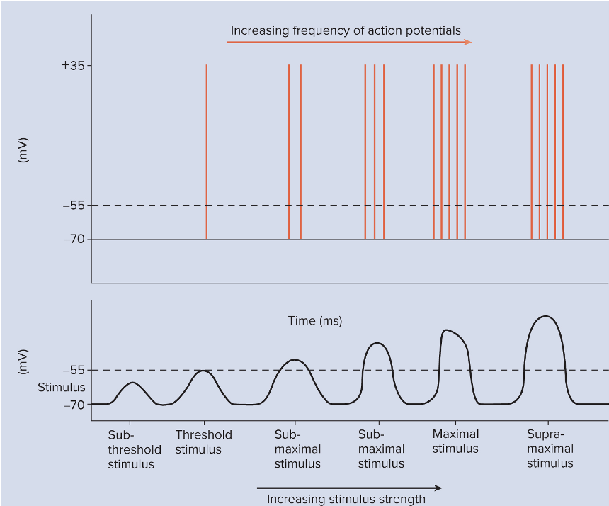
stimulus strength and AP frequency
Strength of Ap doesn’t change w increase in strength of stimulus BUT frequency of action potential can inc. to a point
target of AP will interpret the higher frequency as a stronger stimulus which could lead to a stronger contraction/secretion
target of AP will interpret the higher frequency as a stronger stimulus which could lead to a stronger contraction/secretion
48
New cards
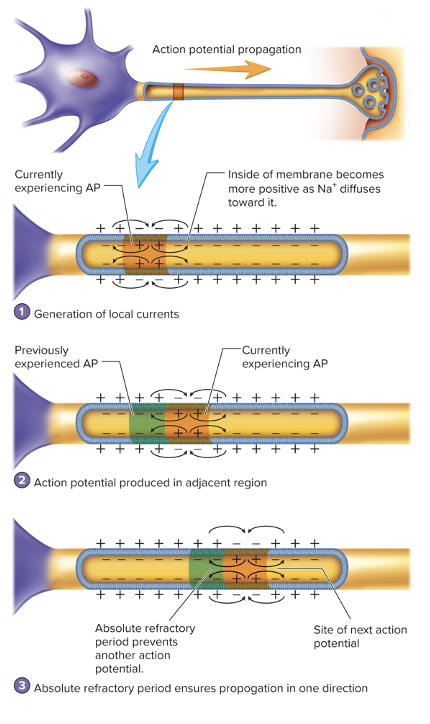
AP Propagation in a unmyelinated axon
AP is propagated more slowly
Na+ diffuses into membrane and creates a local current that travels down the axon
Na+ diffuses into membrane and creates a local current that travels down the axon
49
New cards
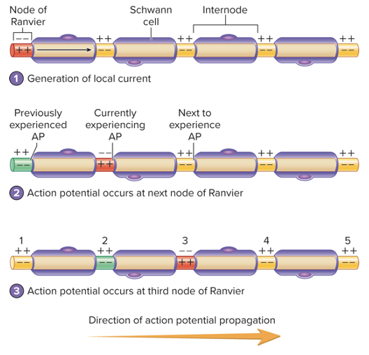
saltatory conductions
AP at a node of Ranvier generates local currents
myelin sheath insulates and forces current from node to node
Na+ channels are highly concentrated
myelin sheath insulates and forces current from node to node
Na+ channels are highly concentrated
50
New cards
The synapse
junction between two cells
where action potentials in one cell causes an action potential in another cell
where action potentials in one cell causes an action potential in another cell
51
New cards
Types of cells in a synapse
presynaptic (signal to synapse)
postsynaptic (receiving signal)
postsynaptic (receiving signal)
52
New cards
types of synapses
electrical (gap junction→ cardiac/smooth)
chemical (neurotransmitters→ skeletal)
chemical (neurotransmitters→ skeletal)
53
New cards
Electrical Synapses
connected by gap junctions
graded current flow between adjacent cells
cardiac and smooth muscle (where contractility is important)
graded current flow between adjacent cells
cardiac and smooth muscle (where contractility is important)

54
New cards
Connexons
protein tubes in cell membrane
55
New cards
chemical synapses
components: presynaptic terminal, synaptic cleft, postsynaptic membrane
neurotransmitters released by AP
AP causes Ca2+ to enter and neurotrasmitter to be released from vesicles (voltage-gated)
neurotransmitter binds to receptor on ligand gated ion channels
neurotransmitters released by AP
AP causes Ca2+ to enter and neurotrasmitter to be released from vesicles (voltage-gated)
neurotransmitter binds to receptor on ligand gated ion channels
56
New cards
Steps in a chemical synapse
1. AP opens V-gated Ca channels
2. Ca into cell and bind to vesicles and they release neurotransmitter
3. neurotransmitter diffuse across cleft
1. nt. bind to receptors and creates a graded potential in membrane
57
New cards
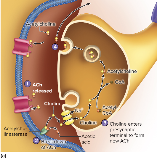
Removal of neurotransmitter from synaptic cleft (ACh)
1. Act released from receptors
2. Ach-esterase splits ACh into choline and acetic acid
3. choline is taken up by presynaptic terminal and binds with acetyl-CoA to reform acetylcholine
1. other molecule diffuse into ECF
58
New cards
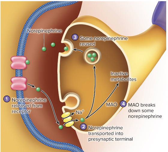
Removal of Neurotransmitter from Synaptic Cleft (norepinephrine; NE)
1. NE released from cleft
2. taken up by presynaptic terminal
3. repackaged into synaptic vesicles
1. enzyme monoamine oxidase (MAO) breaks down some molecules of NE
59
New cards
Specificity of receptor molecules in synapses
neurotransmitter only fits one receptor
only effects cells with receptors specific to them
some neurostransmitters are excitatory and/or are inhibitory
some can also attach to the presynaptic terminal and modulate its own release
only effects cells with receptors specific to them
some neurostransmitters are excitatory and/or are inhibitory
some can also attach to the presynaptic terminal and modulate its own release
60
New cards
Neuromodulators
released from neurons to pre/post synaptic membrane to synaptically influence the likelyhood of an action potential
ex. decrease the chance of excitatory NT release and prevent the generation of a AP
ex. decrease the chance of excitatory NT release and prevent the generation of a AP
61
New cards
Neurotransmitters and Neuromodulators
chemical messengers secreted by neurons
neurons can secrete more than 1 type (100 diff ones)
neurons can secrete more than 1 type (100 diff ones)
62
New cards
criteria to be a neurotransmitter
synthesized by neuron and stored in synaptic vesicles in presynaptic terminals
AP must stimulate its exocytosis
must bind to receptor in postsynaptic membrane
must evoke a response
AP must stimulate its exocytosis
must bind to receptor in postsynaptic membrane
must evoke a response
63
New cards
classification of neurotransmitter is based on
chemical structure
effect on postsynaptic membrane
mechanism of action at the targy
effect on postsynaptic membrane
mechanism of action at the targy
64
New cards
Ionotropic effect of neurotransmitters
bind to ion channels
65
New cards
metabotropic effect on neurotransmitters
binding to G-protein-linked receptors
66
New cards
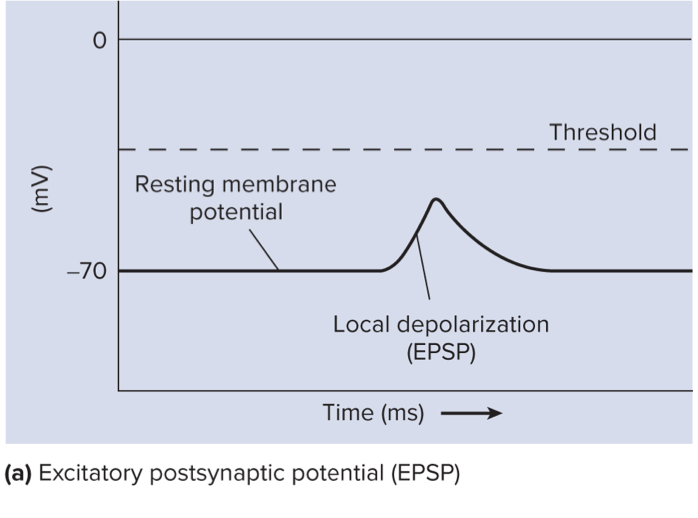
Excitatory postsynaptic potential
\
\
* Depolarization occurs and response is stimulatory.
* Depolarization might reach threshold producing an action potential and cell response.
* Depolarization might reach threshold producing an action potential and cell response.
67
New cards
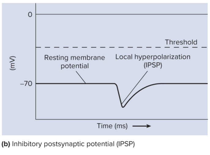
Inhibitory postsynaptic potential (IPSP)
hyper polarization and response is inhibitory
decrease likelihood of action potential by moving membrane potential farther from threshold
decrease likelihood of action potential by moving membrane potential farther from threshold
68
New cards
Axoaxonic synapses
Neuromodulator
axon of one near synapses with the presynaptic terminal (axon) of another
common in CNS
axon of one near synapses with the presynaptic terminal (axon) of another
common in CNS
69
New cards
Presynaptic inhibition
neuromodulator
reduction in amount of neurotransmitter released from presynaptic terminal
reduction in amount of neurotransmitter released from presynaptic terminal
70
New cards
presynaptic facilitation
neuromodulator
amount of neurotransmitter released from presynaptic terminal increases
amount of neurotransmitter released from presynaptic terminal increases
71
New cards
Spatial summation
AP 1 and 2 cause the production of graded potential at 2 different dendrites
summate at the trigger sone to produce a graded potential that exceeds threshold resulting in an action potential
summate at the trigger sone to produce a graded potential that exceeds threshold resulting in an action potential
72
New cards
Temporal Summation
two AP arrive in close succession at the presynaptic membrane
1st causes production of a graded potential that does not reach threshold at the trigger zone. The second action potential results in the production of a second graded potential that summates with the first to reach threshold, resulting in the production of an action potential.
1st causes production of a graded potential that does not reach threshold at the trigger zone. The second action potential results in the production of a second graded potential that summates with the first to reach threshold, resulting in the production of an action potential.