Anatomy and Physiology I - Lab LESSON ONE
 Call Kai
 Learn
Learn Practice Test
 Spaced Repetition
 Match
Match Flashcards
 Knowt Play
1/71

There's no tags or description
Looks like no tags are added yet.
Name | Mastery | Learn | Test | Matching | Spaced | Call with Kai | Chat |
|---|
No analytics yet
Send a link to your students to track their progress
72 Terms
Anatomical Orientation Equivalent: Superior
Cephalad (HEAD)
Anatomical Orientation Equivalent: Posterior
Dorsal
Anatomical Orientation Equivalent: Anterior
Ventral
Anatomical Orientation Equivalent: Inferior
Caudal (towards the tail/bottom end)
Frontal Plane
Divides the body into a front and back

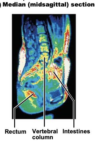
Median (Midsagittal) Plane
Divides the body in the middle, with a left and right side
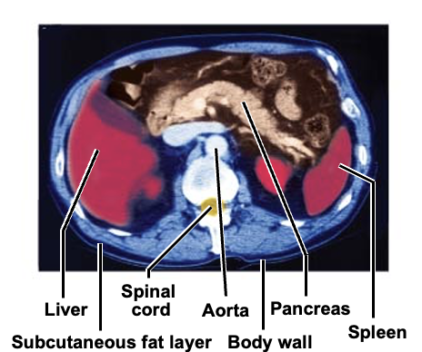
Transverse Plane
Divides the body into a top and bottom half
What does the dorsal body cavity contain?
Cranial cavity + vertebral cavity
What does the cranial cavity contain?
Brain
What does the vertebral cavity contain?
Spinal cord
What does the ventral body cavity contain?
Thoracic + abdominopelvic cavities
What does the thoracic cavity contain?
Heart (pericardial) + lungs (pleural)
What does the abdominal cavity contain?
Digestive viscera
What does the pelvic cavity contain?
Urinary bladder, reproductive organs, rectum
What is a synovial cavity? Where is it located?
Cavity located in a joint between neck vertebrae
Found in ALL synovial joints, not just joins between vertebrae
What is the ventral body cavity lined with?
Epithelial cells
What are the layers of the serosa/serous membrane?
Parietal serosa - lines the cavity WALL
Visceral serosa - covers the surface of the organs within the cavity
Why are serous membrane important?
Membrane compartmentalizes various organs to prevent the spread of infection from one organ to another
What are the 4 abdominopelvic quadrants?
Right upper quadrant
Right lower quadrant
Left upper quadrant
Left lower quadrant
Right hypochondriac region
Liver, gallbladder, diaphragm
Right lumber
Ascending colon, small intestine
Right iliac
Cecum, appendix
Epigastric
Liver, stomach diaphragm
Umbilical
Small intestine, transverse colon
Hypogastric
Small intestine, sigmoid colon, rectum, urinary bladder
Left hypochondriac
Diaphragm, spleen, stomach
Left lumber
Small intestine, descending colon
Left iliac
Initial segment of sigmoid colon
Microscope component: Arm
Connects the base and head of the microscope
Microscope component: mechanical stage
Consists of spring clips or clamps to hold a slide in position
Microscope component: mechanical stage CONTROL
Moves the mechanical stage and slide held by the mechanical stage
Microscope component: stage
Platform that a slide rests on
Microscope component: coarse adjustment knob
Used for initial focusing of slide image
Microscope component: fine adjustment knob
Used for precise focusing of slide image after coarse focusing has been completed
Microscope component: base
Supports the microscope
Microscope component: light control
Turns on/off power to the light source
Microscope component: ocular lenses
Eye pieces used to view the magnified image from the slide
Microscope component: rotating nosepiece
Contains objective lenses, allowing for changing of lenses for variable magnification of slide image
Microscope component: objective lenses
Used to magnify the slide sample image
Microscope component: condenser
Small substage lens that concentrates the light on the specimen
Microscope component: substage light
Light source for microscope
If a mirror is used instead of a light, it reflects light from an external source
What does the plasma membrane consist of? What regions are polar and nonpolar?
Phospholipid bilayer:
Polar/hydrophilic HEADS
Non-polar/hydrophobic TAILS
What is the Fluid Mosaic Model?
Explains that there are proteins and cholesterol that ‘float’ in the phospholipid bilayer that are good for structural integrity
When hot, membrane becomes more fluid; when cold, membrane stiffens
Integral membrane proteins
Proteins that stick through the entire membrane
Peripheral membrane proteins
Proteins that only stick out on one side of the membrane
Nucleus
Contains DNA in a thread-like form (chromatin)
During cell division, chromatin condenses and coils into a tightly packed form called chromosomes that are bound by a porous double-layered nuclear envelope
Nucleolus
Assembly site for ribosomes (PIT OF NUCLEUS)
Plasma membrane
Separates external environment from internal cell
Is selectively permeable**
Cytoplasm/Cytosol
Fluid INSIDE of the cell that suspends the organelles
Ribosomes
Composed of RNA and proteins - are sites of protein synthesis
Endoplasmic Reticulum
System of membranous tubules + cisternae for the synthesis and transport of proteins
Rough ER - has ribosomes
Smoother ER - no ribosomes
Golgi Apparatus
Flattened sacs that package material
Can become part of plasma membrane, a lysosome, or be secreted from cell depending on the packaged material
Lysosomes
Membrane-bound sacs containing digestive enzymes that digest worn-out cell structures and foreign substances
Peroxisomes
Sacs that contain oxidases that use oxygen to detoxify harmful substances
Mitochondria
Rod-shaped organelles that produce energy in the form of ATP
Centrioles
Rod-shaped bodies used in cell division
Cytoskeletal Elements
Parts that form the internal scaffolding that supports and moves the substances within the plasma membrane
AKA the “INTERSTATE”
What are the 3 parts of the cytoskeletal system?
Microtubules
Intermediate filaments
Microfilaments
Cytoskeletal elements: microtubules
Maintain cell shape, suspend organelles, transport substances, involved in cell division
Cytoskeletal elements: intermediate filaments
Provide support to the cell
Cytoskeletal elements: microfilaments
Support the plasma membrane and cell mobility
What are the 2 components of a cell’s life cycle?
Interphase
Cell division
Interphase
Cell grows as usual and prepares itself for cell division by duplicating its DNA
NOT A PART OF CELL DIVISION ONLY PREPARATION
Cell Division
Cell duplicates itself to yield 2 genetically identical daughter cells
Involves mitosis + cytokinesis
Mitosis
Nuclear division of a cell (nucleus divides)
Cytokinesis
Cell undergoes cytoplasmic division (divides the cell’s internal contents into 2 halves) to form 2 identical daughter cells W
What does cytokinesis start through?
Formation of a cleavage furrow by actin microfilaments during late anaphase throughout telophase
Early Prophase
DNA chromatin shortens into chromosomes with a centromere in the middle
Nucleolus disappears
Centrioles split into centrosomes; each one moves towards opposite ends of the cell
Late Prophase
Nuclear envelope disappears
Mitotic spindles/microtubules form from the centrosomes and attach to centromere of chromosomes
Metaphase
Chromosomes line up at center of the cell + attach onto the mitotic spindles of centrioles at opposite sides of cell
Anaphase
Centromeres split + chromosomes migrate to opposite ends towards centrioles
Telophase
DNA uncoils back into chromatin form
Nuclear envelope reappears around chromatin mass
Nucleolus reappears within nucleus
Spindles break down and disappear