Anatomy Lec 1
1/41
Earn XP
Description and Tags
USC Summer Anatomy
Name | Mastery | Learn | Test | Matching | Spaced |
|---|
No study sessions yet.
42 Terms
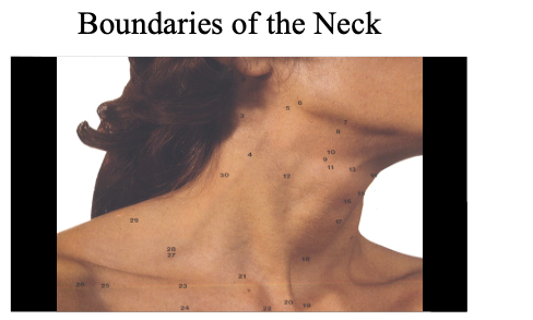
Boundaries of the neck: ______ Aspect & _______ Aspect. & contents
Boundaries of the Neck
•Anterior Aspect:
Superior: Inferior border of the mandible
Inferior: Superior surface of the manubrium and clavicle
•Posterior Aspect:
Superior: Superior nuchal line
Inferior: Horizontal line between C7 and T1
Platysma muscle:
Describe appearance
innervated by…
covers ______ aspect of ______
origin:
insertion:
Function:
paperthin ; superficial muscle of the neck responsible for facial expression.
cervical branch of facial nerve (CNVII)
covers (entire) anterior aspect of neck
origin: deltoid + Pectoralis fascia
insertion: inferior border of mandible and skin/hypodermis
depression of lower lip +
corner of mouth.
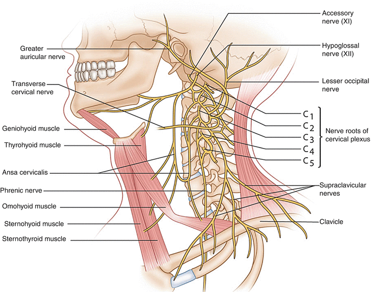
What is a cervical Plexus?
a network of nerves located in the neck that provides both sensory and motor innervation to the head, neck, and upper chest. It's formed by the anterior rami of spinal nerves C1-C4.
This plexus is essential for various functions, including sensation in the neck and upper chest and controlling muscles in the neck, including the diaphragm.
Cervical Plexus involves both _____ and _______ innervation
motor and sensory
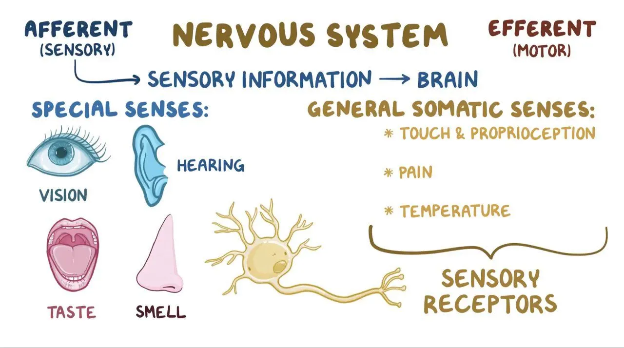
What does somatosensory mean? I mean like what is it?
special senses - vision, hearing, taste, and smell. + And general somatic senses, involved in the sense of touch, proprioception, pain, and temperature.

__,__,__, & form the cervical plexus and are __________.
-C1, C2, C3, and most of C4 form
the cervical plexus
-Somatosensory
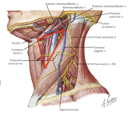
Nerve Innervation on Front of Neck (3) + Their corresponding C letters
Great Auricular Nerve (C2 & C3)
Transverse Cervical Nerve (C2 , C3)
Supraclavicular nerve (C2 & C4)
-anterior (medial)
-Middle (intermediate
-Posterior (lateral)
Red=great auricular
Blue=Transverse Cervical
Green=Supraclavicular
Great Auricular Nerve emerges from ….
emerges from posterior border of sternocleidomastoid muscle
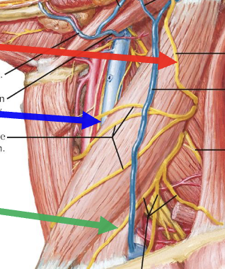
Ansa Cervicalis: is a _____ _____…
Location
description of what it looks like
has 2 ______ formed by __,__ ,& __. They are called: _____ & _______. first one travels with ___and second one is from ___ ___.
is a _______ nerve
Function:
innervations
The ansa cervicalis is a nerve loop located in the neck that is part of the cervical plexus
anterior OR IN carotid sheath IM NOT SURE (fascia that protects the big 3
-Common Carotid artery
-internal jugular vein
-Vagus Nerve)
U shaped
roots, C1,C2,C3, superior, inferior, CN XI, Cervical Region
somatoMOTOR nerve
Motor innervation of sternohyoid, sternothyroid, omohyoid (NOT thyrohyoid, its innervated by nerve to thyrohyoid)
same as 5 hehe
number 1
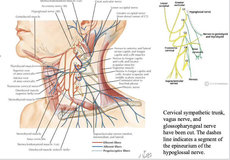
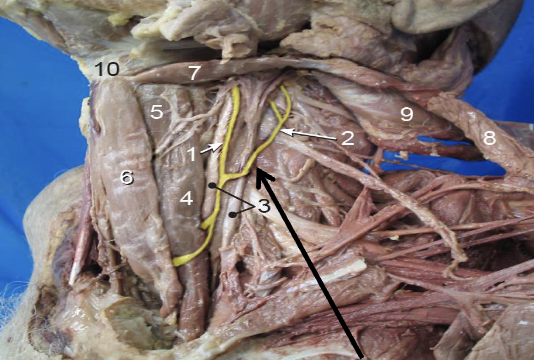
Accessory nerve (CN __)
__ components of fibers: __& __
function: innervation of ___ & ___
CN XI
2, cranial root, spinal root
-Sternocleidomastoid muscle (scm)
-trapezius muscle
Best way to find accessory nerve?
Cut SCM 4-5 cm from its origin of sternum part, move the scm muscle flap upwards, find the border of mandible, and move over a little bit and u find it
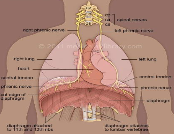
Phrenic Nerve cervical components __, _, & _
Motor function?
Sensory Function?
C3, C4, C5
Motor: Diaphraghm
345 keep the diaphragm alive
Sensory: Mediastinal pleura and pericardium of the heart
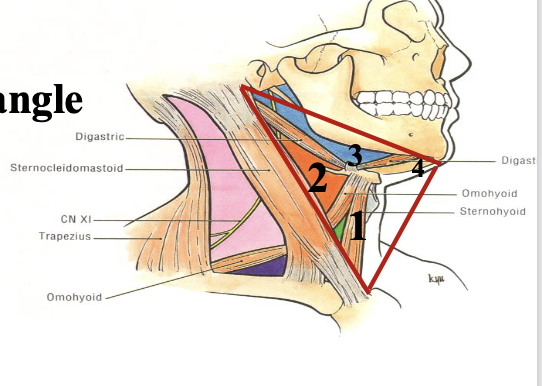
What are the 3 boundaries for the anterior triangles of the neck?
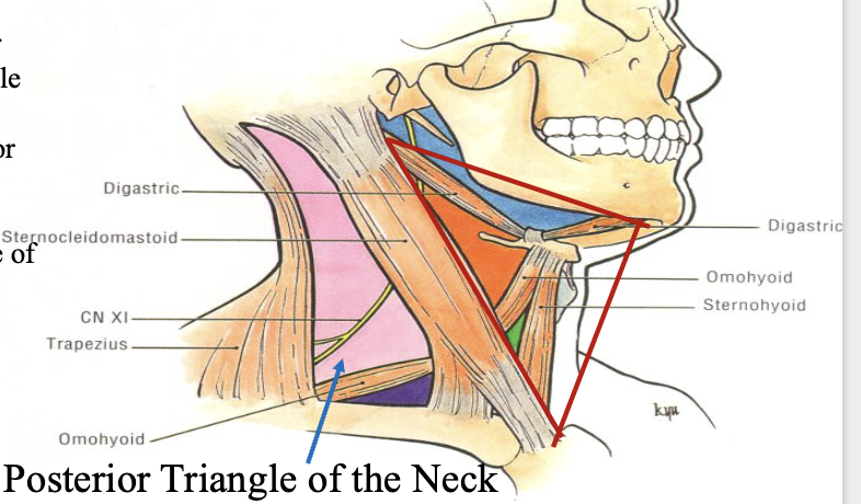
RED TRIANGLE = Anterior Triangle
Top part: inferior border of mandible
front part: midline of neck
back part: Anterior border of SCM
Anterior Triangle of Neck has ____ subtriangles.
They are:
4
Muscular Triangle
Carotid triangle
Submandibular Triangle
Submental Triangle
Boundaries of Muscular Triangle:
Top: ___________________
lower: _____________________
Anterior: _____________________
What are the contents/what is contained within this triangle? What is each of these contents innervated by? All found within the ______ region. They are also known as ____ muscles bc they are somewhat _____.
Top: superior belly of omohyoid
lower: lower half- anterior border of SCM
Anterior: Midline
sternohyoid (ansa)
sternothyroid (ansa)
thyrohyoid (C1 via CN XII)
omohyoid (ansa)
Infrahyoid Region
stripe? vertical up and down
what is the name of CN XII and what is it innervated by
Hypoglossal nerve innervated by C1
What are the muscles that are above the hyoid bone called
Suprahyoid muscles
sternohyoid;
originates in the ——- and goes into the ———-
the longest?
originates in the sternum and goes into the hyoid
_______ + _______ muscles help to suspend the hyoid bone
infrahyoid + suprahyoid

Gotta learn how to label these. Make a purposegames lol
what is a good spot for opening an emergency airway?
Median Cricothyroid ligament
The median cricothyroid ligament sits between the ___ & ___
thyroid cartilage and cricoid cartilage
Where would we do a tracheotomy? where is this location hiding behind?
2nd and 3rd ring of trachea, isthmus portion of thyroid gland
hypoglossal nerve CN __
XII
Thyroid Gland; dev starts from __ __
dev starts from oral cavity
where?
foramen of secum?
What will we not find in many patients because it gives rise to the thyroglossal gland and then disappears? But in some patients it can remain and turn into a ___
thyroglossal duct, cyst
thyroglossal duct descend into ——- neck region then expands to —— side
anterior
lateral
in order to do tracheotomy u have to pull what aside in order to access the trachea
the isthmus of the thyroid, bc it covers the 2nd and 3rd rings of trachea
Carotid Triangle Boundaries?
Boundaries:
Superior: Posterior belly of digastric muscle
Inferior: Superior belly ofomohyoid
Posterior: Anterior border of SCM
Anterior belly of digastric arises from (which also gives rise to __ & __) during dev.
1st pharyngeal arch
mandible & maxilla
Associated with the 1st pharyngeal arch is the ____ nerve (CN __). Thats why the anterior belly of digastric receives motor innervation from this nerve.
trigeminal (CN V)
The posterior belly of digastric is derived from the __ __ __ , which is associated with/innervated by the ___ nerve (CN __).
2nd pharyngeal arch
facial (CN VII)
In dissection, how do we get to the contents of the carotid traingle?
First, cut the SCM, push that flap back.
Then, will need to cut the carotid sheath.
What is the carotid Sheath? What is inside it?
fascia covering that protects the big 3
-Common Carotid artery
-internal jugular vein
-Vagus Nerve
Contents of Carotid triangle
Contents:
a. common carotid artery - internal carotid artery - external carotid artery
b. internal jugular vein
c. vagus N (CN X)
d. ansa cervicalis
**Carotid Sinus and Carotid Body
Common carotid artery arises from ___ part of ___, ascends, then when it gets to the level of about __, most common area to bifurcate;tunring into __ and __ carotid
lower
neck
C4/or thyroid cartilage level
external
internal
How do we tell the internal carotid apart from the external carotid?
the internal carotid doesnt branch out vs external branches out into many branches. ECA more medial
Function of Carotid Sinus + Nerve Number? why important
baroreceptors - BP regulator, CN IX
Ex: If someone is losing a lot of blood, senses that and prioritizes brain
Function of Carotid body + Nerve Number? Why important?
Chemoreceptor, CN IX
senses level of oxygen and CO2 in blood