OPT 210 Slit Lamp Settings & Anterior Chamber Depth Techniques
1/70
There's no tags or description
Looks like no tags are added yet.
Name | Mastery | Learn | Test | Matching | Spaced |
|---|
No study sessions yet.
71 Terms
What is the purpose of the diffuse slit lamp setting?
assessment of adnexa, lids, lashes, and conjunctiva
What illumination is used for the diffuse slit lamp setting?
moderate
What magnification is used for the diffuse slit lamp setting?
10x
What beam height is used for the diffuse slit lamp setting?
full
What beam width is used for the diffuse slit lamp setting?
full
What angle/displacement is used for the diffuse slit lamp setting?
0° (click stop)
What is the purpose of the parallelpiped slit lamp setting?
assessment of conjunctiva, cornea, lens, and more (surface irregularities)
What illumination is used for the parallelpiped slit lamp setting?
moderate
What maginification is used for the parallelpiped slit lamp setting?
10x or 16x
What beam height is used for the parallelpiped slit lamp setting?
full
What beam width is used for the parallelpiped slit lamp setting?
2 mm
What angle/displacement is used for the parallelpiped slit lamp setting?
45-60° (perpendicular to surface plane, must be able to see cross section)
What is the purpose the optic section slit lamp setting?
assessment of corneal layers and more (thickness and uneven-ness of the structure so helpful to identify where the issue is)
What illumination is used for the optic section slit lamp setting?
moderate to high
What magnification is used for the optic section slit lamp setting?
16x
What beam height is used for the optic section slit lamp setting?
full
What beam width is used for the optic section slit lamp setting?
as narrow as possible
What angle/displacement is used for the optic section slit lamp setting?
45-60° (perpendicular to the surface plane; must be able to see cross section on clear structures)
What is the purpose of the van herick slit lamp technique?
estimation of angle
What illumination is used for the van herick slit lamp technique?
moderate to high
What magnification is used for the van herick slit lamp technique?
16x
What beam height is used for the van herick slit lamp technique?
full
What beam width is used for the van herick slit lamp technique?
between optic section and parallel piped
What angle/displacement is used for the van herick slit lamp technique?
60°
What is the purpose of the tonometry (GAT) slit lamp technique?
intraocular pressure measurement
What illumination is used for the tonometry (GAT) slit lamp technique?
high with cobalt blue filter
What magnification is used for the tonometry (GAT) slit lamp technique?
10x or 16x
What beam height is used for the tonometry (GAT) slit lamp technique?
full
What beam width is used for the tonometry (GAT) slit lamp technique?
widest
What angle/displacement is used for the tonometry (GAT) slit lamp technique?
45-60°·
What is the purpose of the gonioscopy slit lamp technique?
visualization of the angle and insertion of the iris and localized funduscopy
a visualization of the angle 360° using a contact lens/mirror
What illumination is used for the gonioscopy slit lamp technique?
moderate
What magnification is used for the gonioscopy slit lamp technique?
10x to orient, 16x to evaluate angle
What beam height is used for the gonioscopy slit lamp technique?
full (scan), reduce height when scanning to ensure not to enter pupil
What beam width is used for the gonioscopy slit lamp technique?
1-3 mm wide, short enough to avoid pupil
What angle/displacment is used for the gonioscopy slit lamp technique?
click stop
What is the purpose of the funduscopy slit lamp technique?
posterior segment evaluation with 90D, 78D, or other non contact lens
What illumination is used for the funduscopy slit lamp technique?
low to moderate
What maginifcation is used for the funduscopy slit lamp technique?
10x (16x for ONH w/ 90D)
What beam height is used for the funduscopy slit lamp technique?
approximately double the ONH
What beam width is used for the funduscopy slit lamp technique?
approximately double the ONH
What angle/displacement is used for the funduscopy slit lamp technique?
click stop
What is the cobalt blue filter used for?
enhances view or fluorescein dye in the tear film and is typically used for staining, corneal/conjunctival epithelium integrity and goldmann tonometry
What is the red free filter used for?
green filled circle filter is used to enhance the view of blood vessels and hemorrhages (and to determine if retinal pigment is choroidal or retina/RPE)
What is the neutral density filter used for?
decreases maximum brightness for photosensitive patients
What is the heat absorbing filter used for?
built into most lamps and used for decreasing patient discomfort
What is the grey filter used for?
circle with thick line that decreases maximum brightness for photosensitive patients
What is the yellow filter used for?
for good contrast enhancement when using fluorescein and cobalt blue filter (especially for rigid CL)
What is the diffuser used for?
general overall observations of the eye and adnexa
What are alternative methods to anterior chamber depth techniques?
-van herick plus
-anterior chamber/angle OCT (cross section of anterior seg structures)
-ultrasound biomicroscopy OCT (cross section scan of anterior seg structures including behind iris)
-shadow test (gross estimation)
Describe the set up of the van herick technique.
-placed on peripheral most cornea (temporal and nasal)
exactly at 60°
-optic section
What measurement is taken from the van herick method?
anterior chamber to cornea ratio (compare corneal thickness (first beam width) to anterior chamber depth (shadow between illuminated cornea and iris))
What is meant by a grade 4 for the van herick technique?
1:1 ratio; occludability is unlikely
What is meant by a grade 3 for the van herick technique?
1:2 ratio; occludability is unlikely
What is meant by a grade 2 for the van herick technique?
1:4 ratio; occludability is possible and gonio must be performed
What is meant by a grade 1 for the van herick technique?
<1:4 ratio; occludability is very likely and gonio must be performed
How are van herick results recorded?
written as patients right to left, 4 numbers are recorded separated by x or /
ex: OD 2x2, OS 2x2
How is the van herick plus set up?
short, vertical slit beam not reaching the pupil with light beam @ 30°
How is the van herick plus performed?
evaluate inferior angle as estimate of peripheral anterior chamber depth
How is van herick plus graded?
similarly to van herick
I= <1/4
II= 1/4-1/2
III= >1/2-1
IV= >1
What is the anterior chamber/angle OCT used for?
for anterior segment abnormalities or for patients that cannot tolerate gonio
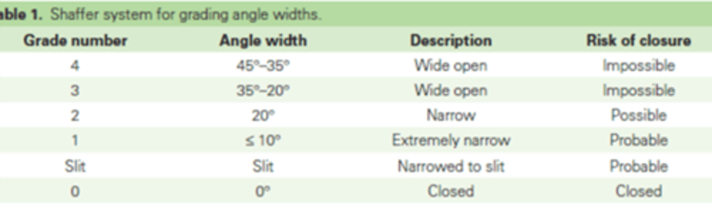
What is the Shaffer system?
system for grading angle widths
What is ultrasound biomicroscopy used for?
for anterior segment abnormalities especially posterior to the iris (has low resolution)
When is the shadow test performed?
when unable to perform van herick or gonio (it is less accurate)
What is the method of the shadow test?
shine penlight or transilluminator laterally to asses anterior chamber depth
What is a grade 4 for shadow test?
no shadow
What is a grade 3 for shadow test?
2/3 illuminated
What is a grade 2 for shadow test?
1/3 to 2/3 illuminated
What is a grade 1 for shadow test?
<1/3 illuminated
What is the purpose of non contact funduscopy?
to evaluate the posterior vitreous and retina, especially the posterior pole
Describe the set up of noncontact funduscopy.
-white light
-red free enhancement filter
-0° displacement
-parallelpiped or wider of moderate height and width
-low to medium illumination
-10x mag
-16x mag for ONH with 90D