Non Alcoholic Liver Disease
 Call Kai
 Learn
Learn Practice Test
 Spaced Repetition
 Match
Match Flashcards
 Knowt Play
1/55

There's no tags or description
Looks like no tags are added yet.
Name | Mastery | Learn | Test | Matching | Spaced | Call with Kai | Chat |
|---|
No analytics yet
Send a link to your students to track their progress
56 Terms
Autoimmune Liver disease defn
Chronic inflammatory condition of liver associated with unknown etiology, autoantibodies and a genetic predisposition
4 main sub types of Autoimmune Liver Disease
Autoimmune hepatitis (AIH)
Primary biliary cholangitis (cirrhosis) (PBC) (bile ducts)
Primary sclerosing cholangitis (PSC) (bile ducts)
Autoimmune overlap syndromes (AIH-PBC) (AIH-PSC)
Name 6 Features common to all Autoimmune Liver diseases
Associated with the presence of autoantibodies (against self antigens)
Usually increased serum IgG +/- IgM
May be associated with AI diseases at other sites – thyroid, adrenal,
Typical inflammatory cells- lymphocytes, plasma cells +/- lymphoid aggregates
General- immune mediated liver damage
May respond to immune suppression
How many types of Autoimmune Hepatitis (AIH) are there
Type 1, 2a, 2b, 3
Targeted cells in AIH
hepatocyte
AIH is more common in what gender
female
Name the 2 main autoantibodies associated with Type 1
ANA, ASM
Name the 2 main autoantibodies associated with Type 2a
ANA, ASM
Name the 2 main autoantibodies associated with Type 2b
LKM-1
Name the 2 main autoantibodies associated with Type 3
Anti-SLA (soluble liver antigen)
Which immunoglobulin will have changed levels in AIH (up/down)
Increased IgG
Which liver enzyme will have changed levels in AIH (up/down)
Aminotransferases increased
What are the 2 genetic predispositions for AIH
HLA DR3, DR4
Treatment for AIH
Responds to immune suppression (steroids)
slide 8-9
Is Primary Biliary Cholangitis (PBC) autoimmune
Yes
2 risk factors for Primary Biliary Cholangitis (PBC)
F>>M 9:1
40-60yrs
What antibody is associated with PBC
Anti-mitochondrial antibodies (AMA) are present in >95% cases
What tissue is targeted in PBC
small bile ducts in liver
Pathology of PBC
Progressive bile duct damage → fibrosis
Cirrhosis develops quite late
2 symptoms of PBC
jaundice, pruritis (bile salts)
2 biochemical changes in PBC
Raised Alk Phophatase
Raised bilirubin
What immunoglobulin is affected by PBC & how
Raised Ig especialy IgM
3 histological features of PBC
Portal tract inflammation- lymphos and plasma cells +/- granulomas.
Hallmark = destruction of small (40-80um) bile ducts, inflammation, swelling in bile ducts may be focal hence need good Bx
Late stage → fibrosis-micronodular cirrhosis
slide 14
What is Primary Sclerosing Cholangitis (PSC)
Chronic disease of liver associated with fibrosis and inflammation in bile duct wall- intermittent areas of fibrosis and narrowing along bile duct
What is the eventual result of the progression of PSC
Eventual replacement of bile ducts with cord of fibrous tissue
Does PSC affect intra/extra hepatic bile ducts
Both
2 Risk factors for PSC
Associated with IBD especially UC
M>F (2-3:1)
How does PSC present
Cholangitis- recurrent inflammation due to obstruction
Cholestasis due to obstruction- jaundice
Fatigue, upper abdominal pain,intermittent/progressive jaundice
Usually <50 years at presentation, can present in adolescence
What is found in serology for PSC
pANCA + in >80% of cases (not specific)
What is found in radiology for PSC
ERCP imaging of biliary tree shows a ‘beaded’ biliary- narrowing along bile duct

What is found in histology for PSC
Inflammation in portal tract, damage to bile duct, progressive loss of bile ducts, peri-ductal ‘onion-skin’ fibrosis pattern (late stage)
(Early histological changes very non-specific)
Complications of PSC
Develop progressive fibrosis of liver with decreased function- liver cirrhosis, failure
10-20% develop cholangiocarcinoma after many years
Treatment for PSC
No effective treatment for PSC
Cirrhosis/failure stage-Transplantation
There is still a risk of recurrence of PSC with transplant
What is Alpha 1 anti-trypsin deficiency
Hereditary metabolic disease of liver
What is abnormal in Alpha 1 anti-trypsin deficiency
Abnormality on chromosome 14-protease inhibitor Pi
slide 21-25
What is Haemochromatosis
Deposition of iron in liver, pancreas, testes
What causes Haemochromatosis
Genetics
Inheritance pattern of Haemochromatosis
Autosomal recessive
Which chromosome, gene & homo/heterozygotes are associated with Haemochromatosis
Chromosome 6
Commonest C282Y gene
Homozygotes
Which gender is more at risk for Haemochromatosis
Male
Age of presentation for Haemochromatosis
Presents 40-60 yrs in men, later in women (periods → later presentation)
Where is iron initially deposited in Haemochromatosis
Peri-portal hepatocytes → then other hepatocytes
What does severe (grade 3–4) iron deposition look like
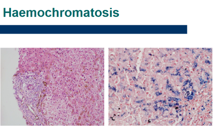
Coarse iron granules within hepatocytes
What stain is used to demonstrate iron?
Perls stain, which stains iron blue.
How does iron overload cause liver damage
Iron → irritation/injury → fibrosis → nodule formation → micronodular cirrhosis
3 steps to Haemochromatosis Diagnosis
1. High iron saturation in serum
2. Grade 3-4 iron on liver biopsy
3. Genetic abnormality (C282Y or H63D gene)
2 treatments for Haemochromatosis
1. Iron removal- venesection (therapeutic phlebotomy)
2. Transplantation- late stage liver failure
What is done first when Haemochromatosis is diagnosed
Must screen family as identification of cases can allow earlier intervention (phlebotomy) and decrease cirrhosis
Possible complication of Haemochromatosis
Haemochromatosis patients x200 increased risk of hepatocellular carcinoma (hepatocellular carcinoma surveillance in these patients)
Risk is associated with cirrhosis
True/False Hepatitis A virus (HAV) is only seen as acute hepatitis
True
Is HAV a DNA/RNA virus
RNA
How is HAV spread
spread fecal-oral- person to person contact or contaminated water
(decreased incidence with increased hygiene in countries)
slide 32