INTRO PT 1: Planes, Positions, Directional Terms, Transducers, Knobology & Quadrants
 Call Kai
 Learn
Learn Practice Test
 Spaced Repetition
 Match
Match Flashcards
 Knowt Play
1/92

There's no tags or description
Looks like no tags are added yet.
Name | Mastery | Learn | Test | Matching | Spaced | Call with Kai | Chat |
|---|
No analytics yet
Send a link to your students to track their progress
93 Terms
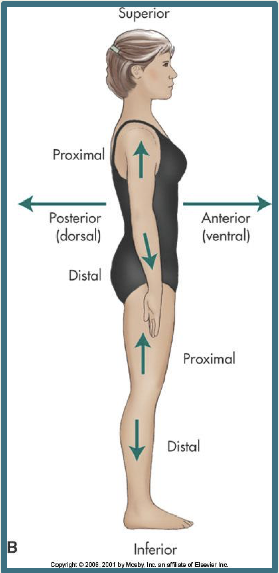
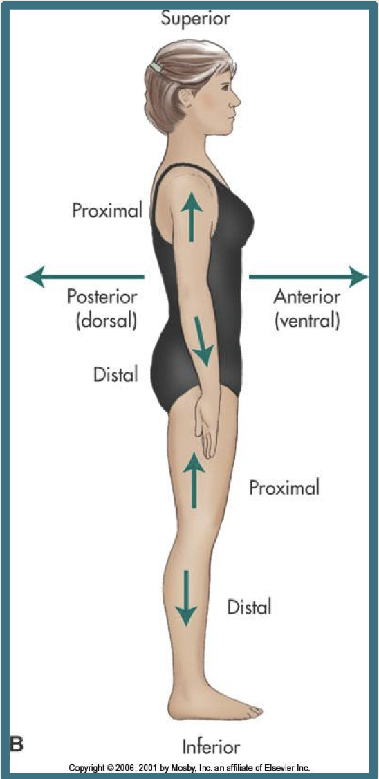
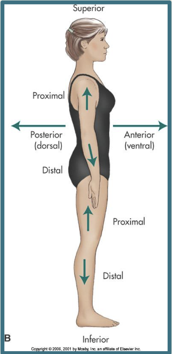
Anterior/ventral
towards the front surface of the body or body part
ex: The aorta is anterior to the vertebral column
Contralateral
on the opposite side of the body

deep
away from the surface of the body or structure
ex: your aorta and IVC are deep structures
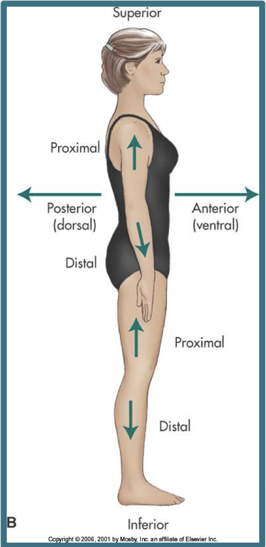
distal
farther away from the attachment point or origin of an extremity to the trunk of the body
ex: the urethra is distal to the urinary bladder

inferior/caudal
towards the feet or away from the head
ex: The uterus is inferior to the gallbladder.

ipsilateral
on the same side of the body
lateral
away from the midline of the body or pertaining to the side; situated at or on the side
ex: the right kidney is lateral to the left lobe of the liver
medial
towards the midline of the body or organ
ex: the left lobe of the liver is medial to the right lobe of the liver

posterior/dorsal
towards the back surface of the body
ex: The right kidney is posterior to the gallbladder

proximal
towards the origin or attachment of a structure to the trunk
ex: the ureter is proximal to the urinary bladder

superficial
closer to the surface of the body or structure
ex: your skin is superficial to your muscles

superior/cephalic
towards the head or higher in the body
ex: The Liver is superior to the urinary bladder.

Midsagittal Plane
divides the body between equal left and right halves
Anatomic Position
Anatomic position assumes that the body is standing erect, the eyes are looking forward, and the arms are at the sides with the palms and toes directed forward.

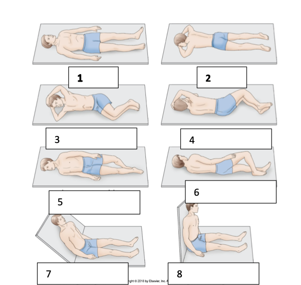
Label these
supine
prone
right lateral decubitus
left lateral decubitus
right posterior oblique
left posterior oblique
sitting semi-erect
sitting erect
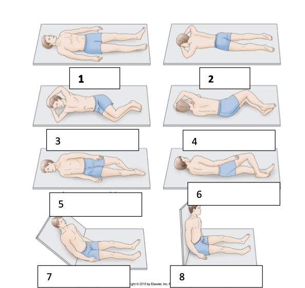
Supine
Lying on the back (1)

Prone
Lying face down (2)

Right lateral decubitus (RLD)
lying the right side (3)

left lateral decubitus (LLD)
lying on the left side (4)

Right posterior oblique (RPO)
Lying semi-supine, rolled up on the right side slightly, and elevating the left side (5)
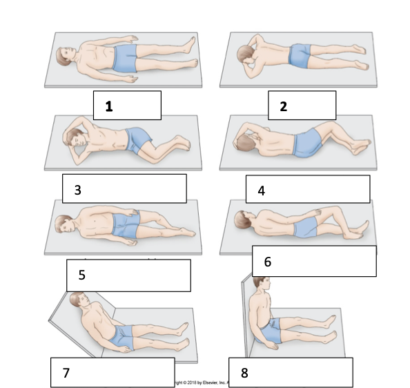
Left posterior oblique (LPO)
Lying semi-supine, rolled up on the left side slightly, and elevating the right side (6)
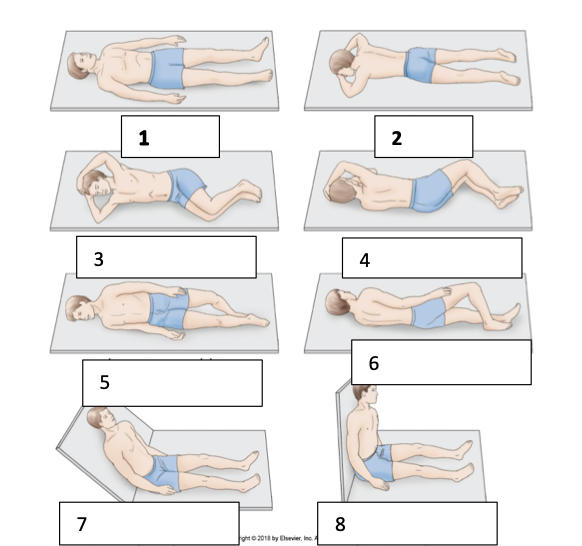
Left anterior oblique (LAO)
Lying semi-prone, rolled up on the left side slightly, and elevating the right side (7)

right anterior oblique (RAO)
Lying semi-prone, rolled up on the right side slightly, and elevating the left side (8)

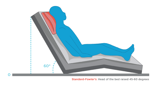
Fowler Position
Various inclined positions in which the head of the bed is raised (varies from low to high); the head of the bed is typically raised 18-20 in above the heart with the knees elevated
Trendelenburg
Person in the supine position with the feet raised higher than the level of the head at an angle of 15-30 degrees

The ____ side of the patients body is aligned with the observer’s ____
right, left
if the patient is supine, the orientation is as if the observer were looking at the patient from the ____
feet

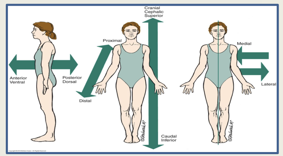
Label This
Anterior / Ventral
Posterior / Dorsal
Distal
Proximal
cranial / cephalic / superior
caudal / inferior
medial
lateral
Transverse Plane
divides superior and inferior
screen displays right to left
Abbreviations: TRV & TRANS

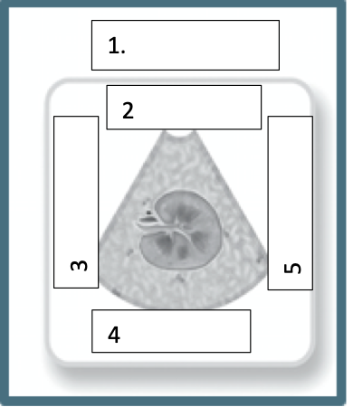
Label this
sonogram
anterior
medial
posterior
lateral

Sagittal / Longitudinal Plane
divides into right and left
screen displays superior to inferior
abbreviations: SAG, LONG
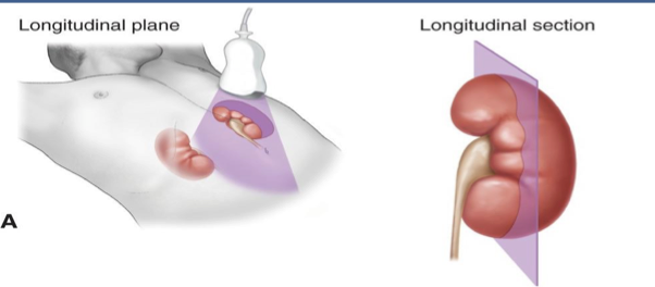

Label this:
sonogram
anterior
cephalic/superior
posterior
caudal/inferior

Coronal Plane
divides into anterior and posterior
abbreviations: COR

Transducers
used to obtain an image of the human body using ultrasound
can convert energy from one form to another
operates using the pulse-echo principle
transducers come in different shapes and sizes for different examination types
Pulse-Echo Principle
transducer uses the pulse-echo principle
the crystal inside of the transducer produces the ultrasound wave once pulsed with electricity
the wave is sent into the body and returns to the transducer
the strength of the echo is displayed on the systems monitor

what is this
linear array transducer

what is this
curvilinear array transducer
Transducer Manipulation
requires the use of acoustic gel
so the ultrasound waves can pass through the body
acts as a lubricant for ease of transducer manipulation
For large organs (i.e liver), drastic movement may be needed
for smaller structures, fine-tuned movement is needed to enhance visualization
transducer may need manipulation to obtain an optimal acoustic or sonographic window
PRACTICE!
Transducer Orientation
All transducers have a reference point to show the monographer the correct transducer orientation
may be called the index or the notch and may be represented by a groove, light, or manufacturer symbol on one part of the transducer
essential to obtaining correct anatomic orientation

Name these transducer positions
perpendicular —- transducer is straight up and down
subcostal —- the transducer is angled superiorly just beneath the inferior costal margin
intercostal —- transducer is between the ribs. It can be perpendicular, subcostal, or angled
angled —- transducer is angled superiorly, inferiorly, or right and left laterally at varying degrees.
rotated —- transducer is rotated varying degrees to oblique the scanning plane

Sweep Scanning motion
requires the transducer to remain in one area while using a large wrist motion with the probe perpendicular to the skin surface to sweep through the abdomen

Slide Scanning Motion
Used when the transducer is physically moved along the abdomen, such as longitudinal movement to follow the course of the abdominal aorta into the bifurcation of the iliac arteries

Rock/Pivot Scanning motion
Once an area of interest is located, the monographer may pause over the structure and slowly “rock” or pivot the transducer back and forth or up and down to image the area completely or to follow the anatomical structure
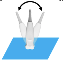
Fan Scanning Motion
smaller version of sweep motion, which is used when the transducer is minutely swept, pivoting on a point of interest.
may be useful in superficial structures such as the breast when differentiation is needed between the duct and the invasive lesion within the duct

rotate scanning motion
motion is useful to navigate between the ribs or to change from transverse to longitudinal planes, where the transducer is held in one area and rotated 90 degrees to the opposite plane
also used for smaller increments as the transducer is slowly turned around the area of interest

Care and Cleaning of transducers
dont drop transducers or roll machine over the cords
report any cracks in transducer to supervisor
never be autoclaved
should be cleaned with manufacturer-recommended disinfectant before and after each examination
transducers used for intracavity purposes may be disinfected with a glutaraldhyde-based solution
Brightness Mode (B-Mode imaging)
displays the returning echoes as dots of varying brightness related to the strength of the echo
Realtime Imaging
ability of the machine to gather information and display it on the screen almost instantaneously
What does echocardiography use?
A-Mode (amplitude mode)
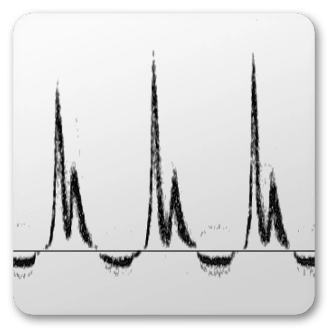
Doppler Technology
the effect of differing frequencies with motion
continuous-wave doppler
pulsed-wave doppler
color doppler imaging
power doppler
Continuous-Wave Doppler
a doppler device that utilizes two elements— one element that continuously sends ultrasound waves into the body and another element to continuously listen for the returning signal

Pulsed-Wave Doppler
intermittently sends sounds and pulses to listen to signals
sonographers can select depth in which the vessel will be scanned with doppler
Color Doppler Imaging
Doppler shift information presented as a color or hue superimposed over a greyscale image

Power Doppler
Amplitude mode of Doppler where it is not the shift itself that provides the signal but rather the strength (amplitude) of the shift

Knobology
the study of knobs or the functionality of controls
Acoustic Power
control that relates to the amount of energy the transducer is emitting into the patient
Annotation Keys
letters of the keyboard that allow for the labeling of images, similar to the display on a computer keyboard
B-Color
colorizes the greyscale of images
Body Markers
tool used for labelling different anatomic structures or for identifying where the transducer is placed on a body part
Calipers
measures structures during a sonographic examination; can measure the depth of structures as well
Cine (Cine Loop)
a collection of image frames stored temporarily in the system’s memory before the image is frozen
Depth
varies the deepness of the echoes that are displayed on the monitor
Dual image
allows the display to be split into two separate images
Ellipse
allows for the measurement of round structures
Field of View (extended field of view)
ability for sonographers to make maximal use of the screen’s potential resolution while simultaneously displaying all of the relevant areas
Freeze
allows all data to be started or stopped
HD Zoom
allows for magnification of the image with an adjustment made for resolution, offering true magnification
Image Store/print
allows for images to be stored within the hard drive of the machine
Multihertz
allows the user to choose between different sending frequencies of the transducer.
In general, increasing the frequency improves resolution while using less penetration
Overall Gain
adjusts the brightness or the degree of echo amplification of the entire image
Time Gain Compensation
controlled by adjusting the slide pots which alter the brightness of an image at a specific depth
Tissue Equalization
allows for system-controlled image optimization for B-Mode and spectral Doppler
Trace
allows for a structure to be traced and the distance measured
Trackball
allows for the control of the cursor for text, cine looping, etc.
Transductor Select
permits the activation of different transducers
M-Mode
motion mode
demonstrates the motion of structures along a single scan line
used in obstetrics to demonstrate the movement of the fetal heart
Pulsed-Wave Doppler
activates and often controls the gain of the PW Doppler.
It can adjust the sample gate or sample volume (area where info is being collected with a PW doppler), baseline, scale, and angle of insonation
Transmit Zone / Focus
enhances the resolution of an area in the image by electronic focusing.
adjusted by moving the indicator (caret)
Primary Imaging Controls
Depth
Gain
Time Gain Compensation
Annotation
Focal Zone
Section Width
Zoom
Color
Doppler
Measurements (calipers)
Attenuation
the weakening of sound as it travels through a medium
Basic Operation
enter patient name and ID
select appropriate transducer
place transducer in patient with a generous amount of coupling gel
adjust imaging depth to cover the area of interest
adjust TGC and overall gain to achieve the desired image
Picture Archiving and Communication System
images viewed and stored on a computer and/or network server
digital format
allows for improved image resolution
software controlled
Hospital Information System (HIS)
allows the sonographer communication with the patient’s medical records
Radiology Information System (RIS)
provides images and reports from other imaging examinations
Subcostal Plane
the upper horizontal line joins the lowest point of the costal margin on each side of the body

Intertubercular Plane
the lowest horizontal line joins the tubercles on the iliac crests

Transpyloric Plane
the horizontal plane passes through the pylorus, duodenal junction, neck of the pancreas, and hilum of the kidneys

Abdominal Quadrents
Right Upper Quadrant (RUQ)
Left Upper Quadrant (LUQ)
Right Lower Quadrant (RLQ)
Left Lower Quadrant (LLQ)
the quadrant of determined by the midsagittal and transverse planes that pass through the umbilicus

What is in the RUQ?
right lobe of liver
gallbladder
pylorus
first 3 parts of duodenum
head of pancreas
right adrenal gland
right kidney
right hepatic flexure
superior part of ascending colon
right half of transverse colon
What is in RLQ?
cecum
appendix
most of ileum
inferior part of ascending colon
right ovary
right uterine tube
ureter
right spermatic cord
What is in LUQ?
left lobe of liver
spleen
most of stomach
jejunum and proximal ileum
body and tail of pancreas
left adrenal gland
left kidney
left scenic flexure
superior part of descending colon
left half of transverse colon
what is in LLQ?
sigmoid colon
inferior part of descending colon
left ovary
left uterine tube
left ureter
left spermatic cord
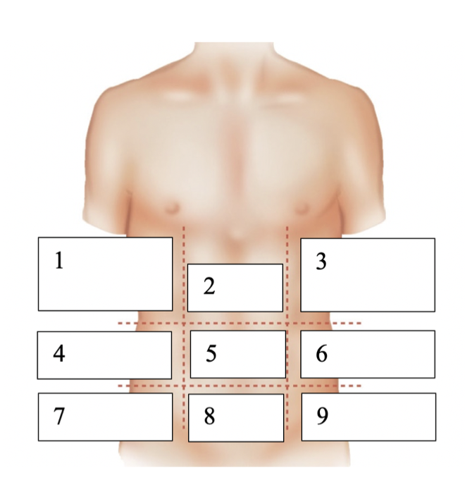
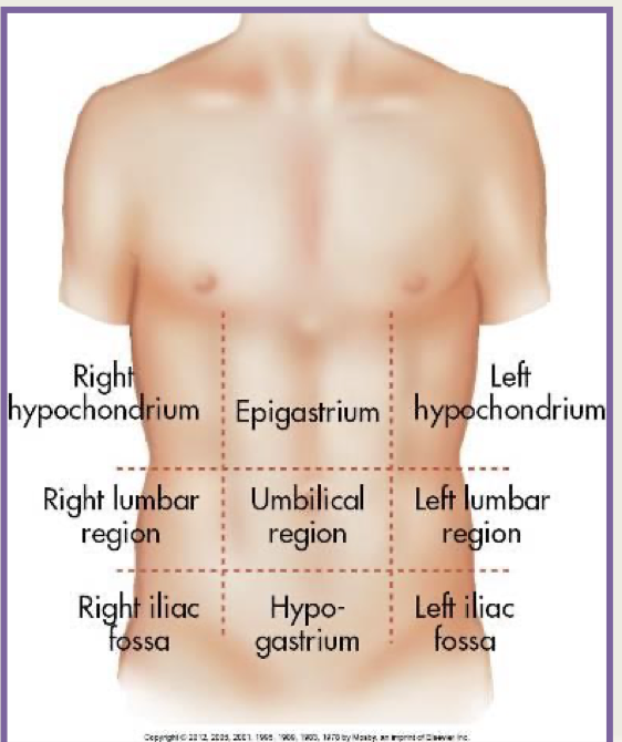
Name these regions
right hypochondrium
epigastrium
left hypochondrium
right lumbar region
umbilical region
left lumber region
right iliac fossa
hypogastrium
left iliac fossa